ABSTRACT
Background: CD8+T suppressor cells may play a role
in immunoregulation. Recent studies have characterized this population by the lack of the CD28 molecule. These CD8+CD28–T cells differ phenotypically and
functio-nally from CD8+ CD28 + T cells. Little is known about CD8+ CD28–cells in atopy. Our aim was to analyze the
phenotype and functional properties of CD8+ CD28–
T cells in atopic and non-atopic individuals.
Methods: Peripheral blood mononuclear cells (PBMC) were obtained after density gradient centrifu-gation. CD8+ CD28–and CD8+ CD28 + T cells were
isolated using immunomagnetic beads. Relative per-centages of these cells and expression of several phenotypic markers were analyzed by flow cytome-try. Proliferation was assessed by thymidine incorpo-ration in isolated populations and in co-cultures with PBMC usingDermatophagoides pteronyssinus as stimulus. Cytokine synthesis was evaluated in culture supernatants by cytometric bead array.
Results: The relative percentages of CD8+CD28–T
cells and their phenotypic expression in atopic and non-atopic volunteers were not significantly different. However, CD8+ CD28–T cells showed greater
prolif-eration than did CD8+CD28+T cells when stimulated
withD. pteronyssinus, although cytokine synthesis patterns were similar. CD8+CD28–co-cultures with
PBMC showed greater proliferation than CD8+CD28+ T cell co-cultures, but cytokine synthesis patterns were not different.
Conclusions: Our data confirm phenotypic and functional differences between CD28+and CD28–T
cells, irrespective of atopic status. Purified human CD8+CD28–T cells, freshly isolated from peripheral
blood, do not have suppressor properties on aller-gen-specific proliferation or on cytokine synthesis in PBMC.
Key words: Allergy; Atopy; CD8+CD28–T cells;
Hu-man.
INTRODUCTION
T cells that use non-cytolytic mechanisms to down-regulate the immune response (suppressor T cells or Ts) are thought to play an essential role in control-ling reactivity to foreign antigens and inducing toler-ance to self antigens1. Nevertheless, more than
three decades since their discovery, an understand-ing of the mechanisms whereby suppressor cells ex-ert their activity is still incomplete.
O R I G I N A L A R T I C L E S
Functional and phenotypic characterization
of CD8
+
CD28
+
and CD28
–
T cells in atopic individuals
sensitized to Dermatophagoides pteronyssinus
O. Lourençoa, A.M. Fonsecaa, A. Paivab, F.A. Arosacand L. Taborda-Barataa,d
aCICS. Centro de Investigação em Ciências da Saúde. University of Beira Interior. Covilhã. Portugal. b
Histo-compatibility Centre. Coimbra. Portugal. cInstitute for Molecular and Cell Biology (IBMC). Porto. Portugal. dCova
da Beira Hospital. Covilhã. Portugal.
Correspondence: L. Taborda-Barata
Department of Medical Sciences Faculty of Health Sciences University of Beira Interior Avenida Marques d’Avila e Bolama 6200-001 Covilhã. Portugal Tel:+ 351 275319881 Fax:+ 351 275319883 E-mail: [email protected]
Phenotypically, CD8+Ts are characterized by the lack of the CD28 co-stimulatory molecule2. CD8+CD28–
T cells are thought to arise from CD8+CD28+T cells that have proliferated several times since they have shorter telomeres3and share oligoclonal expansions4.
In vitro, CD8+CD28–T cells arise from CD8+CD28+
T cells repeatedly stimulated in the presence of inter-leukin (IL)-25. In contrast, IL-4 can block this
differenti-ation6.
Recent evidence has been accumulating to show that CD8+CD28–T cells can inhibit T helper cell
activa-tion and proliferaactiva-tion in mitogen and antigen-driven responses7-9.In vitro, antigen-specific CD8+CD28–T
suppressor cells have been generated by multiple rounds of stimulation of human CD8+CD28+T cells with APC either from an allogeneic or a xenogeneic donor8,10. The generated cells expressed Foxp311, a
gene related with regulatory function, and could in-hibit proliferation of CD4+T cells interacting directly
with the APC used for priming12,13.
Non-antigen specific CD8+CD28–T cells with
sup-pressor activity have also been generated from CD8+CD28– T cells in the presence of IL-2 and
IL-1014. These suppressor cells inhibited both
anti-gen-specific CD4+ T cell proliferation and cellular cytotoxicity by secreting cytokines such as IFN-␥, IL-6 and IL-1015,16. Defects in this antigen-nonspecific
suppression have been described in multiple sclero-sis 15 and in systemic lupus erythematosus pa-tients17, and are primarily seen in chronic progressive
situations.
Atopic allergic diseases are immune disorders caused by anomalous, Th2 cell-dominated responses to otherwise innocuous substances, such as proteins from house dust mite or grass or tree pollen18. Many
authors believe that atopy and allergic diseases are the result of inadequate or impaired inhibition of al-lergen-specific T helper-type responses by the regu-latory T cells19, or even a consequence of decreased
frequencies of those cells in atopic individuals20.
There is a growing body of evidence which suggests that CD8+T cells play an important part in regulating the IgE response to non-replicating antigens21,22.
Nonetheless, the presence of CD8+suppressor T cells in human atopic patients is not yet confirmed.
MATERIALS AND METHODS Subjects
Peripheral blood was obtained from 30 non atopic and 32 atopic (with allergic rhinitis) adult volunteers (Caucasian, non-smokers), matched for age and gen-der. Atopic volunteers were recruited from the
aller-gy clinic of the Cova da Beira Hospital and non atopic volunteers were recruited among the staff of the Hospital and the University.
Atopy was assessed by positive skin prick tests and specific IgE levels toDermatophagoides ptero-nyssinus (Der p). Volunteers who received immu-notherapy or were on systemic medication were ex-cluded. Pregnant or breastfeeding women and all volunteers with disease affecting the immune sys-tem were also excluded. Informed written consent was signed by all the volunteers. The study was ap-proved by the Ethics Committee of Centro Hospitalar Cova da Beira.
Monoclonal antibodies
The following monoclonal antibodies (mAbs) were used: anti-CD3 mAb conjugated with APC was pur-chased from Pharmingen (San Diego, CA, USA), anti-CD8 mAb conjugated with PerCp was purchased from Becton Dickinson (San José, CA, USA), anti-CD28 mAb conjugated with FITC was purchased from Pharmingen, anti-TCR␣, -TCR␥␦, -CD25, -CTLA-4, -HLA-DR, -CD56, -CD94, -CD158a, -CD161, -NKB1 mAb conjugated with PE were all purchased from Pharmingen, anti-V␣24 mAb conjugated with PE was purchased from Serotec (Oxford, UK). Iso-type-matched, PE-conjugated control mAbs IgG1, IgG2a, and IgM were purchased from Pharmingen.
Blood preparation and antibody staining
Freshly collected peripheral blood mononuclear cells (PBMC) were stained either from whole blood after lysis of the erythrocytes (10 mM Tris, 0.15 M NH4Cl, pH= 7.4). Staining was performed at 4 °C for
30 min in staining solution (PBS, 0.2 % bovine serum albumin (BSA), 0.1 % NaN3) in round-bottomed
mi-crotiter plates (Greiner, Nürtingen, Germany) with ≈ 0.5× 10⁶ cells/well. After staining, the cells were washed, resuspended in 500l PBS and acquired in a FACSCalibur Flow cytometer (Becton Dickinson).
Flow cytometry analysis
Data were collected on 20,000 cells/sample using FACSCalibur flow cytometer equipped with an argon ion laser and a red diode laser, for quantification of CD28+and CD28–T cells. Anti-CD8, -CD3, and
-CD28 monoclonal mAb were used to define the CD3+CD8+CD28+and CD28–T cell populations. T
scat-tering properties and on CD3 staining. Positively and negatively stained populations were calculated by quadrant dot plot analysis determined by isotype controls.
For phenotypic analysis of the defined subpopula-tions, 30,000 events were collected per sample. Flu-orescence dot plots and histograms were analysed using cytological software (Cell Quest Pro, Becton Dickinson). Phenotypic analysis of the TCR␣ and TCR␥␦ was also performed on the isolated fractions of CD3+CD8+CD28+and CD28–T cells.
Cell isolation
Peripheral blood mononuclear cells (PBMC) from the volunteers were separated from peripheral blood (120 ml) by centrifugation over Lymphoprep. Lym-phocytes were further purified by allowing the cells to adhere to plastic for 1 h at 37 °C, 5 % CO2. CD8+T
cells were isolated from the supernatant by negative selection using magnetic beads (Miltenyi Biotec, Ber-gisch Gladbach, Germany). To separate CD28+and CD28–T cells from CD8+cell suspensions, cells were
incubated with CD28-FITC mAb (Pharmingen) and then with anti-FITC microbeads (Miltenyi Biotec). Pu-rity of the fractions was greater than 85 % as evalu-ated by flow cytometry analysis. The adherent frac-tion was collected as well and used as APC in cell cultures after mitomycin treatment.
Cell Cultures
Responder cells were cultured at a concentration of 0.5× 10⁶ cells/ml for 6 days in RMPI 1640 com-plete medium [RPMI 1640, 2 mM L-glutamine, 1 % antibiotic/antimicotic (Sigma-Aldrich, St Louis, MO, USA), and 10 % foetal calf serum (Biochrom, Berlin, Germany)] in 96-well flat-bottomed plates (Nunc, Roskilde, Denmark) with or without APC and 10 g/ml purified Der p extract (a kind gift from Dr. Joost Van Neerven, The Netherlands). For posi-tive control, cells were incubated with 0.5 g/ml OKT3 (eBiosciences, San Diego USA) or with 5g PHA (Sigma-Aldrich) for 3 days in the same con-ditions as forDer p. Fourteen hours before the end of the culture [3H] thymidine was added to the wells
(1Ci/well). [3H] thymidine (Amersham
Bioscien-ces, Uppsala, Sweden) incorporation was determin-ed by scintillation counts in a TopCount (Perkin Elmer, MA, USA) and results were expressed as counts per minute (cpm). Mean cpm of the triplicate cultures and standard error of the mean were cal-culated.
Culture conditions were as follows: CD8+CD28+ cells alone, CD8+CD28–cells alone, CD8+CD28+cells
+ APC + Der p, and CD8+CD28–cells+ APC + Der p.
Cell co-cultures
PBMC (0.5× 10⁶ cells/ml) were incubated in dif-ferent proportions (1:1, 2:1, 4:1) with freshly isolat-ed CD8+CD28+and CD8+CD28–T cells for 6 days in
96-well flat-bottomed plates with and without 10g/ml Der p extract. Tritiated thymidine (1 Ci/ well) was added 14 hours prior the end of the cultu-re. Cells were harvested on fiber filters, and in-corporated thymidine was determined by scintilla-tion counting.
Cytokine production
For soluble cytokine detection in the culture su-pernatants, the human Th1/Th2 cytokine bead array kit was used, according to the manufacturer’s in-structions (Becton Dickinson).
Statistical analysis
Statistical analysis of the results was performed using Minitab 14 statistical Software. Mann Whitney U test was used to assess differences between atopic and non atopic volunteers. Wilcoxon signed rank test was used to assess differences within the same population. A p value of less than 0.05 was considered significant.
RESULTS
First, we evaluated the frequency of CD8+CD28–
T cells in whole blood from 22 atopic and 18 non atopic volunteers (age 22-42) by flow cytometry. We observed that the relative percentages were not significantly different (p= 0.80), with 41 ± 14.3 and 40± 13.4 % of CD8+CD28–T cells in atopic and non
atopic volunteers respectively. CD28–T cells were
larger, morphologically more complex and expressed CD3 with higher mean fluorescence intensity (MFI) than CD28+T cells (1048 vs 978).
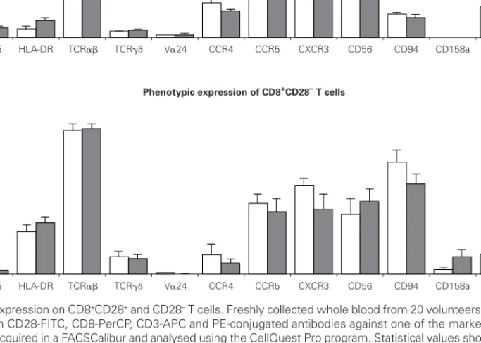
We then evaluated the expression of several phe-notypic markers on CD3+CD8+CD28+ and CD28–
PBMC subpopulations in whole blood from ten atopic and ten non atopic volunteers (fig. 1). Phenotypic ex-pression was similar in both of these groups. Howev-er, taking all the data togethHowev-er, more CD8+CD28+T
cells than CD8+CD28–T cells expressed CD25 (5 %
vs. 1.7 %) and CTLA-4 (5.5 % vs. 4 %). The reverse was true for HLA-DR expression (7.7 % vs. 29.7 %). Next, we examined the expression of TCR␣, TCR␥␦ and TCRV␣24 in CD8+CD28+and CD28–T
cells. As expected, the majority of CD8+T cells ex-pressed a TCR␣ (87 % of CD8+CD28+and 83 % of CD8+CD28–T cells). Interestingly, when the
expres-sion of TCR␥␦ was examined, we found a higher per-centage of CD28–T cells than CD28+T cells
express-ing that receptor (11 % vs. 4 %). TCR␣ and TCR␥␦ expression was also studied in the magnetically iso-lated CD28+and CD28–cells fractions and the results
obtained were not significantly different (data not shown).
Interestingly, the relative frequency of all the phe-notypic markers related to NK cells studied (CD56, CD94, CD158a, and NKB1) was higher on CD28–T
cells than on CD28+cells. CD94 expression was con-siderably higher on CD8+CD28–T cells when
com-pared to CD8+CD28+T cells (63 % vs. 11 %).
Expres-sion of CD161 was low (17 % vs. 14 % for CD28+and
CD28–, respectively) and that of CD158a and NKB1
was very low (under 10 % for both populations).
Ex-pression of chemokine receptors was also different, with the CXCR3 relative percentage being higher on CD28+than on CD28–cells (69.6 % vs. 49.1 %).
How-ever, no difference was observed in terms of CCR5 expression between these two cell subsets. On the other hand, a higher percentage of CD28+ than CD28–T cells expressed the CCR4 chemokine
recep-tor (21.3 % vs. 18.4 %).
In order to assess the proliferative response to al-lergen of CD8+CD28+and CD28–T cells, we set up
cultures of isolated fractions with and without APC andDer p. As shown in figure 2, contrary to what oc-curred with unstimulated cells, CD8+CD28–T cells
proliferated when stimulated withDer p, displaying a significantly higher level (p= 0.001) of proliferation as compared with CD8+CD28+T cells in atopic and non atopic individuals. However, the proliferation level was not significantly different between atopic and non atopic volunteers. We also stimulated some fractions with OKT3 and, in this case, CD8+CD28–T cells
proli-ferated less significantly (p= 0.003) than CD8+CD28+
T cells (data not shown).
Cytokine synthesis was studied using the cyto-metric bead array (CBA) in culture supernatants col-Figure 1.—Phenotypic expression on CD8+CD28+and CD28–T cells. Freshly collected whole blood from 20 volunteers (10 atopic and 10 non
atopic) was stained with CD28-FITC, CD8-PerCP, CD3-APC and PE-conjugated antibodies against one of the markers under study. Thirty thousand events were acquired in a FACSCalibur and analysed using the CellQuest Pro program. Statistical values shown (mean and s.e.m.) were obtained using the Minitab 14 program.
100 60 50 40 30 20 10 90 80 70 0 CTLA-4 CD25 HLA-DR
Phenotypic expression of CD8+CD28+ T cells
TCR␣ TCR␥␦ V␣24 CCR4 CCR5 CXCR3 CD56 CD94 CD158a CD161 NKB1
Atopic Non atopic
100 60 50 40 30 20 10 90 80 70 0 CTLA-4 CD25 HLA-DR
Phenotypic expression of CD8+CD28– T cells
lected on day 3. None of the cultures synthesized IL-5, IL-4 or IL-2, but all theDer p -stimulated cultures synthesised IFN-␥, TNF-␣ and IL-10. However, we did not observe statistically significant differences either between the cultures with CD8+CD28+and CD28–T cells or between atopic and non atopic
indi-viduals (fig. 3).
In order to study the potential suppressor proper-ties of the isolated subpopulations, we performed co-cultures with PBMC at different ratios. None of the isolated subpopulations was able to suppress the allergen-specific PBMC proliferation, since we ob-served an increase in cpm counts in co-cultures (fig. 4).
Cytokine synthesis was also evaluated in the co-cul-ture supernatants on day 3 and on day 5, by CBA. Syn-thesis of IL-5, IL-4 and IL-2 was very low in all the cul-tures. When cytokine synthesis in the co-cultures was compared between days 3 and 5, only IFN-␥ levels in-creased. There were no significant differences in cy-tokine synthesis either between cultures from atopic and non atopic or between co-cultures with CD8+CD28–or CD8+CD28+T cells (fig. 5).
DISCUSSION
This is the first study on suppressor function of CD8+CD28–T cells in allergy. Previous studies on the
regulation of atopic allergy mainly included natural T
regulatory cells (CD4+CD25+) and showed inconclu-sive results23,24. We believe that this study can
en-hance the knowledge and shed some light on the functional properties of the CD8+CD28–T cells and
their implication in the development of atopy. We observed that CD8+CD28–T cells are not
phe-notypically different between atopic and non atopic individuals. This may imply that atopy is not associ-ated with a specific CD8+T cell phenotype. However, it is important to mention that our data on phenotyp-ic differences between CD28+and CD28–
subpopu-lations is in line with other reports in health and dis-ease25,26. Moreover, the increased expression of the
NK cell related receptors in CD8+CD28–T cells, in a
context where co-stimulation is not present, may be important towards limiting T cell cytolytic respons-es, and act as a form of “regulation”.
Classical antigen-presentation studies showed that MHC class I molecules present peptides derived from proteins synthesized within the cell, whereas MHC class II molecules present exogenous proteins captured from the environment. Emerging evidence indicates, however, that dendritic cells have a spe-cialized capacity to process exogenous antigens into the MHC class I pathway27. According to our
expec-tations, unstimulated cells did not proliferate. Inter-estingly enough, CD8+CD28–but not CD8+CD28+T
cells proliferated in response toDer p, in the pres-ence of APCs. This fact suggests that CD28–T cell
proliferation is not impaired in spite of the absence of CD28 co-stimulation28and that CD8+CD28–T cells
re-spond to common aeroallergens. This results should be considered with caution as the two subsets of CD8+T cells studied here are oligoclonal 4 with re-spect to their TCR, and the use of this allergen might stimulate an insignificant minority of clones and lead to inconclusive results. For this reason, we per-formed cell cultures for 3 days with OKT3 to confirm whether multi-clonal cellular responses were differ-ent in both subpopulations. Results show a prolifera-tion impairment in the CD8+CD28–T cells which
cor-roborates previous results29,25. This implies that our
results must be confirmed by performing the same studies with another allergen.
When cytokine production was analysed, no de-tectable IL-5, IL-4 or IL-2 production was induced by the allergen in CD28+or CD28–T cells. On the other
hand, IFN-␥, TNF-␣ and IL-10 were synthesized at similar levels by both isolated populations. Other inves-tigators mention that a subpopulation of suppressor CD8+CD28–T cells synthesises IL-10, thus
mediat-ing the suppressive effect14. Moreover, Seneviratne
et al. linked low levels of IL-10 with severe atopic dis-ease30. Since both our subsets produced IL-10, we
performed the co-culture studies in order to evalu-Figure 2.—CD8+CD28–T cells proliferate more than CD8+CD28+
T cells when stimulated withDer p. Isolated CD8+CD28–and
CD8+CD28+T cells from 8 atopic and 10 non atopic volunteers
were incubated for 6 days in 96-well flat-bottomed plates with and without 10g/ml Der p extract. Tritiated thymidine (1 Ci/well) was added 14 h prior to the end of the culture. Cells were har-vested on fiber filters, and incorporated thymidine was deter-mined by scintillation counting. Results show thymidine incorpo-ration (cpm, mean± s.e.m.). Wilcoxon signed ranked test was used for comparison between conditions.
CD8+CD28- + APC + Derp CD8+CD28 -CD8+CD28+ + APC + Derp CD8+CD28+ 0 500 1,000 * p=0.001 cpm counts 1,500 2,000 2,500
ate the “regulatory” capacity of these cells. The pro-duction of cytokines varied between donors, but fol-lowed similar patterns. The failure to detect the oth-er cytokines (IL-5, IL-4, IL-2) in the supoth-ernatants does not necessarily imply that they are not synthesized but may also suggest that they are immediately used as autocrine factors.
Finally, we performed co-culture studies and obser-ved that freshly isolated CD8+CD28+or CD8+CD28–
T cells did not show suppressive properties, and were not able to inhibit allergen-driven PBMC proliferation or cytokine synthesis. These results are in line with previ-ous reports namely by Suciu-Foca and Filaci groups31
where suppressor CD8+CD28–T cells were only
gener-Figure 3.—Cytokine synthesis is similar for both isolated populations. Isolated CD8+CD28–and CD8+CD28+T cells from 4 atopic and 3 non
atopic volunteers were incubated in 96-well flat-bottomed plates with and without 10g/ml Der p extract. On day 3, culture supernatants were collected and cytokine synthesis (IFN-␥, TNF-␣, IL-10, IL-5, IL-4, IL-2) was evaluated by cytometric bead array, following the manu-facturer’s protocol. Values shown are mean± sem. Mann-Whiney U test was used for comparison between atopic and non atopic and Wilcoxon signed rank test was used for comparison between conditions.
500 400 300 200 100 25 20 15 10 5 0 pg/ml IFN-␥ TNF-␣ IL-10 Day 3 Atopic
IL-5 IL-4 IL-2 CD8+ CD28+ CD8+ CD28– CD8+ CD28+ + APC + Derp CD8+ CD28– + APC + Derp 500 400 300 200 100 25 20 15 10 5 0 pg/ml IFN-␥ TNF-␣ IL-10 Day 3 Non atopic
IL-5 IL-4 IL-2 CD8+ CD28+
CD8+ CD28–
CD8+ CD28+ + APC + Derp CD8+ CD28– + APC + Derp
A B
Figure 4.—CD8+CD28–co-cultures with PBMC proliferate more than CD8+CD28+co-cultures. Isolated CD8+CD28–and CD8+CD28+T cells
from 8 atopic and 6 non atopic volunteers were incubated in different proportions with PBMC for 6 days in 96-well flat-bottomed plates with and without 10g/ml Der p extract. Tritiated thymidine (1 Ci/well) was added 14 h prior to the end of the culture. Cells were harvested on fiber filters, and incorporated thymidine was determined by scintillation counting. Results show thymidine incorporation (cpm, mean± s.e.m.). Wilcoxon signed ranked test was used for comparison between conditions.
PBMC + CD8+28- + Derp (4:1) PBMC + CD8+28+ + Derp (4:1) PBMC + CD8+28- + Derp (2:1) PBMC + CD8+28+ + Derp (2:1) PBMC + CD8+28- + Derp (1:1) PBMC + CD8+28+ + Derp (1:1) CD8+28- + APC + Derp CD8+28- + APC + Derp PBMC- + Derp CD8+28 -CD8+28 -PBMC 0 2,000 4,000 * * * * cpm counts 6,000 8,000 10,000 12,000 14,000 16,000 18,000
ated after multiple rounds of stimulation of PBMCs with allogeneic13, xenogeneic8or antigen-pulsed32
autolo-gous APC.
However the presence of CD8+CD28–suppressor
T cellsin vivo was observed in transplanted patients without rejection33. These facts may imply a need for
an elevated and sustained contact with the antigen (hence the multiple rounds of stimulation) in order to generate antigen-specific suppressor cells. It would be interesting to further study suppressive properties of CD8+CD28–T cells, by using allergen-specific T cell
lines developed from atopic and non atopic volun-teers.
In summary, in the present study we have shown that atopy is not associated with altered relative per-centages or specific phenotypes in CD8+CD28+or CD28–human T cells. Freshly immunomagnetically
isolated CD8+CD28+or CD28–human T cells have
dis-tinct phenotypes and, although sharing similar cy-tokine production patterns, they proliferate at different levels to common stimuli. Both subpopulations show
similar proliferation capacity in atopic and non atopic individuals and do not have any suppressor capacity.
ACNOWLEDGEMENTS
The authors would like to thank all the volunteers without whom this study would have been impossi-ble, and Dr. Joost Van Neerven for kindly providing theDer p extract.
O.L. is the recipient of a fellowship from the Portu-guese Foundation for Science and Technology (FCT) (BD16448/2004).
REFERENCES
1. Jiang H, Chess L. An integrated view of suppressor T cell sub-sets in immunoregulation. J Clin Invest. 2004;114:1198-208. 2. Suciu-Foca N, Manavalan JS, Cortesini R. Generation and function of antigen-specific suppressor and regulatory T cells. Transpl Immunol. 2003;11:235-44.
Figure 5.—Cytokine synthesis is similar in all the co-cultures stimulated withDer p. Isolated CD8+CD28–and CD8+CD28+T cells from 4 atopic
and 3 non atopic volunteers were incubated in a proportion of 1:1 with PBMC in 96-well flat-bottomed plates with and without 10g/ml Der p extract. On day 3 and day5, culture supernatants were collected and cytokine synthesis (IFN-␥, TNF-␣, IL-10, IL-5, IL-4, IL-2) was evaluated by cytometric bead array, following the manufacturer’s protocol. Values shown are mean± sem. Mann-Whiney U test was used for com-parison between atopic and non atopic volunteers and Wilcoxon signed rank test was used for comcom-parison between conditions.
1,500 1,250 1,000 750 500 250 5 0 pg/ml IFN-␥ TNF-␣ IL-10 Day 3 Atopic
IL-5 IL-4 IL-2 PBMC PBMC + Derp PBMC + CD8+ CD28+ + Derp PBMC + CD8+ CD28– + Derp 1,500 1,250 1,000 750 500 250 5 0 pg/ml IFN-␥ TNF-␣ IL-10 Day 3 Non atopic
IL-5 IL-4 IL-2 PBMC PBMC + Derp PBMC + CD8+ CD28+ + Derp PBMC + CD8+ CD28– + Derp 2,500 2,250 2,000 1,250 1,000 750 500 250 5 0 pg/ml 10 15 1,500 1,750 IFN-␥ TNF-␣ IL-10 Day 5 Non atopic
IL-5 IL-4 IL-2 PBMC PBMC + Derp PBMC + CD8+ CD28+ + Derp PBMC + CD8+ CD28– + Derp 2,500 2,250 2,000 1,250 1,000 750 500 250 5 0 pg/ml 10 15 1,500 1,750 IFN-␥ TNF-␣ IL-10 Day 5 Atopic
IL-5 IL-4 IL-2 PBMC PBMC + Derp PBMC + CD8+ CD28+ + Derp PBMC + CD8+ CD28– + Derp A C D B
3. Batliwalla FM, Rufer N, Lansdorp PM, Gregersen PK. Oligo-clonal expansions in the CD8(+)CD28(-) T cells largely explain
the shorter telomeres detected in this subset: analysis by flow FISH. Hum Immunol. 2000;61:951-8.
4. Mugnaini EN, Spurkland A, Egeland T, Sannes M, Brinchmann JE. Demonstration of identical expanded clones within both CD8+CD28+and CD8+CD28–T cell subsets in HIV type
1-in-fected individuals. Eur J Immunol. 1998;28:1738-42. 5. Borthwick NJ, Lowdell M, Salmon M, Akbar AN. Loss of
CD28 expression on CD8(+)T cells is induced by IL-2 receptor gamma chain signalling cytokines and type I IFN, and increas-es susceptibility to activation-induced apoptosis. Int Immunol. 2000;12:1005-13.
6. Labalette M, Leteurtre E, Thumerelle C, Grutzmacher C, Tourvieille B, Dessaint JP. Peripheral human CD8(+)CD28(+)T lymphocytes give rise to CD28(–)progeny, but IL-4 prevents
loss of CD28 expression. Int Immunol. 1999;11:1327-36. 7. Arosa FA. CD8+CD28–T cells: certainties and uncertainties of
a prevalent human T-cell subset. Immunol Cell Biol. 2002;80: 1-13.
8. Ciubotariu R, Colovai AI, Pennesi G, Liu Z, Smith D, Berlocco P, et al. Specific suppression of human CD4+Th cell
respons-es to pig MHC antigens by CD8+CD28–regulatory T cells. J
Im-munol. 1998;161:5193-202.
9. Ciubotariu R, Vasilescu R, Ho E, Cinti P, Cancedda C, Poli L, et al. Detection of T suppressor cells in patients with organ allo-grafts. Hum Immunol. 2001;62:15-20.
10. Ciubotariu R, Li J, Colovai AI, Platt JL, Cortesini R, Suciu Foca Cortesini N. Human xenospecific T suppressor cells inhibit T helper cell proliferation to porcine aortic endothelial cells, and NF-kappaB activity in porcine APC. Hum Immunol. 2001;62: 470-8.
11. Scotto L, Naiyer AJ, Galluzzo S, Rossi P, Manavalan JS, Kim-Schulze S, et al. Overlap between molecular markers ex-pressed by naturally occurring CD4+CD25+regulatory T cells
and antigen specific CD4+CD25+and CD8+CD28–T suppressor
cells. Hum Immunol. 2004;65:1297-306.
12. Liu Z, Tugulea S, Cortesini R, Lederman S, Suciu-Foca N. Inhi-bition of CD40 signaling pathway in antigen presenting cells by T suppressor cells. Hum Immunol. 1999;60:568-74. 13. Liu Z, Tugulea S, Cortesini R, Suciu-Foca N. Specific
suppres-sion of T helper alloreactivity by allo-MHC class I-restricted CD8+CD28–T cells. Int Immunol. 1998;10:775-83.
14. Filaci G, Fravega M, Negrini S, Procopio F, Fenoglio D, Rizzi M, et al. Nonantigen specific CD8+T suppressor lymphocytes
originate from CD8+CD28–T cells and inhibit both T-cell
prolif-eration and CTL function. Hum Immunol. 2004;65:142-56. 15. Balashov KE, Khoury SJ, Hafler DA, Weiner HL. Inhibition of T
cell responses by activated human CD8+T cells is mediated
by interferon-gamma and is defective in chronic progressive multiple sclerosis. J Clin Invest. 1995;95:2711-9.
16. Filaci G, Fravega M, Fenoglio D, Rizzi M, Negrini S, Viggiani R, et al. Non-antigen specific CD8+T suppressor lymphocytes.
Clin Exp Med. 2004;4:86-92.
17. Filaci G, Bacilieri S, Fravega M, Monetti M, Contini P, Ghio M, et al. Impairment of CD8+T suppressor cell function in
pa-tients with active systemic lupus erythematosus. J Immunol. 2001;166:6452-7.
18. Kay AB. Allergy and allergic diseases. Second of two parts. N Engl J Med. 2001;344:109-13.
19. Ling EM, Smith T, Nguyen XD, Pridgeon C, Dallman M, Arbery J, et al. Relation of CD4+CD25+regulatory T-cell suppression of allergen-driven T-cell activation to atopic status and expression of allergic disease. Lancet. 2004;363:608-15.
20. Akdis M, Verhagen J, Taylor A, Karamloo F, Karagiannidis C, Crameri R, et al. Immune responses in healthy and allergic in-dividuals are characterized by a fine balance between aller-gen-specific T regulatory 1 and T helper 2 cells. J Exp Med. 2004;199:1567-75.
21. Holmes BJ, MacAry PA, Noble A, Kemeny DM. Antigen-spe-cific CD8+T cells inhibit IgE responses and interleukin-4 pro-duction by CD4+T cells. Eur J Immunol. 1997;27:2657-65.
22. Yang M, Cao Y, Mohapatra SS. CD8+T cells inhibit
immuno-globulin E synthesis in low responder SJL/J mice. Immunolo-gy. 1998;93:230-7.
23. Romagnani S. Regulatory T cells: which role in the pathogenesis and treatment of allergic disorders? Allergy. 2006;61:3-14. 24. Umetsu DT, Akbari O, Dekruyff RH. Regulatory T cells control
the development of allergic disease and asthma. J Allergy Clin Immunol. 2003;112:480-7; quiz 8.
25. Azuma M, Phillips JH, Lanier LL. CD28–T lymphocytes.
Antige-nic and functional properties. J Immunol. 1993;150:1147-59. 26. Speiser DE, Valmori D, Rimoldi D, Pittet MJ, Lienard D, Cerun-dolo V, et al. CD28-negative cytolytic effector T cells frequent-ly express NK receptors and are present at variable propor-tions in circulating lymphocytes from healthy donors and melanoma patients. Eur J Immunol. 1999;29:1990-9. 27. Carbone FR, Bevan MJ. Class I-restricted processing and
pre-sentation of exogenous cell-associated antigenin vivo. J Exp Med. 1990;171:377-87.
28. Wang B, Maile R, Greenwood R, Collins EJ, Frelinger JA. Naive CD8+T cells do not require costimulation for
prolifera-tion and differentiaprolifera-tion into cytotoxic effector cells. J Immu-nol. 2000;164:1216-22.
29. Hombach A, Sent D, Schneider C, Heuser C, Koch D, Pohl C, et al. T-cell activation by recombinant receptors: CD28 co-stimulation is required for interleukin 2 secretion and recep-tor-mediated T-cell proliferation but does not affect receptor-mediated target cell lysis. Cancer Res. 2001;61: 1976-82. 30. Seneviratne SL, Jones L, King AS, Black A, Powell S,
McMi-chael AJ, et al. Allergen-specific CD8(+)T cells and atopic
dis-ease. J Clin Invest. 2002;110:1283-91.
31. Filaci G, Suciu-Foca N. CD8+T suppressor cells are back to the
game: are they players in autoimmunity? Autoimmun Rev. 2002;1:279-83.
32. Jiang S, Tugulea S, Pennesi G, Liu Z, Mulder A, Lederman S, et al. Induction of MHC-class I restricted human suppressor T cells by peptide primingin vitro. Hum Immunol. 1998;59:690-9. 33. Sindhi R, Manavalan JS, Magill A, Suciu-Foca N, Zeevi A.
Re-duced immunosuppression in pediatric liver-intestine trans-plant recipients with CD8+CD28–T-suppressor cells. Hum