BJRS
RADIATION SCIENCES
07-02A (2019) 01-10ISSN: 2319-0612
Patient dose reduction by changing the amount of
18F-FDG
radiopharmaceutical injected
F. G. Paiva
a; P. C. Santana
b; A. P. Mourão Filho
ca Departamento de Engenharia Nuclear, Universidade Federal de Minas Gerais, CEP: 31270-901, Belo Horizonte,
MG, Brasil
b Departamento de Anatomia e Imagem/ Faculdade de Medicina, Universidade Federal de Minas Gerais, CEP:
30123-970, Belo Horizonte, MG, Brasil
c Centro de Engenharia Biomédica, Centro Federal de Educação Tecnológica de Minas Gerais, CEP: 30421-169, Belo
Horizonte, MG, Brasil
ABSTRACT
Images of Positron Emission Tomography (PET) associated with Computed Tomography (CT) have important diagnos-tic applications, mainly for oncology. These compound tomographic devices allow the overlapping of functional images obtained from the administration of radiopharmaceuticals and anatomical images generated by X-ray beam attenuation. This work evaluated the impact of reducing the effective dose by reducing the activity injected into the patient using the ICRP 106 biokinetic model. The activity to be injected may vary according to the patient mass and the detector sensitiv-ity. In this work was used the fixed mass of Alderson phantoms, as a standard adult, this mass is 73.5 kg for the male, and 50 kg for the female. Different values of activity to be injected were simulated, from 0.07 mCi to 0.15 mCi per kg, and with 10 mCi fixed, protocol used in some services. Thus, for the acquisition of PET scans, any reduction of the administered activity implies a proportional reduction of the effective dose in patient. The effective dose may vary up to 114% altering the injected activity between 0.07 and 0.15 mCi. The fixed value of 10 mCi is between these variations.
It is expected that the PET/CT scans protocols are changed at the end of the study, so that the absorbed and effective dose received by the patient decreases without losing image quality.
1. INTRODUCTION
Since its introduction in 1998, the combination of Positron Emission Tomography (PET) and Com-puted Tomography (CT) - PET/CT, has received great attention in the medical community. For the first time patients can be examined with both CT and PET in a single examination [1-3]. These compound tomographic devices allow the overlapping of functional images obtained from the ad-ministration of radiopharmaceuticals and anatomical images generated by X-ray beam attenuation [4].
The clinical applications of PET/CT have been expanding mainly in oncologic diagnosis and man-agement leading to the increasing demand for PET/CT studies [5,6]. However, PET/CT examina-tions, especially those that include diagnostic CT, result in increased patient radiation exposure compared with a single CT or PET examinations. The effective dose is a combination of the dose from PET and the dose from CT [7] and it is calculated as the weighted average of the mean ab-sorbed dose to the various body organs and tissues, where the weighting factor is the radiation det-riment for a given organ as a fraction of the total radiation detdet-riment [8].
The principle of PET technology is based on the coincidence detection of two 511 keV gamma pho-tons emitted from the decay of a positron-emitting radionuclide injected into the patient, when oc-curs an electron-positron annihilation [9]. Fludeoxyglucose (18F-FDG), a glucose analog, is a tracer of positron-emitting cell metabolism, and is currently the most widely used for image aquisition [10]. The PET detectors convert the high energies of the detected photons into electrical signals treated by mathematical algorithms to obtain PET image [11]. This conversion efficiency is known as sensitivity (the number of counts per unit of time detected by the device per unit of activity of source) and depends on the geometrical characteristics of the system and detection mode, i.e., it can be different for each equipment [12].
The amount of photons produced is proportional to the quantity of radiopharmaceutical activity administered to the patient. The photons that emerge from the patient are captured externally by a set of scintillator detectors in a circular arrangement [13]. The amount of activity injected varies according to the patient mass and with the detector sensitivity [14].
The PET technique is relatively new in Brazil, where the first PET apparatus was installed in the early 2000s. For that reason, specific Brazilian standards related to quality control procedures and medical exam protocols are yet to be established, such as the case of recommended “doses” or ac-tivities to be administered to patients [14]. Several studies have been carried out by nuclear medi-cine societies and their collaborators, trying to standardize protocols for the acquisition and inter-pretation of PET/CT examinations with 18F-FDG. However, the guidelines indicate for the calcula-tion of the activity, only weight and age variables (separated for adults and children) without con-sidering or qualifying parameters of image quality related to the equipment.
In this work the simulation of the final effective dose was proposed according to the activity of the radiopharmaceutical injected into the patient. The activity to be injected may vary according to the patient mass and the detector sensitivity. The impact of reducing the effective dose by reducing the activity injected into the patient was evaluated using the model proposed by the International Com-mission on Radiological Protection (ICRP) number 106, entitled “Radiation Dose to Patients from Radiopharmaceuticals biokinetic model” [15].
2. MATERIALS AND METHODS
The fixed mass of Alderson phantoms, Figure 1, was used as a worldwide standard adult; it is 73.5 kg for the male and 50 kg for the female. Different values of activity to be injected were simulated, from 0.07 mCi to 0.15 mCi per kg; and with 10 mCi fixed, protocol used in some services.
Figure 1: Female and male Alderson phantoms
Source: Radpro, 2017 [16]
2.1. 18F-FDG biokinetic model proposed by ICRP 106
ICRP 106 provides biokinetic models for the distribution of some radiopharmaceuticals, including the 18F-FDG, which has a more realistic model. According this publication, after intravenous ad-ministration of 18F-FDG, the most part of the compound is quickly distributed into the body with a biological half-life less than one minute. However, there are some components with a half-life up to 90 minutes [15].
The biokinetic model proposed by ICRP 106 shows the initial absorption percentage of 18F FDG in the heart (4%), brain (8%), liver (5%), lungs (3%) and other tissues (80%) as a function of the in-jected activity. Retention in these source organs is considered to be full. A 30% fraction of other organs and tissues is considered to be excreted in the urine with a biological half-life of 12 minutes (25%) and 90 minutes (75%), according to the kidney-bladder model [15].
The 18F-FDG has higher affinity for organs with high glucose uptake such as brain and heart, in addition to organs of excretion such as the bladder. Therefore, the radiopharmaceutical will contrib-ute to higher levels of radiation in several organs outside the region of diagnostic interest.
2.2. Effective Dose calculation
According to the model proposed by ICRP 106 [15], coefficients ( ) are used for calculate the amount of absorbed dose in the organs (DT) from the radioactive Activity (A) injected and, thus,
determine the Effective Dose (E) to the patient. Using the tissue or organ weighting factor (WT), the
radiation type weighting factor (WR), the quantity of injected radionuclide (18F) and the patient age,
according to Equations 1 and 2.
DT = . A (1)
E = ∑[WT . ∑(DT . WR)] (2)
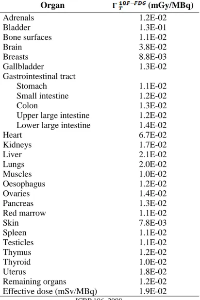
The coefficient ( ) for each organ are presented in the Table1.
Values of activity were simulated from 0.07 mCi to 0.15 mCi, and with 10 mCi, based in protocols used in some services. The value of activity was multiplied by the phantom mass and this value converted to MBq. It was multiplied by the coefficient ( ) and the effective dose was found.
Table 1: Absorbed dose per unit activity administered (mGy/MBq)
Organ (mGy/MBq)
Adrenals 1.2E-02
Bladder 1.3E-01
Bone surfaces 1.1E-02
Brain 3.8E-02
Breasts 8.8E-03
Gallbladder 1.3E-02
Gastrointestinal tract
Stomach 1.1E-02
Small intestine 1.2E-02
Colon 1.3E-02
Upper large intestine 1.2E-02
Lower large intestine 1.4E-02
Heart 6.7E-02 Kidneys 1.7E-02 Liver 2.1E-02 Lungs 2.0E-02 Muscles 1.0E-02 Oesophagus 1.2E-02 Ovaries 1.4E-02 Pancreas 1.3E-02
Red marrow 1.1E-02
Skin 7.8E-03 Spleen 1.1E-02 Testicles 1.1E-02 Thymus 1.2E-02 Thyroid 1.0E-02 Uterus 1.8E-02
Remaining organs 1.2E-02
Effective dose (mSv/MBq) 1.9E-02
ICRP 106, 2008
3.
RESULTS AND DISCUSSION
The effective dose results are show in Table 2. The effective dose may vary up to 114% altering the injected activity between 0.07 and 0.15 mCi., The effective dose using fixed 10 mCi is between these values, closer to the bigger ones. The required amount of activity to be injected varies accord-ing to the detector sensitivity, i.e., varies in each equipment. A reduction in injected activity may
imply an increase in examination time, which may cause motion artifacts in the image. The reduc-tion of activity should take into considerareduc-tion in addireduc-tion to patient exposure, the quality of the im-age. The professionals, according to the patient’s profile, should evaluate the feasibility of this re-duction.
It is also important to emphasize the need for the activity injected into the patient to be proportional to their weight, and not a fixed value for all patients. Thus, the patient receives the necessary dose for the accomplishment of its exam, without causing unnecessary exposure.
Table 2: Effective Doses (mSv) Activity per kg
(mCi)
Total Injected Activity con-sidering Alderson mass
(mCi) Effective Dose (mSv) Man Woman 0.07 5.15 3.62 2.46 0.08 5.88 4.13 2.81 0.09 6.62 4.65 3.16 0.10 7.35 5.17 3.52 0.11 8.09 5.68 3.87 0.12 8.82 6.20 4.22 0.13 9.56 6.72 4.57 0.14 10.29 7.23 4.92 0.15 11.03 7.75 5.27 - 10.00 7.03 7.03
4. CONCLUSION
In PET scans, any reduction of the administered activity implies a proportional reduction of the ef-fective dose in patient. In oncological studies, in which the patient undergoes a series of control tests, the search for techniques that allow the reduction of the administered dose of radiation has great relevance. The principle of optimization governs this study. Doses must be as low as reasona-bly achievable, i.e. all tools that reduce doses to the patient without generating diagnostic damage should be used. In addition, sensitivity tests of the detector should be periodically performed for greater use in the capture of the photons and consequently smaller injected activities. It is expected
that the PET/CT scans protocols be revised at the end of the study, so that the absorbed and effec-tive dose received by the patient decreases.
ACKNOWLEDGMENT
The authors are thankful to FAPEMIG and CAPES for the financial support.
REFERENCES
1. CZERNIN, J; SCHELBERT, H. PET/CT imaging: facts, opinions, hopes, and ques-tions. Journal of Nuclear Medicine, v. 45, n. 1 suppl, p. 1S-3S, 2004.
2. HAINER, J. General concepts of PET and PET/CT imaging. Case-Based Approach to PET/CT in Oncology, ed. Victor H. Gerbaudo. Published by Cambridge University Press. Cam-bridge University Press 2012.
3. BAR-SHALOM, R. et al. Clinical performance of PET/CT in evaluation of cancer: addi-tional value for diagnostic imaging and patient management. Journal of nuclear medicine, v. 44, n. 8, p. 1200-1209, 2003.
4. FLUX, Glenn et al. The impact of PET and SPECT on dosimetry for targeted radionuclide therapy. Zeitschrift für Medizinische Physik, v. 16, n. 1, p. 47-59, 2006.
5. CZERNIN, J.; ALLEN-AUERBACH, M.; SCHELBERT, H.R. Improvements in cancer staging with PET/CT: literature-based evidence as of September 2006. Journal of Nuclear Medi-cine, v. 48, n. 1 suppl, p. 78S-88S, 2007.
6. GAMBHIR, Sanjiv S. et al. A tabulated summary of the FDG PET literature. Journal of nuclear medicine, v. 42, n. 5 suppl, p. 1S-93S, 2001.
7. HUANG, Bingsheng; LAW, Martin Wai-Ming; KHONG, Pek-Lan. Whole-body PET/CT scanning estimation of radiation dose and cancer risk. Radiology, v. 251, n. 1, p. 166-174, 2009.
8. MCCOLLOUGH, Cynthia H.; SCHUELER, Beth A. Calculation of effective dose. Medical physics, v. 27, n. 5, p. 828-837, 2000.
9. CAMARGO, Edwaldo E. Experiência inicial com PET/CT. Radiologia Brasileira, v. 38, n. 1, p. 0-0, 2005.
10. QUINN, Brian et al. Radiation dosimetry of 18F-FDG PET/CT: incorporating exam-specific parameters in dose estimates. BMC medical imaging, v. 16, n. 1, p. 41, 2016.
10. OLIVEIRA, S.M.V. et al. Protocol for 18F-FDG quantification in pet-ct whole-body ex-ams. Thyroid, v. 20, n. 4.3, p. 0.6, 2010.
11. ROBILOTTA, Cecil Chow. A tomografia por emissão de pósitrons: uma nova modalidade na medicina nuclear brasileira. Panam Salud Publica, v. 20, n. 2/3, p. 135, 2006.
12. NAGAKI, A.; ONOGUCHI, M.; MATSUTOMO, N. Patient Weight–Based Acquisition Pro-tocols to Optimize18F-FDG PET/CT Image Quality. Journal of nuclear medicine technology, v. 39, n. 2, p. 72-76, 2011.
13. ALESSIO, A. M. et al. Weight-based, low-dose pediatric whole-body PET/CT proto-cols. Journal of Nuclear Medicine, v. 50, n. 10, p. 1570-1578, 2009.
14. OLIVEIRA, Cássio Miri et al. Suggestion of a national diagnostic reference level for 18F-FDG/PET scans in adult cancer patients in Brazil. Radiologia Brasileira, v. 46, n. 5, p. 284-289, 2013.
15. ICRP- INTERNATIONAL COMMISSION ON RADIOLOGICAL PROTECTION. The 2007 Recommendations of the International Commission on Radiological Protection. Radiation Dose to Patients from Radiopharmaceuticals. ICRP, Publication 106, 2008.
16. RADPRO, Radiation Protection for the Radiation Professionals. Avaible at: <https://www.radpro-int.com/>. Last accessed: 29 Nov. 2017.