Vol.45, n. 3 : pp. 309-315, September 2002
ISSN 1516-8913 Printed in Brazil BRAZILIAN ARCHIVES OF BIOLOGY AND TECHNOLOGY
A N I N T E R N A T I O N A L J O U R N A L
The Influence of Ca
2+on Gluconeogenesis Stimulation by
Glucagon in the Liver of Arthritic Rats
Ana M. Kelmer-Bracht*; Zélio Fedatto-Júnior; Emy L. Ishii-Iwamoto; Silvana M. Caparroz-Assef and Adelar Bracht
Universidade Estadual de Maringá; Laboratório de Metabolismo Hepático; 87020-900; Maringá - PR - Brazil
ABSTRACT
Ca2+ participates in the stimulation of hepatic gluconeogenesis by glucagon and there is evidence that Ca2+ fluxes are modified in arthritic rats. These findings raise the question whether hepatic gluconeogenesis in arthritic rats responds differently to glucagon and Ca2+. The experimental system was the isolated perfused rat liver. In the presence of Ca2+, stimulation of hepatic gluconeogenesis by glucagon in arthritic rats was equal to that in normal rats in absolute terms, but higher in relative terms (104.5 and 45.2%, respectively). In the absence of Ca2+, however, stimulation of hepatic gluconeogenesis by glucagon in arthritic rats was smaller in both absolute and relative terms (18.5 and 41.9%, respectively). It can be concluded that the Ca2+-dependent component of gluconeogenesis activation by glucagon is more important in arthritic than in normal rats.
Key words: Arthritis, liver, gluconeogenesis, glucagon.
* Author for correspondence
INTRODUCTION
The adjuvant-induced arthritis is an experimental immunopathology in rats which shares many features of human rheumatoid arthritis (Rosenthale and Capetola, 1982). For this reason it is one of the most widely used models for evaluation of anti-inflammatory and antirheumatic drugs (Rainsford, 1982; Billingham, 1983). Furthermore, this model has also been used for studying metabolic alterations induced by rheumatoid arthritis. Caparroz-Assef et al., 1998 and Fedatto Jr. et al., 1999, for example, have recently shown that livers from adjuvant-induced arthritic rats present lower rates of gluco-neogenesis from a variety of substrates, including lactate plus pyruvate. It was also shown that glucose uptake is
increased in livers from arthritic rats due to higher glucokinase activities (Fedatto Jr. et al., 2000).
Inflammation also affects Ca2+ fluxes in
hepatocytes. Barrit and Whitehouse (1977), for example, found that mitochondria isolated from inflamed rats exhibit a decreased ability to retain
accumulated Ca2+ and a more rapid and greater
degree of Ca2+ induced osmotic swelling.
More-over, Somasundaram and Sadique (1986) found that mitochondria from inflamed rats exhibit a reduction in Ca2+ uptake.
Ca2+ movements and redistributions, on the other hand, are frequent and important events during hormone actions. Even glucagon, a hormone that acts predominantly via cyclic AMP, is also
influenced by Ca2+. The latter seems to be
perfused rat liver the Ca2+ dependencies of the simultaneous gluconeogenesis and respiration stimulations by glucagon are strongly reduced at a lactate to pyruvate ratio of 100. At the more physiologic lactate to pyruvate ratio of 10, the Ca2+ dependencies of gluconeo-genesis and respiration stimulations are less pronounced, but still evident. It should be mentioned that glucagon increases the activity of several mitochondrial dehydrogenases and other enzymes, an action that is believed to be
caused by Ca2+ accumulation (McCormack, 1985;
Quinlan and Halestrap, 1985; Walajtys-Rhode et al., 1992; Deaciuc et al., 1992).
Both phenomena, changes in Ca2+ movements in
the liver of arthritic rats and the Ca2+ dependence of the glucagon action, immediately raise the
question about the role of Ca2+ in hepatic
gluconeogenesis stimulation by glucagon under the former conditions. An answer to such a question can only be obtained by experimental means. For this reason, we have decided to measure gluconeogenesis from lactate plus pyruvate in livers from arthritic rats, in the
presence and absence of Ca2+. These experiments
should bring an answer to the question whether
Ca2+ affects, or not, the action of glucagon on
hepatic gluco-neogenesis in arthritic rats.
MATERIALS AND METHODS
Materials. The liver perfusion apparatus was built in the workshops of the University of Maringá. Crystalline glucagon was purchased from “Eli Lilly do Brasil”. All enzymes and coenzymes used in the enzymatic assays were purchased from “Sigma Chemical Co.” (St. Louis, USA). All other chemicals were from the best available grade (98-99.8% purity).
Animals. Male albino rats (Wistar), weighting
200-250 g, were fed ad libitum with a standard
laboratory diet (Purina, São Paulo, Brazil). For the induction of adjuvant arthritis, the animals were injected in the left hind paw with 100 µl of
heat inactivated Mycobacterium tuberculosis
suspended in mineral oil at a concentration of 0.5% (w/v). Two weeks after the induction of the disease, the animals showing characteristic arthritic lesions were selected for the experiments (Pearson, 1956). Rats with similar ages served as
controls. All rats were starved for 24 hours before the surgical removal of the liver.
Liver perfusion. For the surgical procedure the rats were anesthetized by intraperitoneal injection of sodium pentobarbital (50 mg/kg). Hemoglobin-free, non-recirculating perfusion was done. The surgical technique was that one described by Scholz and Bücher (1965). After cannulation of the portal and cava veins the liver was positioned in a plexiglass chamber. The flow was maintained constant by a peristaltic pump. The perfusion fluid was Krebs/Henseleit-bicarbonate buffer (pH 7.4), saturated with a mixture of oxygen and carbon dioxide (95:5) by means of a membrane oxygenator with simultaneous temperature adjustment at 37°C.
Ca2+-free perfusion. For performing Ca2+-free
perfusion, the intracellular Ca2+ pools were
exhausted. The following procedure was adopted.
Livers were pre-perfused with Ca2+-free
Krebs/Henseleit-bicarbonate buffer containing 0.2 mM ethylenediamine tetraacetate (EDTA). In order to ensure maximal depletion of the intracellular Ca2+ pools, phenylephrine (2 µM) was infused repeatedly (3 times) during short periods of 2 minutes, with intervals of 5 minutes. According to Reinhart et al. (8), this procedure
depletes the intracellular Ca2+-pools which are
normally mobilized when hormones are infused.
Analytical. Samples of the effluent perfusion fluid were collected according to the experimental protocol and analyzed for their glucose contents by means of a standard enzymatic procedure (Berg-meyer and Bernt, 1974) The oxygen concentration in the outflowing perfusate was monitored con-tinuously, employing a teflon-shielded platinum electrode adequately positioned in a plexiglass chamber at the exit of the perfusate (Kelmer-Bracht et al., 1984).
Calculations and treatment of data. The mean rates of glucose production (Gs) during substrate infusion and before glucagon infusion were calculated according to the following equation:
0 1
b t
t s
s
t t
dt ] G ) t ( G [ G
1
0
− −
In equation [1] t0 represents the time in which substrate infusion was started and t1 the time at which glucagon infusion was initiated, Gs(t) are the rates of glucose production and Gb the small basal rate of glucose release (before substrate infusion). The mean rates of glucose release
during glucagon infusion (Gg) were calculated
according to the relation:
s 1
2 b t
t g
g G
t t
dt ] G ) t ( G [ G
2
1 −
− −
=
∫
[2]In equation [2] Gg(t) are the experimental rates of glucose release during glucagon infusion, t1 the time at which glucagon infusion was initiated and t2 the time at which glucagon infusion was stopped.
The integrals in equations [1] and [2] were calculated analytically after fitting the experi-mental data to cubic spline functions. These
calculations were done by means of the scientist
program from MicroMath Scientific Software. The statistical significance of the differences between parameters was evaluated by means of the Student-Newman-Keuls test after variance analysis; p < 0.05 was adopted as a criterion of significance.
RESULTS
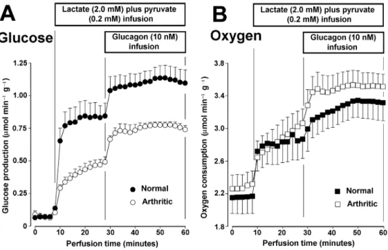
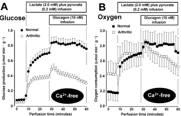
Figures 1 and 2 illustrate the experimental protocol and the results obtained in the perfusion experiments. Figure 1 shows the glucose produc-tion and oxygen uptake measurements when the normal Krebs/Henseleit-bicarbonate buffer was employed; and Figure 2 illustrates the same measurements in
the Ca2+-free experiments. As revealed by the
horizontal bars, the substrates (lactate plus pyruvate) were infused alone during 20 minutes.
Figure 1 - Gluconeogenesis and oxygen uptake changes due to lactate + pyruvate and glucagon in perfused livers from normal and arthritic rats perfused with medium containing Ca2+.Livers from fasted rats were perfused with the complete Krebs/Henseleit-bicarbonate buffer, containing 2.5 mM CaCl2, as described in Materials and Methods.
Figure 2 - Gluconeogenesis and oxygen uptake changes due to lactate + pyruvate and glucagon in perfused livers from normal and arthritic rats perfused with Ca2+-free medium.Livers from fasted rats were perfused with the Ca2+-free Krebs/Henseleit-bicarbonate buffer. Depletion of the intracellular Ca2+ pools was accomplished as described in Materials and Methods. L-Lactate and pyruvate and glucagon were infused at the times and concentrations indicated on the top of each graph. Samples of the effluent perfusate were taken for the measurement of glucose. Oxygen uptake was monitored by means of a platinum electrode. The data of normal and arthritic rats are both the means of 5 liver perfusion experiments. Vertical bars represent mean standard errors.
After this time, glucagon was infused during 30 minutes in the presence of these substrates. A lactate to pyruvate ratio of 10 was choosen because this is a more physiologic condition.
Figure 1A allows to compare the responses of glucose production and oxygen uptake in the
presence of Ca2+ of both livers from normal and
livers from arthritic rats. All rats were fasted for a period of 24 hours before the perfusion experiments. Under these conditions the glycogen levels are low and the contribution of glyco-genolysis to glucose release is minimal (Scholz and Bücher, 1965). This is revealed by the very small rates of glucose release in the pre-perfusion period (0 to 8 minutes). Glucose production in the liver of normal rats raised more rapidly after initiation of substrate infusion than in livers from arthritic rats. At the end of 20 minutes hepatic gluconeogenesis in normal rats was clearly superior to that in arthritic rats. This observation confirms previous findings (Fedatto Jr. et al., 1999). The introduction of glucagon at 28 minutes
produced clear increases in glucose production in the normal as well as in the arthritic condition. Oxygen uptake, as revealed by Figure 1B, was similar in livers from normal and arthritic rats, although the latter presented a small tendency toward higher values. Under both conditions the infusion of substrates and the subsequent infusion of glucagon produced increments in oxygen uptake. Especially in the presence of glucagon, there was a tendency toward higher values in livers from arthritic rats but, unlike to what occurred with gluconeogenesis, the sizes of the standard errors do not allow a clear definition of the differences.
Figure 2 shows the time-courses of glucose production and oxygen uptake in livers from normal and arthritic rats in the absence of Ca2+.
Absence of Ca2+ means perfusion with no added
Table 1 - Mean rates of glucose production in livers from normal and arthritic rats: the action of glucagon in the presence and absence of Ca2+. The mean rates were calculated as described in Materials and Methods. The experimental conditions were those described in the legends to Figures 1 and 2.
Gluconeogenesis (µmol min−1 g−1) Animal condition Ca2+ in the
perfusate Before glucagon infusion (
G
s)Increment due to glucagon infusion (
G
g)
Percent stimulation
caused by glucagon
Normal (n=5) 2.5 mM 0.700±0.093*,†,¶ 0.317±0.025*,† 45.2
Normal (n = 5) Ca2+-free 0.510±0.057†,‡ 0.214±0.007†,¶,§ 41.9
Arthritic (n=5) 2.5 mM 0.330±0.044* 0.345±0.024‡,¶ 104.5
Arthritic (n=5) Ca2+-free 0.286±0.028‡,¶ 0.053±0.012*,‡,§ 18.5
In each column the symbols *, †, ‡, ¶ and § represent pairs of values differing statistically from each other according to the Student-Newman-Keuls test (p < 0.05).
The response of the liver of arthritic rats, however, was again lower, especially to glucagon, where it was clearly a transient one. Oxygen uptake of livers from arthritic rats also presented a transient response to glucagon, as revealed by Figure 2B. Figures 1 and 2 allow to appreciate the time courses of the changes produced by substrates and glucagon on gluconeogenesis in a qualitative way. A more rigorous quantitative analysis, however, can be done by means of the data in Table 1. In this table the mean rates of glucose production, calculated as described in Materials and Methods, are listed in addition to the percent stimulation caused by glucagon. In normal rats, hepatic gluconeogenesis was smaller in the absence of Ca2+, the same occurring with the increment due to glucagon, an observation which confirms previous results of our laboratory (Silva et al., 1997). In proportional terms, however, the increment caused by glucagon was similar. In arthritic rats, besides confirming the smaller rates of gluconeogenesis when compared to the normal state, a phenomenon that was already evident in Figures 1 and 2, Table 1 also shows that the action of glucagon was different in the presence and absence of Ca2+. In the presence of Ca2+ the greatest mean increment was found in comparison with all other conditions. In the absence of Ca2+, on the other hand, livers from arthritic rats produced the smallest mean increment caused by glucagon. The latter is valid in absolute as well as in relative terms.
DISCUSSION
The main conclusion allowed by the observa-tions of the present work is that stimulation of hepatic gluconeogenesis by glucagon is rendered more dependent on Ca2+ in arthritic than in normal rats. The following combination of observations leads to this conclusion: a) hepatic gluconeo-genesis stimulation by glucagon in the arthritic condition was superior in relative terms to the normal condition in the presence of Ca2+; b) in the absence of Ca2+, however, stimulation of gluco-neogenesis by glucagon in the arthritic condition was the smallest one, in relative as well as in absolute terms.
It should be added that the kinetics of gluconeogenesis changes was accompanied by a similar kinetics in the oxygen uptake changes. When the gluconeogenesis changes were only transient, as it happened in the arthritic condition
and in the absence of Ca2+, the oxygen uptake
changes followed a similar pattern. It is usually accepted that the increase in oxygen uptake due to glucagon infusion under conditions of gluco-neogenesis is a consequence of the increased energy demands (Zwiebel and Scholz, 1986). Nonwithstanding, dissociations between both variables have been reported under some special conditions, as for example, in the presence of diltiazem (Bracht et al., 1999).
believed that gluconeogenesis stimulation by glucagon depends largely on oxygen uptake
stimulation via a Ca2+-dependent stimulation of
mitochondrial dehydrogenases (McCormack, 1985; Quinlan and Halestrap, 1985; Walajtys-Rhode et al., 1992; Deaciuc et al., 1992). In the arthritic condition, as already mentioned in the
Intro-duction, the capacity of accumulating Ca2+
intramitochondrially is reduced (Barrit and White-house, 1977; Somasundaram and Sadique, 1986). The apparent contradiction that hepatic gluco-neogenesis stimulation by glucagon in arthritic rats is even more dependent on Ca2+ may be related to
the redox potential of the NAD+/NADH couple in
these animals. Derbocio (1999) has shown that the
mitochondrial NADH/NAD+ ratio in the liver of
arthritic rats is approximately half that of normal rats, meaning thus a much more oxidized state. This could be the result of a diminished substrate supply or an enhanced activity of the respiratory chain. The latter is improbable, because respiration in the arthritic condition is not substantially different from that of the normal condition. Consequently, the reduced substrate supply is more probable. This means also lower substrate concentrations for the various dehydrogenases that, in turn, require a more pronounced stimulation when glucagon is introduced in order to cope with the necessities of gluconeogenesis. This is a situation which could explain the more
pronounced dependence on Ca2+ in livers from
arthritic rats, in spite of the fact that the capacity of accumulating this cation is reduced, although not eliminated (Barrit and Whitehouse, 1977; Soma-sundaram and Sadique, 1986). In the absence of
Ca2+, glucagon would be unable to induce the
generation of sufficient extra amounts of reducing equivalents in order to enhance respiration and gluconeogenesis.
The latter seems to be a plausible explanation, but one should not exclude the possibility that the phenomenon detected in this work depends also on cytosolic or intracellular membrane-bound factors. There are several circulating and cellular factors that are changed in the arthritic condition as, for example, tumour-necrosis factor alfa (TNFα), interleukin and stress hormones (Lee et al., 1987; Hellerstein et al., 1989; Roubenoff et al., 1994; Roubenoff et al., 1997). These and other factors could be modifying enzyme activities. For example, an increased activity of both phosphofructokinase and fructose bisphosphate phosphatase has been demonstrated in cultured
myocytes treated with the cytokine cachetin/TNFα (Zentella et al., 1993). These enzymes are among those that are controlled by glucagon. Conse-quently, the possibility of a differential action of glucagon on enzymatic activities changed during the arthritic condition should not be ruled out and deserves further investigations.
RESUMO
O Ca2+ participa do estímulo da neoglicogênese
hepática pelo glucagon e há também indicações de
que fluxos de Ca2+ são modificados em ratos
artríticos. Estes dados permitem levantar a questão de se a neoglicogênese hepática em ratos artríticos responde diferentemente ao glucagon e ao Ca2+. O sistema experimental foi o fígado em perfusão
isolada. Na presença de Ca2+, o estímulo da
neoglicogênese hepática pelo glucagon em ratos artríticos foi quase igual a aquele encontrado em ratos normais em termos absolutos, mas maior em termos relativos (104,5 e 45,2%, respectiva-mente). Na ausência de Ca2+, no entanto, o estímu-lo da neoglicogênese hepática peestímu-lo glucagon em ratos artríticos foi menor em termos absolutos e relativos (18,5 e 41,9%, respectivamente). Pode-se
concluir que o componente Ca2+-dependente do
estímulo da neoglicogênese pelo glucagon é mais importante em ratos artríticos do que em ratos normais.
REFERENCES
Barrit, G. J. and Whitehouse, M. W. (1977), Patho-biodynamics: effect of extrahepatic inflammation on calcium transport and drug metabolism by rat liver mitochondria in vitro. Biochemical Medicine,
17, 99-115.
Bergmeyer, H. U. and Bernt, E. (1974), Glucose determination with glucose oxidase and peroxidase. In- Bergmeyer, H. U. (ed.). Methods of Enzymatic Analysis. Academic Press : New York. pp. 1205-1215. Billingham, M. E. J. (1983), Model of arthritis and the
search for antirheumatic drugs. Pharmacology and Therapeutics, 21, 389-428.
Caparroz-Assef, S. M.; Bersani-Amado, C. A.; Nascimento, E. A.; Kelmer-Bracht, A. M. and Ishii-Iwamoto, E. L. (1998), Effects of the nonsteroidal anti-inflammatory drug nimesulide on energy meta-bolism in livers from adjuvant-induced arthritic rats. Research Communications in Molecular Pathology and Pharmacology,99,93-116.
Deaciuc, I. V.; D’Souza, N. B. and Miller, H. I. (1992), A novel mechanism for Ca2+ dependent regulation of hepatic gluconeogenesis: Stimulation of mito-chondrial phosphoenolpyruvate synthesis by Ca2+.
International Journal of Biochemistry, 24, 129-132. Derbocio, A. M. (1999), Efeitos metabólicos do
zimo-sano no fígado de rato em perfusão isolada. M. Sc. Thesis, Maringá, Brazil.
Fedatto Jr., Z.; Ishii-Iwamoto, E. L.; Amado, C. B.; Vicentini, G.; D'Urso-Panerari, A.; Bracht, A. and Kelmer-Bracht, A. M. (1999), Gluconeogenesis in the liver of arthritic rats. Cell Biochemistry and Function, 17, 271-278.
Fedatto Jr., Z.; Ishii-Iwamoto, E L.; Amado, C. B.; Maciel, E. R. M.; Bracht, A. and Kelmer-Bracht, A. M. (2000), Glucose phosphorylation capacity and gly-colysis in the liver of arthritic rats. Inflam-mation Research, 49,128-132.
Hellerstein, M. K.; Meydani, S. N.; Meydani, M.; Wu, K. and Dinarello, C. A. (1989), Interleukin-1 induced anorexia in the rat: influence of prosta-glandins.
Journal of Clinical Investigation, 84, 228-235. Kelmer-Bracht, A. M.; Ishii, E. L.; Andrade, P. V. M.
and Bracht, A. (1984), Construction of a liver perfusion apparatus for studies on metabolic regula-tion and mechanisms of drug action.
Arquivos de Biologia e Tecnologia, 27, 419-438. Lee, J. C.; Dimartino, M. J.; Votta, B. J. and Hanna, N.
(1987), Effect of auranofin treatment on aberrant splenic interleukin production in adjuvant arthritic rats. Journal of Immunology, 139, 3268-3274. McCormack, J. G. (1985), Studies on the activation of
rat liver pyruvate dehydrogenase and 2-oxo-glutarate dehydrogenase by adrenaline and gluca-gon.
Biochemical Journal, 231, 597-608.
Pearson, C. M. (1956), Development of arthritis, periarthritis and periostitis in rats given adjuvants.
Proceedings of Experimental Biology and Medi-cine,
91,95-101.
Quinlan, P. T. and Halestrap, A. P. (1986), The mechanism of the hormonal activation of respira-tion in isolated hepatocytes and its importance in the regulation of gluconeogenesis. Biochemical Journal, 236, 789-800.
Rainsford, K. D. (1982), Adjuvant polyarthritis in rats: is this model a satisfactory model for screening anti-arthritic drugs? Agents and Actions, 12, 452-458. Reinhart, P. H.; Taylor, W. M. and Bygrave, F. L.
(1982), Calcium ion fluxes induced by the action of α-adrenergic agonists in perfused rat liver.
Biochemical Journal,208, 619-630.
Rosenthale, M. E. and Capetola, R. J. (1982), Adjuvant arthritis: immunopathological and hyperalgesic features. Federation Proceedings, 41, 2577-2582. Roubenoff, R.; Roubenoff, R. A.; Kehayias, J. J.;
Zhuang, H.; Dawson-Hughes, B.; Dinarello, C. A. and Rosenberg, I. H. (1994), Rheumatoid ca-chexia: cytokine-driven hypermetabolism accom-panying reduced body cell mass on chronic inflam-mation.
Journal of Clinical Investigation,93, 2379-2386. Roubenoff, R.; Freedman, L. M.; Smith, D. E.; Abad, L.
W.; Dinarello, C. A. and Kehayias, J. J. (1997) Adjuvant arthritis as a model of inflammatory cachexia. Arthritis and Rheumatism,40, 534-539. Scholz, R. and Bücher, T. (1965), Hemoglobin-free
perfusion of rat liver. In- Chance, B.; Estabrook, R. W. and Williamson, J. R. (eds.). Control of Energy Metabolism. Academic Press: New York. pp. 393-414. Silva, A. C. M.; D’Ávila, R. B.;Ferrari, A. G.;
Kelmer-Bracht, A. M.; Constantin, J.; Yamamoto, N. S. and Bracht, A. (1997), Brazilian Journal of Medical and Biological Research, 30, 827-836
Somasundaram, S. and Sadique, J. (1986), The role of mitochondrial calcium transport during inflamma-tion and the effect of anti-inflammatory drugs.
Biochemical Medicine and Metabolic Biology, 36, 220-230.
Walajtys-Rhode, E.; Zapatero, J.; Moehren, G. and Hoek, J. B. (1992), The role of the matrix calcium level in the enhancement of mitochondrial pyruvate car-boxylation by glucagon pretreatment. Journal of Biological Chemistry,267, 370-379.
Zentella, A.; Manogue, K. and Cerami, A. (1993), Cachetin/TNF mediated lactate production in cultured myocytes is linked to activation of a futile substrate cycle. Cytokine,5, 436-444.
Zwiebel, F. and Scholz, R. (1986), The perfused liver system in the study of gluconeogenesis. In- Kraus-Friedman, N. (ed.). Hormonal Control of Gluconeogenesis. CRC Press : Boca-Raton. pp. 97-110.