Vol.50, Special Number : pp.199-207, September 2007
ISSN 1516-8913 Printed in Brazil BRAZILIAN ARCHIVES OF BIOLOGY AND TECHNOLOGY
A N I N T E R N A T I O N A L J O U R N A L
Technetium-99m-labeled Stealth pH-sensitive Liposomes: A
New Strategy to Identify Infection in Experimental Model
Vildete Aparecida Sousa Carmo1, Mônica Cristina de Oliveira1, Luciene das Graças Mota2, Luís Paulo Freire2, Raphael Ligório Benedito Ferreira2 and Valbert Nascimento Cardoso2*
1
Departamento de Produtos Farmacêuticos; 2Departamento de Análises Clínicas e Toxicológicas; Faculdade de Farmácia; Universidade Federal de Minas Gerais; Avenida Antônio Carlos, 662; Pampulha; 31270-901; cardosov@farmacia.ufmg.br; Belo Horizonte - MG - Brasil
ABSTRACT
The diagnosis of inflammatory and infectious processes is an important goal in medicine. The use of radiopharmaceuticals for identification of inflammation and infection foci has received considerable attention. The aim of this work was to evaluate the uptake and the imaging potential of stealth pH-sensitive liposomes radiolabelled with 99mTechnetium (99mTc) to identify infection sites in mice. The liposomes containing glutathione were labeled with 99mTc-Hexamethylpropyleneamine oxime (HMPAO) complex. The 99mTc-labeled stealth pH-sensitive liposomes (99mTc-SpHL) were injected in mice bearing infection in the right thigh muscle induced by Staphylococcus aureus. Biodistribution studies and scintigraphic imaging were performed at different times after injection of radiopharmaceutical. The 99mTc-SpHL was significantly uptaken by abscess when compared to the respective control. The abscess was visualized as early as 0.5 hours after injection of 99mTc-SpHL becoming more prominent with the time. These results indicate that99mTc-SpHL is a promising radiopharmaceutical for visualizing infection foci in patients.
Key words: 99mTechnetium, pH-sensitive liposomes, infection imaging, scintigraphy
INTRODUCTION
Nuclear medicine imaging allows in vivo detection of inflammatory and infectious diseases in various parts of the body by the intravenous injection of radiolabelled substances and by the external detection of radioactivity using the gamma camera.* The sensitivity of this technique usually allows detection of physiopathological processes in the initial stages before the development of anatomical alterations detectable by conventional radiographic techniques and before the clinical onset of the disease (Signore et al., 2002). The radiopharmaceuticals most commonly used for
* Author for correspondence
imaging of inflammation and infection are
67Gallium-citrate (67Ga-citrate) and 99m
Considering the disadvantages these radiopharmaceuticals efforts has been devoted to the search of new agents for scintigraphic imaging which allows for the quick and efficient identification of inflammatory and infectious foci, with a high level of sensibility and specificity (Van Eerd et al., 2005; Love and Palestro, 2004; Laverman et al., 1999).
Stealth liposomes radiolabelled composed generally of egg phosphathidylcholine, cholesterol, and hidrophylic polymers anchored
phospholipids (mPEG2000-DSPE) have been
studied as imaging agents for the investigation of inflammatory and infectious processes. However, in the clinical studies using these liposomes formulation was observed that in some patients presented hypersensitivity reactions attributed the complement system activation; thus, becomes necessary to investigate a new lipid composition aimed to avoid this inconvenient (Szebeni, 2005; Moghimi and Szebeni, 2003; Brouwers et al.,
2000; Dams et al., 2000; Boerman et al., 2000; Devine and Bradley, 1998).
The pH-sensitive liposomes are designed to promote efficient release of entrapped agents in response to low pH of pathological tissues, such as inflamed and infected areas; thus they are also potential candidates for the preparation of radiopharmaceuticals (Simões et al., 2004). We have developed a new radiopharmaceutical based on stealth pH-sensitive liposomes labeled
99m
Technetium–hexametylpropylene amine oxime (99mTc-SpHL). These liposomes are composed of dioleoylphosphatidylethanolamine (DOPE), cholesterylhemisuccinate (CHEMS), and
methoxypoly(ethylene glycol)2000
-distearoylphosphatidylethanolamine (mPEG2000
-DSPE). DOPE has a strong propensity to form a nonbilayer structure due to its cone-shape geometry, and a weakly acidic amphiphile, such as CHEMS, confers stability to the bilayer phase at neutral pH. These lipids provide electrostatic repulsions, which decrease DOPE intermolecular interactions, thus preventing the appearance of fusogenic properties. However, under acidic conditions, such as in the inflammation and infection regions, the CHEMS molecules becomes partially protonated, thus losing its negative charge and, therefore, its ability to stabilize the bilayers of the vesicles with a subsequent release of trapped radioactive markers. In this study, 99mTc-SpHL was investigated as a useful radiopharmaceutical to image experimental infection foci.
MATERIALS AND METHODS
Methoxypoly(ethylene glycol)2000
-distearoylphosphatidylethanolamine (mPEG2000
-DSPE) and dioleoylphosphatidylethanolamine (DOPE) were supplied by Lipoid GmbH. Cholesteryl hemisuccinate (CHEMS) and glutathione (GSH) were purchased from the Sigma Chemical Company. 99mTc was obtained from a molybdenum generator (IPEN/Brazil). All other chemicals and reagents used were commercially available in analytical grades. Male Swiss mice were allowed free access to a standard laboratory pellet diet with water ad libitum.
Preparation of liposomes
Liposomes encapsulating glutathione were prepared using a procedure based on polycarbonate membrane extrusion, as described previously (Laverman et al., 1999). Briefly, chloroform aliquots of DOPE, CHEMS and mPEG2000-DSPE (total lipid concentration 40 mM;
molar ratio 6.5:3.0:0.5) were transferred to a round bottom flask and a lipid film was formed by rotary evaporation under reduced pressure (Buchi R215, Switzerland). The lipid film obtained was hydrated in 50 mM glutathione in HEPES buffer (10 mM Hepes, 135 mM NaCl, 5 mM EDTA at pH 7.4). The suspension of liposomes obtained was submitted to a filtration through 0.4µ, 0.2µ, and 0.1µ polycarbonate membranes (10 cycles for each) using a medium pressure extruder (Lipex Biomembranes Inc., Canada). Unencapsulated glutathione was separated from the liposomes by ultracentrifugation (Ultracentrifuge SORVALL Ultra 80, USA) at 150.000g at 4 °C for 90 minutes. The mean diameter of the liposomes containing GSH was determined by unimodal analysis by the quasi-elastic light scattering, at 25 °C, and at an angle of 90°. The size measurement was performed in triplicate using the 3000HS Zetasizer equipment (Malvern Instruments, UK). The samples were diluted using a HEPES buffer.
Labeling procedures
The commercially available kit of HMPAO (Ceretec®, Amersham Inc., UK) was labeled with
99m
organic phase (Barthel et al., 1999). Radiolabeling of the stealth pH-sensitive liposomes containing glutathione was performed as described previously by Phillips et al. (1992) with slight modifications. Preformed liposomes were labeled by transporting
99m
Tc as a lipophilic 99mTc-HMPAO complex through the bilayer. The liposomes were incubated for 30 minutes at 370C with freshly prepared
99mTc-HMPAO (1 MBq/µmole phospholipids).
Lipophilic HMPAO carries the 99mTc into the liposomes where it interacts with encapsulated glutathione resulting in its conversion to hydrophilic 99mTc-HMPAO becomes irreversibly trapped in the internal aqueous phase of the liposomes (Ballinger et al., 1988; Philips et al.,
1992). Unencapsulated 99mTc-HMPAO was
removed by gel filtration on a Sephadex G-25 column using HEPES buffer pH 7.4 as an eluent. Labeling efficiencies were checked by determining the activity before and after column separation of the 99mTc-SpHL using a dose calibrator (Capintec
CRC.15R, USA).The 99mTc-SpHL, after
purification, was administered immediately into infection bearing mice.
Mouse model of infection
Swiss male mice (approximately 23-25g in weight) were used for in vivo studies. Focal infection in the right thigh muscle was induced by intramuscular injection with 2 x 10⁷ colony forming units (CFU) of Staphylococcus aureus in 0.05 mL suspension of sterile saline. In the left thigh muscle, used as a control, sterile saline was injected. Twenty four hours after the induction of infection, when swelling of the muscle was apparent, mice were injected with 99mTc-SpHL in the tail vein. All protocols were approved by the Ethics Committee for Animal Experiments at the Federal University of Minas Gerais and are in compliance with the guide for the care and use of laboratory animals recommended by the Institute of Laboratory Animal Resources.
Biodistribution studies
After 24 hours of the induction of the infectious foci, 1.5 MBq of 99mTc-SpHL were injected in the tail vein of the mouse (n=3). At 0.5, 2, 4, 8, and 18 hours after radiopharmaceuticals administration, the animals were anesthetized with a mixture of xylazine (7.5 mg/kg) and ketamine (60 mg/kg) and then sacrificed by cervical dislocation. Blood sample, liver, spleen, lungs, kidneys, heart, left thigh muscle, and infected right thigh muscle were
collected. The dissected tissues were weighed and their radioactivity measured using an automatic scintillation apparatus covering an energy window of 70-210 KeV (ANSR-Abott, USA). A standard dosage containing the same injected amount was counted simultaneously in a separate tube, to correct physical decay and to calculate radiopharmaceuticals uptake in each organ. The measured radioactivity in tissues was expressed as percentage of injected radioactivity dose per gram of tissue (%ID/g).
Imaging studies
99mTc-SpHL (25 MBq) was injected in the tail vein
of the Swiss male mice which presented infection sites in the right thigh muscle (n=5). At 0.5, 1, 2, 4, 6, and 18 hours the mice were anesthetized with a mixture of xylazine (7.5 mg/kg) and ketamine (60 mg/kg) and placed in the prone on a gamma camera equipped with a low-energy collimator
high resolution (NucleineTM TH, Mediso,
Hungary). Five-minute static planar images were acquired for all times, except for the time of 18 hours (ten-minute), using a 256 x 256 pixels matrix.
The scintigraphic results were analyzed using regions of interest (ROI) drawn around the infected muscle (abscess), contralateral muscle (background), and the entire body to determine total body counts. The abscess-to-background ratio and activity in the abscess at various times were determined by following equations:
Abscess-to-background ratio = Counts abscess__ Counts background
% Activity in abscess = Counts abscess__ x 100 Counts total body
Statistical analysis
RESULTS
Liposome characterization and labeling procedures
The mean diameter and polidispersity index of the vesicle dispersion was 116.3 ± 3.96 nm and 0.17 ± 0.01, respectively, showing a good homogeneity. The lipophilic level of HMPAO obtained by solvent extraction method was equal to 80%. The
mean labeling efficiency of the 99mTc-SpHL was 77%.
Biodistribution studies
The biodistribution data obtained from ex vivo
counting of dissected tissues of Swiss mice bearing infection in the right thigh muscle induced by S. aureus after administration intravenous of
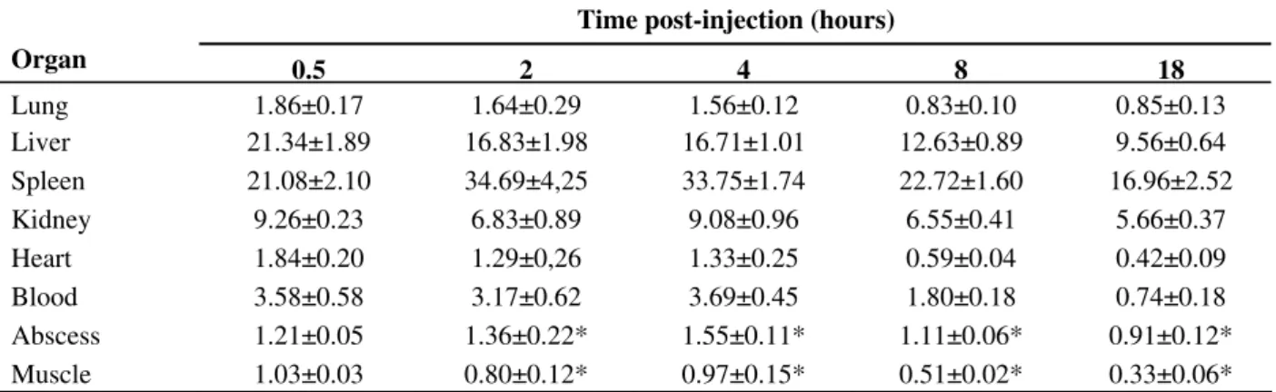
99mTc-SpHL are summarized in the Table 1.
Table 1 - Biodistribution results from 99mTc-SpHL in Swiss mice with infection in the right thigh muscle induced by
S. aureus
Time post-injection (hours)
Organ 0.5 2 4 8 18
Lung 1.86±0.17 1.64±0.29 1.56±0.12 0.83±0.10 0.85±0.13
Liver 21.34±1.89 16.83±1.98 16.71±1.01 12.63±0.89 9.56±0.64
Spleen 21.08±2.10 34.69±4,25 33.75±1.74 22.72±1.60 16.96±2.52
Kidney 9.26±0.23 6.83±0.89 9.08±0.96 6.55±0.41 5.66±0.37
Heart 1.84±0.20 1.29±0,26 1.33±0.25 0.59±0.04 0.42±0.09
Blood 3.58±0.58 3.17±0.62 3.69±0.45 1.80±0.18 0.74±0.18
Abscess 1.21±0.05 1.36±0.22* 1.55±0.11* 1.11±0.06* 0.91±0.12*
Muscle 1.03±0.03 0.80±0.12* 0.97±0.15* 0.51±0.02* 0.33±0.06*
All values are expressed as %ID/g ± s.e.m. for n = 3. The asterisks indicate a statistically significant difference between right thigh muscle (abscess) and left thigh muscle (background) (p<0.05)
The results indicate that 99mTc-SpHL was uptake mainly by the liver and spleen, attaining a maximum peaks at 0.5 hours and 2 hours after administration, respectively. In the liver, the percentage of 99mTc-SpHL uptake was maintained constant up to 4 hours (p>0.05) after injection with a decline of radioactivity level observed at 8 hours (p<0.01). To the spleen the radioactivity level was constant to 2, 4, and 8 hours (p>0.05) has been reduced after this time. In the lung, the maximum %ID/g was attained at 0.5 hours remaining constant until 4 hours after the administration of
99m
Tc-SpHL. Analysis of the blood samples indicated that radioactivity level was maintained constant up to 4 hours (p>0.05). In the times subsequent (8 and 18 hours) the 99mTc-SpHL concentration in the blood was reduced around half to each time (Fig. 1).
The radioactivity level in the kidney presented a fluctuation during the experiment with the maximum retention of 99mTc-SpHL occurred at 0.5 hours and 4 hours followed by a decrease after this time. The abscess radioactivity level did not differ significantly between the times investigated
following 99mTc-SpHL administration. In the left thigh muscle (background) was observed a lower uptake of 99mTc-SpHL to all investigated times, except at 0.5 hours, when compared with the right thigh muscle (p<0.05), indicating that 99mTc-SpHL administration leads to a higher accumulation in the abscess.
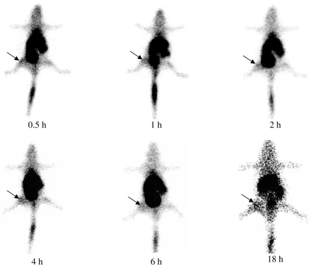
Imaging studies
Figure 1 - Blood clearance of 99mTc-SpHL in Swiss mice bearing infection in the right thigh muscle induced by S. aureus. Average amount of 3 mice is expressed as %ID/g present in blood. Curves represent best nonlinear exponential decay calculated by Prism software program (GraphPad Software, Inc.)
0.5 h 1 h 2 h
4 h 6 h 18 h
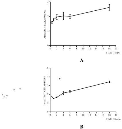
The result of biodistribution observed in the scintigraphic images is in agreement with biodistribution data obtained from excised tissue showing radioactivity high level in the MPS organs. Also, note the high radioactivity concentration in the bladder. Quantitative analysis of the image performed for each time interval is show in the Fig. 2. The abscess-to-background ratio was 1.58 ± 0.08 at 0.5 hours, increasing significantly to 2.62 ± 0.20 at 18 hours after injection of 99mTc-SpHL (p<0.01). The percent activity in abscess was 1.76% ± 0.06% at 0.5 hours increasing significantly to 3.44% ± 0.24 at 18 hours (p<0.01).
These results indicate a preferential accumulation of 99mTc-SpHL in the abscess when compared at uninfected contralateral muscle. Besides, it was observed a better contrast during experiment between the infectious site and the background.
DISCUSSION
It has been demonstrated that radiolabelled liposomes are potentials imaging agents for the diagnosis of inflammatory and infectious sites since they are able to accumulate in this areas (Boerman et al., 2000). The mechanism of accumulation in the infectious sites of radiolabelled liposomes is by leakage of the vesicles through vessels due increased vascular permeability and subsequent phagocythosis by macrophages of the infected tissue (Goins et al., 1993; Erdogan et al., 2000; Laverman et al., 2001). Moreover, in the inflamed tissue the blood vessel presents endothelial junctions allowing the escape of the particles smaller that 200 nm from the blood circulation (Crommelin et al., 1999).
A
B
Figure 3 - Quantitative analysis (mean ± s.e.m.) of the scintigraphic images of mice bearing focal infection injected with 99mTcSpHL and imagined over an 18h period (n=5). A -Abscess-to-Background ratios calculated from ROI image analysis. B - Activity in the abscess determined from ROI image analysis. Symbols different indicate a statistically significant difference (p<0.05)
*
#
*
The results obtained in this work showed small size for the liposomes (116.3 ± 3.96 nm). Studies have demonstrated that small liposomes (100 – 200nm) remain in the circulation for a longer time resulting in an increased accumulation at sites of focal infection (Awasthi et al., 2003; Crommelin et al., 1999; Erdogan et al., 2000; Boerman et al., 1997; Litzinger et al., 1994). In contrast, large liposomes are not retained in infectious sites since that they are rapidly cleared from circulation by the organs of the mononuclear phagocyte system (Oyen et al., 1996). Therefore, the small size of the liposomes used in the study was adequate for scintigraphic detection of infection sites.
The stealth pH-sensitive liposomes containing glutathione encapsulated were labeled with 99m Tc-HMPAO complex. This method provides radiolabelled liposomes whit high efficiency and stable in vivo (Phillips et al., 1992). Nevertheless, the lipophilicity of the 99mTc-HMPAO complex is crucial in the labeling efficiency of the liposomes; this characteristic is that allows its crossing through the liposomal membrane. The lipophilicity level of 99mTc-HMPAO obtained in the work is in agreement with the recommendations of the manufacturer so that promoted a good labeling efficiency (77%) of stealth pH-sensitive liposomes. In addition, we use the isotope 99mTc which present ideal dosimetry and characteristics adequate to scintigraphic imaging of infectious foci (Erdogan et al., 2000).
In this work, a novel liposomal formulation containing DOPE, CHEMS, and mPEG2000-DSPE
in its composition, know as stealth pH-sensitive liposomes, was prepared and evaluated their biodistribution and ability to identify infectious sites. These kinds of liposomes are in and of themselves a targeting strategy for the identification of infection sites as a consequence of their ability to release preferentially the radiotracer in this region due to lower exhibited pH as compared to normal tissue. Moreover, preliminary
in vitro assays performed in our laboratory showed evidences that liposomal formulation investigated in this study is poor activator of complement both classical and alternative pathway (data not shown). The imaging agent for infections diagnosis must to accumulate specifically and rapidly in the foci and clear quickly from the normal tissues to allow visualization of the lesion shortly after injection (Corstens and Van Der Meer, 1999; Rennen et al., 2001). For imaging of a lesion inflammatory or
infectious, the target-to-background ratio must be at least 1.5 to allow the acquisition of scintigraphic imaging with better quality (Phillips, 1999). We demonstrated through biodistribution and imaging studies the tropism of 99mTc-SpHL for infectious foci. The uptake in the infected area was significantly higher than the control thigh muscle. These results were also confirmed by scintigraphic images that showed rapid accumulation of 99m Tc-SpHL in the abscess allowing its visualization within the first 0.5 hours. In addition, the fast clearance of 99mTc-SpHL from non-target tissues contributed to superior delineation of the infectious focus in the times subsequent. This fact can be observed by increase of abscess-to-background ratio that varied from 1.58 ± 0.08 at 0.5 hours to 2.62 ± 0.20 at 18 hours (p<0.01) and also to the values observed of percent activity-to-total body that varied from 1.76 ± 0.06 at 0.5 hours to 3.44 ± 0.24 at 18 hours (p<0.01). These values found are similar those described by others authors, using non-pH-sensitive liposomes to investigate inflammatory and infectious processes in experimental model (Erdogan et al., 2000; Andreopoulos et al., 1997; Oyen et al., 1996; Goins et al., 1993). Thus, the data obtained showed that 99mTc-SpHL accumulates specifically and rapidly in the abscess. In addition, it presents rapid clear in the non-target tissues resulting in a good images quality. In this way, we can
speculate that 99mTc-SpHL present ideal
characteristics to identify inflamed and infected areas.
This observation could be confirmed by size extremely large of the bladder images.
CONCLUSION
This study shows that the 99mTc-SpHL would potentially be available not only as carrier of agents imaging for identify inflammation and infection sites but also for various applications such as drug delivery systems for treatment of the disease.
ACKNOWLEDGEMENTS
The authors would like to thank FAPEMIG, CNPq, and FINEP for their financial support. We also wish to thank Lipoid GmbH for providing materials.
RESUMO
O diagnóstico de processos inflamatórios e infecciosos é um objetivo importante em medicina. O uso de radiofármacos para identificação de focos de inflamação e infecção tem recebido considerável atenção. O objetivo deste trabalho foi avaliar a captação e o potencial de imagem de lipossomas pH-sensíveis furtivos radiomarcados com 99mTecnécio (99mTc) para identificar sítios de infecção em camundongos. Os lipossomas contendo glutationa foram marcados com o complexo 99mTc-hexametilpropilenoamina oxima (HMPAO). Os lipossomas pH-sensíveis furtivos
marcados com 99mTc (99mTc-LpHS) foram
injetados em camundongos com infecção induzida por Staphylococcus aureus no músculo da coxa direita. Estudos de biodistribuição e imagem cintilográfica foram realizados em diferentes tempos após injeção do radiofármaco. Os 99m Tc-LpHS foram captados significativamente pelo abscesso quando comparado ao respectivo controle. O abscesso foi visualizado rapidamente (0,5 horas) após injeção do 99mTc-LpHS tornando-se mais evidenciado com o tempo. Estes resultados
indicam que 99mTc-LpHS é um promissor
radiofármaco para identificação de focos inflamatórios e infecciosos em pacientes.
REFERENCES
Andreopoulos, D.; Kasi, L. P. (1997), 99mTc-labelled diphytanoylphosphatidylcholine liposomes: in vitro
and in vivo studies. J. Microencapsul., 14, 427-436. Awasthi, V. D.; Garcia, D.; Goins, B. A.; Phillips, W.
T. (2003), Circulation and biodistribution profiles of long-circulating PEG-liposomes of various sizes in rabbits. Int. J. Pharm., 253, 121-132.
Ballinger, J. R.; Reid R. H.; Gulenchyn K. Y. (1988), Technetium-99m HM-PAO stereoisomers: differences in interaction with glutathione. J. Nucl. Med., 29, 1998-2000.
Barthel, H.; Kampfer, I.; Seese, A.; Dannenberg, C.; Kluge, R.; Burchert, W.; Knapp, W. H. (1999), Improvement of brain SPECT by stabilization of Tc-99m-HMPAO with methylene blue or cobalt chloride. Nuklearmedizin, 38, 80-84.
Boerman, O. C.; Oyen, W. J.; Van Bloois, L.; Koenders, E. B., Van Der Meer, J. W.; Corstens, F. H. M.; Storm, G. (1997), Optimization of technetium-99m-labeled PEG liposomes to image focal infection: effects of particle size and circulation time. J. Nucl. Med., 38, 489-493.
Boerman, O. C.; Laverman, P.; Oyen, W. J. G.; Corstens, F. H. M.; Storm, G. (2000), Radiolabeled liposomes for scintigraphic imaging. Prog. Lipid Res., 39, 461-475.
Brouwers, A. H.; De Jong, D. J.; Dams, E. T. M.; Oyen, W. J.G.; Boerman, O. C.; Laverman, P.; Naber, T. H. J.; Storm, G.; Corstens, F. H. M. (2000), Tc-99m-PEG-liposomes for the evaluation of colitis in Crohn’s disease. J. Drug Targeting, 8, 225-233. Chianelli, M.; Mather, S. J.; Martin-Comin, J.; Signore,
A. (1997), Radiopharmaceuticals for the study of inflammatory processes: a review. Nucl. Med. Commun., 18, 437-455.
Corstens, F. H. M.; Van Der Meer, J. W. M. (1999), Nuclear medicine’s role in infection and inflammation. Lancet, 354, 765-770.
Crommelin, D. J. A.; Van Rensen, A. J. M. L.; Wauben, M. H. M.; Storm, G. (1999), Liposomes in autoimmune diseases: selected applications in immunotherapy and inflammation detection. J. Control. Release, 62, 245-251.
Devine, D., Bradley, A. (1998), The complement system in liposome clearance: can complement deposition be inhibited? Adv.Drug Deliv. Rev., 32, 19-29.
Erdogan, S.; Ozer, A. Y.; Ercan, M. T.; Hincal, A. A. (2000), Scintigraphic imaging of infections with 99m-Tc-labelled glutathione liposomes. J. Microencapsul.17, 459-465.
Gabizon, A.; Shmeeda, H.; Barenholz, Y. (2003), Pharmacokinetics of pegylated liposomal doxorubicin: review of animal and human studies. Clin. Pharmacokinetics, 42, 419-436.
Goins, B.; Klipper, R.; Rudolph, A. S.; Cliff, R. O.; Blumhardt, R.; Phillips, W. T. (1993), Biodistribution and imaging studies of technetium-99m-labeled liposomes in rats with focal infection. J. Nucl. Med.,
34, 160-2168.
Laverman, P.; Dams, E. T.; Oyen, W. J.; Storm, G.; Koenders, E. B.; Prevost, R.; Van Der Meer, J. W.; Corstens, F. H. M.; Boerman, O. C. (1999), A novel method to label liposomes with 99mTc by the hydrazino nicotinyl derivative. J. Nucl. Med., 40, 192-197.
Laverman P.; Dams, E. T.; Storm, G.; Hafmans, T. G.; Croes, H. J.; Oyen, W. J.; Corstens, F. H. M.; Boerman, O. C. (2001), Microscopic localization of PEG-liposomes in a rat model of focal infection. J. Control. Release, 75, 347-355.
Litzinger, D. C.; Buiting, A. M. J.; Van Rooijen, N.; Huang, L. (1994), Effect of liposome size on the circulation time and intraorgan distribution of amphipathic poly(ethylene glycol)-containing liposomes. Biochim. Biophys. Acta, 1190, 99-107. Love, C.; Palestro, C. J. (2004), Radionuclide imaging
of infection. J. Nucl. Med. Technol., 32, 47-57. Moghimi, S. M.; Szebeni, J. (2003), Stealth liposomes
and long circulating nanoparticles: critical issues in pharmacokinetics, opsonization and protein-binding properties. Prog. Lipid Res., 42, 463-478.
Oyen, W. J.G.; Boerman, O. C.; Storm, G.; Van Bloois, L.; Koenders, E. B.; Claessens, R. A.; Perenboom, R. M.; Crommelin, D. J. A.; Van Der Meer, J. W. M.; Corstens, F. H. M. (1996), Detecting infection and inflammation with technetium-99m-labeled stealth liposomes. J. Nucl. Med., 37, 1392-1397.
Phillips, W. T.; Rudolph, A. S.; Goins, B.; Timmons, J. H.; Klipper, R.; Blumhardt, R. (1992), A simple method for producing a tecnetium-99m-labeled liposome which is stable in vivo. Nucl. Med. Biol.,
19, 539-547.
Phillips, W. T. (1999), Delivery of gamma-imaging agents by liposomes. Adv. Drug Deliv. Rev., 37, 13-32.
Rennen, H. J. J. M.; Boerman, O. C.; Oyen, W. J. G.; Corstens, F. H. M. (2001), Imaging infection/inflammation in the new millennium. Eur. J. Nucl. Med., 28, 241-252.
Rennen, H. J. J. M.; Boerman, O. C., Oyen, W. J. G. (2005), Radiomarcadores para el diagnóstico de infecciones e inflamaciones. In-Martín-Comin, J. (Ed.). Diagnóstico de la inflamación y de la infección en medicina nuclear, pp.53-75.
Signore, A.; D´Alessandria, C., Annovazzi, A.; Scopinaro, F. (2002), Radiolabed cytokines for imaging chronic inflammation. Braz. Arch. Biol. Technol., 45, 15-23.
Szebeni, J. (2005), Complement activation-related pseudoallergy: a new class of drug-induced acute immune toxicity. Toxicology, 216, 106-121.
Simões, S.; Moreira, J. N.; Fonseca, C.; Duzgunes, N.; Lima, M. C. P. (2004), On the formulation of pH-sensitive liposomes with long circulation times. Adv. Drug Deliv. Rev., 56, 947-965.
Van Eerd, J. E. M.; Broekema, M.; Harris, T.D.; Edwards, D. S.; Oyen, W. J. G.; Corstens, F. H. M.; Boerman, O. C. (2005), Imaging of infection and inflammation with an improved 99mTc-labeled LTB4 antagonist. J. Nucl. Med., 46, 1546-1551.