1. Clinic of Rheumatology, University Hospital “St. Marina”, Medical University, Varna, Bulgaria
2. Clinic of Rheumatology, University Hospital “St. Ivan Rilski”, Medical Faculty, Medical University, Sofia, Bulgaria
3. Department of Clinical Immunology, University Hospital Lozenetz, Sofia, Bulgaria
tential prognostic biomarker for cartilage injury in pa-tients with KOA.
Keywords: Osteoarthritis; Knee; Cartilage; Biomar kers; Matrix Metalloproteinases
INTRODUCTION
Matrix metalloproteinases (MMPs) are a large family of extracellular zincdependent endopeptidases that cata -lyze the production and degradation of extracellular matrix (ECM) under both physiological and pathologi -cal conditions1. Seven MMPs have been shown to be expressed under certain circumstances in articular car-tilage, three among them (3, 8, and MMP-9) appearing to be characteristic of pathologic circum-stances only2,3.
MMP-3 is the main MMP family member involved in cartilage degradation and is considered to possess some unique characteristics. Enhanced by interleukin-14, it possesses a broad substrate specificity enabling to be acti ve against types II, III, and IV collagens, gelatin, laminin, proteoglycans, fibronectin, and fibulin-35,6. Additionally, 3 is able to activate 1, MMP--2, MMP-9, and MMP-137,8.
Serum levels of MMP-3 are elevated in a number of inflammatory rheumatic conditions, characterized by joint synovitis, including rheumatoid arthritis, polymyalgia rheumatica, psoriatic arthritis, and acute crystal arthritis, reflecting synovial inflammation9. A recent study has been published to show that MMP3 le -vels may be a prognostic factor for rheumatoid arthri-tis progression10. Serum MMP-3 was closely related to knee joint symptoms in rheumatoid arthritis patients11. Initially, the role of MMP-3, as an important carti-lage-degrading enzyme in knee osteoarthritis (KOA), was suggested in a rat model12. Recent studies found out that this enzyme played an important role in the
Serum levels of matrix metalloproteinase-3 as
a prognostic marker for progression of cartilage
injury in patients with knee osteoarthritis
Georgiev T1, Ivanova M2, Velikova T3, Stoilov R2
ACTA REUMATOL PORT. 2020;45:207-213
ABSTRACT
Objective: To evaluate serum matrix metalloproteinase (MMP)-3 levels as a prognostic marker for the pro-gression of cartilage damage in patients with knee os-teoarthritis (KOA).
Methods: Fifty-six patients who met the ACR criteria for KOA, were included in a one-year observational prospective cli nical study. Complete baseline and fol-low-up data were collected from 50 out of 56 patients. X-ray and magne tic-resonance images were carried out at baseline and after 12 months. They were evaluated according to the Kellgren-Lawrence and Whole-Organ magnetic Resonance iMaging Score (WORMS) semi-quantitative scales, respectively. Progression of carti-lage damage in the medial tibiofemoral compartment was registered at the end of the follow-up using the change in WORMS. Serum levels of MMP-3 were mea-sured during the baseline visit, using enzyme-linked immunosorbent assay.
Results: Significantly higher values of baseline MMP-3 levels were observed in patients with a registered pro-gression of cartilage injury in the medial tibiofemoral compartment of the knee compared with patients with no progression (p = 0.005). Binary logistic regression analysis showed that levels of serum MMP-3 (ng/ml) were an independent predictor of subsequent progres-sion of cartilage injury in the medial tibiofemoral com-partment of the index knee (assessed by MRI) (OR = 1.042, CI 95%: 1.0021.084). Receiver operating cha -racteristic analysis was performed to separate progres-sors from non-progresprogres-sors.
po-pathogenesis of osteoarthritis (OA)13,14. Finally, if we consider the level of genetics, MMP3 polymorphisms may predict the activity and severity of OA15.
MMP-3 levels in moderate and advanced OA are higher than those in early OA and healthy controls14. In addition, MMP-3 levels correlate with the reduction in the volume of the hyaline cartilage over time16and polyarticular involvement17. Although serum MMP-3 concentration was also shown to predict radiographic narrowing of the joint space18, to our knowledge, its prognostic ability was not evaluated by a more sensi-tive imaging technique for reporting the progression of cartilage damage in KOA such as magnetic resonance imaging (MRI). Since semi-quantitative assessment of the joints by expert interpreters of MRI data has in-creased our understanding of the natural history of this complex disease, current Osteoarthritis Research So-ciety International (OARSI) recommendations advo-cate the use of MRI for assessing cartilage morphome-try in trials of OA19.
Therefore, we aimed to explore the predictive va lue of baseline serum MMP-3 levels for the subsequent progression of cartilage damage in the medial tibiofemoral compartment of the knee joint in patients with KOA while controlling for the effects of other po-tential predictors in the model.
MATeRIAlS AND MeTHODS
DeSIgN
One-year observational prospective clinical study PATIeNTS
Fifty-six patients, including 6 males and 50 females, aged 40 to 80 years (62.59 ± 10.11 years) who met the American College of Rheumatology (ACR) criteria for KOA20, were included in this prospective, longitudinal, observational clinical study from 2015 to 2017. Briefly, all the participating patients suffered from sym -ptomatic unilateral or bilateral KOA engaging the me-dial tibiofemoral joint space with a duration of com-plaints of more than 6 months. Patients who met one or more of the exclusion criteria in Table I were not cluded in the study. All patients provided written informed consent prior to clinical, serological, and ima -ging assessments at the baseline visit. The study was approved by the local Medical Ethics Committee.
After clinical examination, laboratory and imaging tests were completed at the baseline visit, and
treat-ment was initiated in line with the local guidelines for the treatment of KOA.
MMP-3 levelS MeASUReMeNT
Serum levels of matrix metalloproteinase 3 were mea-sured during the baseline visit, as a potential prognos-tic biomarker. For this purpose, an enzyme-linked immu nosorbent assay (ELISA) kit (BMS2014/2, Affy -metrix, eBioscience, BenderMed Systems GmbH, Aus-tria) was used, whereas the "biologic detection limit" (calculated as 2.0 SD above the analytic detection li-mit) is 0.008 ng/mL. The ELISA plates were read at 450/630 wavelength. Serum MMP-3 concentrations were determined using a standard curve according to the manufacturer's instructions in nanograms per mil-liliter (ng/ml). The assays were performed blindly, without prior knowledge of the patient’s clinical and ima -ging characteristics. Serum MMP-3 measurement was performed at the same time for all samples.
Serum samples were collected by standard venipuncture with vacuum tubes from overnight-fast-ed knee OA patients who were askovernight-fast-ed to rest at least 30 minutes before blood collection.
IMAge OBTAINMeNT AND INTeRPReTATION Plain radiography. Anteroposterior radiographs of both fully extended knees were carried out at baseline and at the end of the follow-up. For this purpose, a di-gital X-ray machine "GE Precision Rxi" was used. Pa-tients were asked to stand in an upright weight-bear-ing position while beweight-bear-ing radiographed. The images were interpreted by a specialist in imaging diagnostics who stratified the patients according to the Kellgren-Lawrence system into three groups (KLI, KLII, KLIII)21. Magnetic resonance imaging. The magnetic reso-nance images were obtained in the imaging department with magnetic resonance "GE Signa HDxt" with 1.5 T field intensity. MRI scans were carried out at baseline and the end of the follow-up on the symptomatic knee in patients with unilateral KOA, and in case of bilate-ral involvement - on the knee, which showed more advan ced structural damage, according to the Kellgren-Lawrence scale. The scanned knee was called the “in-dex” knee. If both knees showed the same grade of ra-diographic progression, the index knee was chosen based on the patient’s perception of the more painful joint.
Images were evaluated according to the Whole-Or-gan magnetic Resonance iMaging Score (WORMS) by
at least one independent imaging specialist. WORMS is a system for semi-quantitative evaluation of the magne tic resonance characteristics of KOA, namely: loss of hyaline cartilage, subchondral bone marrow le-sions, subchondral cysts, osteophytosis, bone erole-sions, meniscal injury, and synovitis or synovial effusion, as well as damage of the oblique ligaments22. WORMS uses a complex sub-regional division of knee areas. The WORMS method provides a multifunctional full knee assessment for KOA using conventional magnetic res-onance images, shows a high degree of agreement among trained assessors, and is validated in a longitu-dinal study23,24.
In accordance with WORMS, the below definitions were used to interpret the MRI scans and to establish a structural change in tibiofemoral cartilage during the follow-up. Cartilage damage at baseline was present if a focal partial- or full-thickness loss of the cartilage of less than 1 cm in its greatest width was observed or there were areas of diffuse partial or total cartilage loss (WORMS score ≥ 2). If worsening in the WORMS score ( WORMS ≥ 1), reflecting the cartilage injury in the medial tibiofemoral region, was observed after 12 months in the same patient, progression of cartilage damage was registered. These patients were indicated as progressors, while patients with no change or im-provement in the cartilage of the medial tibiofemoral compartment were designated as non-progressors. STATISTICS
The statistical analysis was performed using the SPSS 21 software product. The distribution of the data was calculated by the Shapiro-Wilk test. Descriptive
statis-tics and non-parametric tests were used. Spearman rank analysis was carried out to investigate correlations between variables with nonnormal distribution. Fi -sher’s exact test was used to analyze the presence of a linear relationship between categories. Non-normally distributed variables were compared with the Mann–Whitney U-test. Binary logistic regression has identified the relationship between possible predictors (age, BMI, gender, generalized OA, therapy) and pre-dicted variable (progression of cartilage damage in the medial tibiofemoral compartment). A Cochran-Man-tel-Haenszel test was used to assess the conditional in-dependence of categorical predictors associated with categorical outcomes. A receiver operating characte -ristic (ROC) analysis was conducted to establish the threshold values separating progressors from non-pro-gressors. The area under the curve (AUC) was also cal-culated, as diagnostic tests approaching 1.0 indicated perfect discrimination between groups. The sensitivi-ty and specificisensitivi-ty of the proposed cut-off points for MMP-3 were also measured.
ReSUlTS
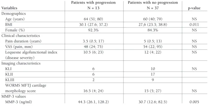
DeMOgRAPHICS AND BASelINe CHARACTeRISTICS Complete baseline and follow-up data were collected from 50 out of 56 (89.2%) patients included in the study. Six patients were excluded from analysis due to lost to follow-up. Cartilage injury progression in the medial tibiofemoral compartment was seen in 13 (26%) participants with KOA. Relevant demographic and clinical features of KOA patients with and without TABle I. exClUSION CRITeRIA fOR THe STUDy
Exclusion Criteria Explanation
End-stage KOA IV KL radiographic stage
Comorbidities Rheumatic diseases: rheumatoid arthritis, psoriatic arthritis, spondyloarthritis, vasculitis and systemic connective tissue diseases, fibromyalgia; decompensated metabolic or cardiovascular disease
Traumatic event Pre-existing intra-articular fracture or documented high-energy trauma of the lower limb OA treatment Systemic glucocorticoids (doses > 7.5 mg) and intra-articular hyaluronan,
glucocorticoids or orthobiotics (e.g. platelet rich plasma) 3 and 6 months prior baseline visit, respectively; treatment with symptomatic slow acting drugs for OA (glucosamine, chondroitin, Avocado/Soybean Unsaponifiables) 6 months prior baseline visit
Deformity of the lower limb Valgus or varus deformity of the knee joints > 20 degrees KL – Kellgren-Lawrence; KOA – knee osteoarthritis; PRP – platelet-rich plasma; OA – osteoarthritis.
compartment by approximately 4% (OR = 1.042, CI 95%: 1.002-1.084, p = 0.04). Potential confounders (age groups, sex, BMI, therapy, gene ralized OA) did not influence the outcome after conducting the MantelHaens -zel test and the partial correlation coefficient.
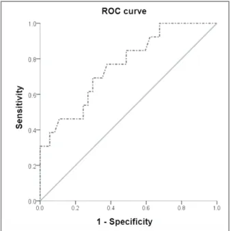
In order to delineate progressors from non-progres-sors by means of the ROC analysis, we used baseline MMP-3 levels (Figure 1). The area under the curve (AUC) for serum MMP-3 levels was 0.761 (95%CI, 0.614-0.910), suggesting a good predictive accuracy of this biochemical test. The ROC curve set high cut--off levels (40.8 ng/ml) with a sensitivity of 69.2% and a specificity of 70.3% for differentiation between pro-gressors and non-propro-gressors. 100% specificity for dis-tiguishing non-progressors from progressors was achieved at biomarker levels below 26.1 ng/ml with a sensitivity of 32.4%.
DISCUSSION
It has already been suggested that higher serum levels advancement of cartilage damage in the medial
tibiofemoral compartment are presented in Table II. Expectedly, progressors have statistically higher body mass index (BMI) than non-progressors (30.1 [27.6; 37.2] vs. 27,6 [23.3; 38.8], p = 0.011). Significantly higher levels of MMP-3 were observed in patients with a progression of cartilage injury in the medial com-partment of the knee (44.3 [26.1, 128.2]) than in pa-tients with no progression (30.7 [12.6; 82.5]) on the 12thmonth (Mann-Whitney U = 114.5, p = 0.005). fOllOw-UP AND TReATMeNT
One-year change in pain and disease severity of the study population, together with treatment during fol-low-up, are shown in Table III dichotomized by the presence and lack of progression in the medial tibiofemoral compartment of the knee joint.
PReDICTIve vAlUe Of BASelINe SeRUM MMP-3 Binary logistic regression analysis showed that each in-crease in MMP-3 with 1 ng/ml inin-creases the chance of progression of cartilage damage in the medial tibiofemoral
TABle II. BASelINe DeMOgRAPHIC, ClINICAl, AND IMAgINg feATUReS Of PATIeNTS wITH PROgReSSION (PROgReSSORS) AND lACk Of PROgReSSION (NON-PROgReSSORS) IN THe MeDIAl TIBIOfeMORAl COMPARTMeNT Of THe kNee jOINT
Patients with progression Patients with no progression
Variables N = 13 N = 37 p-value Demographics Age (years) 64 (51; 80) 60 (40; 79) NS BMI 30.1 (27.6; 37.2) 27,6 (23.3; 38.8) 0.011 Female (%) 92.3% 84.3% NS Clinical characteristics
Pain duration (years) 3.5 (0.5; 17) 5 (0.5; 13) NS
VAS (pain, mm) 48 (24; 75) 54 (22; 95) NS
Lequesne algofunctional index 10.5 (6; 23) 12 (4; 22) NS
(disease severity) Imaging characteristics KLI 6 10 NS KLII 6 17 KLIII 2 9 WORMS MFTJ cartilage morphology score 16.5 (4; 24) 15 (5; 27) NS MMP-3 values MMP-3 (ng/ml) 44.3 (26.1, 128.2) 30.7 (12.6; 82.5) 0.005
BMI = Body Mass Index; GlS = glucosamine sulfate; MMP-3 = matrix metalloproteinase-3; MFTJ = medial femorotibial joint;
KL = Kellgren-Lawrence; NS = not significant; PRP = platelet rich plasma; VAS = visual analogue scale; WORMS = Whole-Organ Magnetic Resonance Imaging Score. The presented data are non-normally distributed and presented as median (min; max)
of MMP-3 and other serum markers are associated with advanced structural changes in patients with KOA and polyarticular involvement of OA2,17,25. One step further would be to understand whether synovial biomarkers and in particular MMP-3, can carry potentially impor-tant information for the prediction of future cartilage injury in OA patients. Logically, higher levels of endo-peptidases would pose a greater risk to more intensive degradation of extracellular matrix components and,
as is the case with MMP-3, activation of other molecu-les from the matrix metalloproteinase family6,7which provides a sound theoretical basis for investigation in this direction. The one-year follow-up period with imaging modalities was in line with the current re-commendations of OARSI for determining the mini-mum time interval for detection and accurate assess-ment of structural changes by X-ray and MRI19. The medial tibiofemoral compartment is the most com-monly affected by OA and more sensitive to change over time in comparison to the other two compart-ments of the knee joint26.
In the study group, MMP-3 levels were associated and were an independent predictor for the progressi-on of structural damage. Any increase in MMP-3 by 1 ng/ml elevated the likelihood of cartilage damage de-terioration by 4%. These results did not depend on BMI, treatment or polyarticular involvement. Based on these data, a ROC curve was constructed which set a value of 40.8 ng/ml as the boundary between the pro-gressors and the non-propro-gressors. Therefore, the pre-dictive value of serum MMP-3 levels for subsequent structural changes suggests that MMP-3 alone or in combination with other prognostic biomarkers may be used as a surrogate indicator for KOA patients with more rapid progression of cartilage damage. Theoreti-cally, patients with higher levels of MMP-3 should be treated more aggressively in the early stages of the di -sease in order to conserve cartilage.
As far as we know, to date, this is the first prospecti -ve observational study that establishes a relationship between MMP-3 levels and the subsequent magnetic resonance progression of cartilage injury in patients fIgURe 1. ROC curve differentiating progressors from
non-progressors using serum MMP-3 (AUC = 0.761) ROC: receiver operating characteristics; MMP-3: matrix metalloproteinase-3; AUC: area under the curve.
TABle III. ONe-yeAR CHANge IN PAIN AND DISeASe SeveRITy AND TReATMeNT DATA Of PATIeNTS wITH PROgReSSION AND lACk Of PROgReSSION IN THe MeDIAl TIBIOfeMORAl COMPARTMeNT Of THe kNee jOINT
Patients with progression Patients with no progression
Variables N = 13 N = 37 P
Change in clinical characteristics
Change in VAS (pain, mm) -10 (-42; 12) -3 (-14; 9) 0.014
Change in Lequesne score -1.5 (-5; 4.5) 0 (-3.5; 2) NS
Treatment
Oral GlS 5 8 NS
Intraarticular hyaluronan 7 24
Intraarticular PRP 1 5
GlS = glucosamine sulfate; NS = not significant; VAS = visual analogue scale. The presented data are non-normally distributed and presented as median (min; max)
with KOA. Nevertheless, an association between struc-tural damage and serum MMP-3 levels was established in 2005 when Lohmander et al. reported that MMP-3 serum concentration could predict radiographic nar-rowing of the joint space observed over a period of 30 months in patients with KOA18. Such a finding was not observed in the present study probably due to the short follow-up period and the small sample size. Serum MMP-3 was also associated with the progression of ra-diographic damage in inflammatory joint diseases such as ankylosing spondylitis, early and advanced rheu-matoid arthritis11,27,28.
Limitations of this study include low sample size and a relatively short follow-up period for registering struc-tural progression in OA. We assume that only 13 pa-tients were classified as progressors, which may repre-sent the main limitation of the work. Since neither any data about the size of a "relevant effect" beforehand were available, nor were much knowledge about the expected variance, our research pretends to be ex-ploratory. It raises the prospect that simple blood tests may be available to predict the progression of the di sease course in patients with KOA. Measuring the le -vels of serum MMP-3 may be even implemented in clinical practice if confirmed in further studies. It may provide additional information to clinicians when mak-ing treatment decisions, since osteoarthritic patients with high cartilage turnover may show increased res -ponsiveness to therapy with cartilage protecting ef-fects29,30. Because our study was limited to an assess-ment of the medial compartassess-ment of KOA and the lateral compartment was not assessed, our results might be somewhat weakened.
CONClUSION
In conclusion, serum MMP-3 levels may serve as a po-tential prognostic biomarker for cartilage injury in pa-tients with KOA, identifying those at higher risk of disea se progression. Further investigations in larger co-horts are warranted before any firm conclusions are made.
CORReSPONDeNCe TO Tsvetoslav Georgiev
Clinic of Rheumatology, University Hospital “St. Marina”, Medical University, Varna
E-mail: tsetso@medfaculty.org RefeReNCeS
1. Fanjul-Fernández M, Folgueras AR, Cabrera S, López-Otín C.
Matrix metalloproteinases: evolution, gene regulation and func-tional analysis in mouse models. Biochim Biophys Acta Mol Cell Res. 2010;1803(1):3-19. PMID: 19631700
2. Chubinskaya S, Kuettner KE, Cole AA. Expression of matrix metalloproteinases in normal and damaged articular cartilage from human knee and ankle joints. Lab Invest 1999;79.12:1669-1677. PMID: 10616215
3. Rose BJ, Kooyman DL. A tale of two joints: the role of matrix metalloproteases in cartilage biology. Dis Markers 2016;2016. PMID: 27478294
4. Cao J, Liu Z, Zhang L, Li J. miR-940 regulates the inflammato-ry response of chondrocytes by targeting MyD88 in os-teoarthritis. Mol Cell Biochem 2019;461(1-2):183–193. PMID: 31435813
5. Nagase H, Ogata Y, Suzuki K, Enghild JJ, Salvesen G. Substrate specificities and activation mechanisms of matrix metallopro-teinases. Biochem Soc Trans 1991 19(3):715–718. PMID: 1783204
6. Djokic J, Fagotto-Kaufmann C, Bartels R, Nelea V, Reinhardt DP. Fibul3,-4, and-5 are highly susceptible to proteolysis, in-teract with cells and heparin, and form multimers. J Biol Chem 2013; 288(31):22821-35. PMID: 23782690
7. Murphy G, Nagase H. Progress in matrix metalloproteinase re-search. Mol Aspects Med 2008; 29:290–308. PMID: 18619669 8. Javaid MA, Abdallah MN, Ahmed AS, Sheikh Z. Matrix metal-loproteinases and their pathological upregulation in multiple sclerosis: an overview. Acta Neurol Belg 2013; 113:381–390. PMID: 24002649
9. Ribbens C, y Porras MM, Franchimont N, Kaiser MJ, Jaspar JM, Damas P, et al. Increased matrix metalloproteinase-3 serum lev-els in rheumatic diseases: relationship with synovitis and steroid treatment. Ann Rheum Dis 2002; 61:161–166. PMID: 11796404
10. Prodanovic SZ, Radunovic G, Babic D, Ristic B, Sefik-Bukilica M, Zlatanovic M, et al. Matrix Metalloproteinases-3 Baseline Serum Levels in Early Rheumatoid Arthritis Patients without Initial Radiographic Changes: A Two-Year Ultrasonographic Study. Med Princ Pract 2018; 27(4):378-86. PMID: 29794470 11. Azukizawa M, Ito H, Nishitani K, Murata K, Okahata A, Tomiza-wa T et al. AB1335 Serum mmp-3 is closely related to knee joint symptoms in rheumatoid arthritis patients: a cross-sectional study from kurama cohort. Ann Rheum Dis 2018; 77:1756-1757. DOI: http://dx.doi.org/10.1136/annrheumdis-2018-eu-lar.5044
12. Pap G, Eberhardt R, Sturmer I. Development of osteoarthritis in the knee joints of Wistar rats after strenuous running exercise in a running wheel by intracranial self-stimulation. Pathol Res Pract 1998; 194(1):41–47. PMID: 9542746
13. Connelly AE, Tucker AJ, Kott LS, Duncan AM, Wright AJ. Serum biochemical markers of joint metabolism and inflammation in relation to clinical symptoms and physical function in adults with symptomatic knee osteoarthritis. Osteoarthritis Cartilage 2014; 22:S66. DOI: https://doi.org/10.1016/j.joca.2014.02.135 14. Li W, Du C, Wang H, Zhang C. Increased serum ADAMTS-4 in knee osteoarthritis: a potential indicator for the diagnosis of os-teoarthritis in early stages. Genet Mol Res 2014; 13(4): 9642-9. PMID: 25501175
15. Tong Z, Liu Y, Chen B, Yan L, Hao D. Association between MMP3 and TIMP3 polymorphisms and risk of osteoarthritis. Oncotarget 2017; 8(48):83563. PMID: 29137364
et al. Decrease in serum level of matrix metalloproteinases is predictive of the disease-modifying effect of osteoarthritis drugs assessed by quantitative MRI in patients with knee osteoarthri-tis Ann Rheum Dis 2010; 69(12):2095-2101. PMID: 20570834 17. Georgiev T, Ivanova M, Kopchev A, Velikova T, Miloshov A, Kurteva E, et al. Cartilage oligomeric protein, matrix metallo-proteinase-3, and Coll2-1 as serum biomarkers in knee os-teoarthritis: a cross-sectional study. Rheumatol Int 2018; 38.5:821-830. PMID: 29164307
18. Lohmander LS, Brandt KD, Mazzuca SA, Katz BP, Larsson S, Struglics A, Lane KA. Use of the plasma stromelysin (matrix metalloproteinase 3) concentration to predict joint space nar-rowing in knee osteoarthritis. Arthritis Rheum 2005; 52(10):3160-7. PMID: 16200596
19. Hunter DJ, Altman RD, Cicuttini F, Crema MD, Duryea J, Eck-stein F, et al. OARSI clinical trials recommendations: knee imag-ing in clinical trials in osteoarthritis. Osteoarthritis Cartilage 2015; 23(5):698-715. PMID: 25952343
20. Altman R, Asch E, Bloch D, Bole G, Borenstein D, Brandt K, et al. Development of criteria for the classification and reporting of osteoarthritis: classification of osteoarthritis of the knee. Arthritis Rheum 1986; 29(8):1039-1049. PMID: 3741515 21. Kellgren JH, Lawrence JS. Radiological assessment of
osteo-arthrosis. Ann Rheum Dis. 1957; 16.4:494. PMID: 13498604 22. Peterfy CG, Guermazi A, Zaim S, Tirman PF, Miaux Y, White D,
et al. Whole-organ magnetic resonance imaging score (WORMS) of the knee in osteoarthritis. Osteoarthritis Cartilage 2004; 12(3):177-190. PMID: 14972335
23. Lynch JA, Roemer FW, Nevitt MC, Felson DT, Niu J, Eaton CB, Guermazi A. Comparison of BLOKS and WORMS scoring sys-tems part I. Cross sectional comparison of methods to assess cartilage morphology, meniscal damage and bone marrow le-sions on knee MRI: data from the osteoarthritis initiative. Os-teoarthritis Cartilage 2010; 18(11):1393-1401. PMID: 20816979
24. Felson DT, Lynch J, Guermazi A, Roemer FW, Niu J, McAlindon T, Nevitt MC. Comparison of BLOKS and WORMS scoring sys-tems part II. Longitudinal assessment of knee MRIs for os-teoarthritis and suggested approach based on their performance: data from the Osteoarthritis Initiative. Osteoarthritis Cartilage 2010; 18(11):1402-1407. PMID: 20851202
25. Takahashi M, Naito K, Abe M, Sawada T, Nagano A. Relation-ship between radiographic grading of osteoarthritis and the bio-chemical markers for arthritis in knee osteoarthritis. Arthritis Res Ther. 2004; 6(3):R208. PMID: 15142266
26. Amin S, LaValley MP, Guermazi A, Grigoryan M, Hunter DJ, Clancy M, et al. The relationship between cartilage loss on mag-netic resonance imaging and radiographic progression in men and women with knee osteoarthritis. Arthritis Rheum 2005; 52(10):3152-9. PMID: 16200595
27. Maksymowych WP, Landewé R, Conner Spady B, Dougados M, Mielants H, van der Tempel H, et al. Serum matrix metalloteinase 3 is an independent predictor of structural damage pro-gression in patients with ankylosing spondylitis. Arthritis Rheum 2007; 56(6):1846-53. PMID: 17530713
28. Houseman M, Potter C, Marshall N, Lakey R, Cawston T, Grif-fiths I, et al. Baseline serum MMP-3 levels in patients with Rheumatoid Arthritis are still independently predictive of ra-diographic progression in a longitudinal observational cohort at 8 years follow up. Arthritis Res Ther 2012; 14(1):R30. PMID: 22314025
29. Georgiev T, Angelov AK. Modifiable risk factors in knee os-teoarthritis: treatment implications. Rheumatol Int 2019; 39(7):1145-1157. PMID: 30911813
30. Christgau S, Henrotin Y, Tanko LB, Rovati LC, Collette J, Bruyère O, et al. Osteoarthritic patients with high cartilage turnover show increased responsiveness to the cartilage pro-tecting effects of glucosamine sulphate. Clin Exp Rheumatol 2004; 22(1):36. PMID: 15005002