1. Reumatologia, Centro Hospitalar e Universitário de Coimbra 2. Reumatologia, Hospital Garcia de Orta
3. Reumatologia, Centro Hospitalar Universitário Lisboa Norte
Keywords: Systemic lupus erythematosus; Ultrasound;
Gastrointestinal involvement.
IntroductIon
Gastrointestinal (GI) complaints are common in Systemic lupus erythematosus (SLE) patients1,2. Early clini cal pre-sentation of lupus enteritis (LE) is unremarkable and non--s pecific, comprising diffuse abdominal cramps or per-sistent pain, nausea, vomiting, fever and diarrhea, making early clinical suspicion of LE difficult to eli cit3,4. Also, glu-cocorticoids and immunosuppressants can mask classical signs of an acute abdomen in SLE patients. When dia -gnosed early, LE usually responds well to treatment with high-dose glucocorticoids, but if left unchecked it can lead to life-threatening complications such as ischemic enteritis, bowel infarction with blee ding and/or perfora-tion and peritonitis.5The aim of this case review is to iden-tify possible strategies for early diagno sis of LE.
Methods
Retrospective analysis of patients with SLE (fulfilling ACR 1997 and/or SLICC classification criteria and reg-istered in the reuma.pt national database) from three tertiary SLE centers, with a clinical diagnosis of LE, be-tween 1999 and 2018, was conducted. The diagnosis was based on clinical and imaging features consistent with LE and exclusion of other causes of GI disorders. Patients with associated antiphospholipid syndrome or with positive antiphospholipid antibodies were ex-cluded. Ultrasound scan was always performed by an experienced radiologist.
results
Seven cases of LE were identified in the participating
cen-ACTA REUMATOL PORT. 2019;44:145-150
How to diagnose lupus enteritis early?
Lessons learned from a multicenter case series
Luís M1, Brites AL1, Duarte AC2, Teixeira V3, Freitas R2, Oliveira-Ramos F3, Macieira C3, Santos MJ2, Inês L1
AbstrAct
Introduction: Lupus enteritis (LE) is a rare,
potential-ly life-threatening manifestation of systemic lupus ery-thematosus (SLE). Early diagnosis is crucial for early treatment and prevention of serious complications such as ischemic enteritis, bowel infarction with bleeding and/or perforation and peritonitis. The objective of this case review is to identify strategies for early diagnosis of LE.
Methods: Retrospective analysis of patients with SLE
(fulfilling ACR 1997 and/or SLICC classification crite-ria) and presenting LE from three tertiary SLE centers was conducted. The diagnosis was based on clinical and imaging features consistent with LE and exclusion of other causes of GI disorders.
Results: We report seven cases of LE (female: 100%;
age range: 16-55 years). All presented with acute onset abdominal pain, nausea and vomiting at the emergen-cy room. Two patients had lupus enteritis as inaugural manifestation of SLE. Of the remaining five, one pre-sented at the previous visit to the lupus clinic with clin-ically active disease and two had serologclin-ically active/ clinically inactive SLE. High anti-dsDNA antibodies and low serum complement were universally present at time of the LE event. Abdominal ultrasound was the first imaging exam to be performed in the emergency room. In all cases it showed bowel wall thickening, di-latation of intestinal segments, increased reflectivity of mesenteric fat and mild ascites, raising the suspicion of LE and immediate start of treatment. These features were later confirmed by CT scan in five patients.
Discussion: Despite being rare, LE must always be
con-sidered in any SLE patient presenting with GI symp-toms. Abdominal ultrasound can be a reliable first line diagnostic tool for LE.
ters. The main characteristics of these patients are summarized in Table I. Each case is presented below. #PAtIent 1
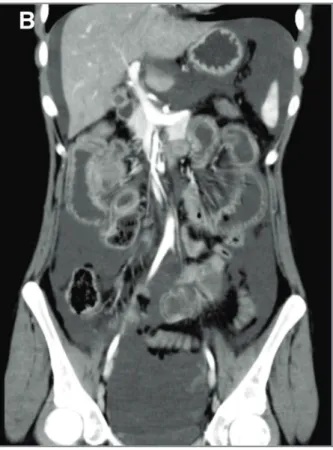
An 18-years-old female was admitted in the emergen-cy room with acute diffuse abdominal pain, fever, nau-sea, vomiting and absence of stools and gas emissions over the previous 4 days. She had been diagnosed with SLE almost one year earlier based on cutaneous, arti -cular, hematological and immunological features. At admission, her abdomen was mildly tender without audible bowel sounds. Laboratory tests showed low complement, new onset anemia, slight hypoalbu-minemia and increased serum ferritin. Erythrocyte sedimentation rate (ESR), C-reactive protein (CRP) and lactate dehydrogenase (LDH) were within normal ranges. An abdominal ultrasound (US) revealed mo -derate ascites and diffused wall thickening of the colon with increased reflectivity of subjacent fat tissue. Abdo -minal computed tomography (CT) clearly showed in-creased wall thickening with abnormal enhancement (target sign) - mainly in the jejunum and ileum but also in the ascending colon - and engorgement of mesenteric vessels. Multiple enlarged mesenteric lymph nodes were also present (Fi gure 1). She was started on IV methylprednisolone (MPDN) 500mg pulses followed by oral prednisone (1mg/kg/day). Twenty-four hours later the symptoms quickly started to resolve and she was discharged 6 days later. After more than a year, the patient remains free of GI symp-toms although soon after the present episode lupus nephritis developed. She was started on induction therapy with mycophenolate mofetil (MMF) 2.5g/d with good response.
#PAtIent 2
A 43yearsold female, with a 10year history of SLE -based on the presence of oligoarthritis, pericarditis, lymphopenia, photosensitive malar rash and im-munological markers - presented with a two-day his-tory of diffuse acute abdominal pain, vomiting and dy-suria. She also described pain and swelling of her left ankle for the last two months. Physical examination showed a tender abdomen and left ankle arthritis. ESR (58 mm/1sthour) and LDH (326 mg/dl) were high while CRP was slightly elevated (0.62 mg/dl). Urinary sediment analysis showed new onset proteinuria (527.5 mg/ 24h). Large ascites and jejunum-ileum wall thickening were seen on US. Abdominal CT scan con-firmed the presence of thickened intestinal loops with
target sign at the left iliac fossa, dilatation of the small bowel lumen and multiple enlarged mesenteric lymph nodes. Additionally, the lateral wall of bladder was also thickened suggesting concomitant involvement of the urinary tract. She was started on IV MPDN 1g pulses followed by oral prednisone 1 mg/kg/day, with com-plete resolution of symptoms within 3 days. At 1-year follow-up, under no therapy apart from hydroxy-chloroquine (HCQ), the patient remained free of ab-dominal complaints or other signs of disease activity. #PAtIent 3
A 16-years-old female had been diagnosed with SLE 3 years earlier, with cutaneous, articular, neurological, hematological, renal and immunological involvement. She suffered from late stage chronic kidney disease and secondary hypertension as a result of a previous severe class IV lupus nephritis. She was admitted to the emer-gency room with acute onset diffuse abdominal pain, nausea and vomiting that started less than 24 hours before. She was hemodynamically unstable, had a ten-der abdomen and no bowel sounds. Laboratory tests showed worsening of renal function and hypoalbu-minemia. Both ESR and CRP were normal. US revealed small bowel wall edema, increased vascularization on Doppler and moderate ascites. She wasn’t submitted to abdominal CT since her renal function didn’t allow the administration of iodate contrast. IV MPDN 1g pulses were immediately started with excellent clinical re-sponse, although she maintained changes in control US 3 days later. After hospital discharge, recurrent self--limited abdominal pain episodes persisted. Two years later, she presented again with abdominal pain, this time more focused on the right iliac fossa, and diar-rhea. US confirmed the presence of thickened and hy-poechogenic ascending colon wall with enhancement of the surrounding fat tissue and associated bilateral hydronephrosis with bladder wall thickening. CT scan showed thickening of small bowel wall, multiple small mesenteric adenopathies, increased number of ob-served mesenteric vessels and mild ascites. She was again treated with IV MPDN 1g pulses, with no relapse until today, after 5 years follow-up. Due to progressive renal function deterioration, she is currently being considered for kidney transplant.
#PAtIent 4
A 40-years-old female, with a 21-year history of SLE with hematological, renal and immunological in-volvement, had been suffering from recurrent episodes
tA b le I. c lI n Ic A l A n d l A b o r A to r y c h A r A ct er Is tI cs , t r eA tM en t A n d f o ll o w -u P P at ie n t 1 P at ie n t 2 P at ie n t 3 P at ie n t 4 P at ie n t 5 P at ie n t 6 P at ie n t 7 A g e (y ea rs ) 1 8 4 3 1 6 4 0 3 6 3 4 1 8 G en d er F F F F F F F D is ea se d u ra ti o n ( y ea rs ) 1 1 0 3 2 1 0 0 1 P re v io u s S L E m an if es ta ti o n s C u ta n eo u s C u ta n eo u s C u ta n eo u s H em at o lo g ic N A N A C u ta n eo u s A rt ic u la r A rt ic u la r A rt ic u la r Im u n o lo g ic A rt ic u la r H em at o lo g ic H em at o lo g ic H em at o lo g ic R en al H em at o lo g ic Im u n o lo g ic Im u n o lo g ic Im u n o lo g ic Im u n o lo g ic S er o so u s N eu ro lo g ic R en al T h er ap y ( m g /d ay ) H C Q 4 0 0 H C Q 4 0 0 H C Q 4 0 0 H C Q 4 0 0 N /A N A N o n e P D N 5 P D N 7 .5 M P D N 4 P D N 5 M M F 2 0 0 0 M M F 2 5 0 0 E x tr a-G I m an if es ta ti o n s L u p u s cy st it is L u p u s cy st it is L u p u s n ep h ri ti s L u p u s cy st it is A rt h ri ti s A rt h ri ti s N ep h ri ti s A rt h ri ti s re la p se A u to im m u n e S er o si ti s h em o ly ti c an em ia SL E D A I (p re vi o u s/ d u ri n g fl ar e) 0 /1 3 4 /1 7 0 /1 3 4 /1 6 -/ 1 8 -/ 1 6 1 2 /3 3 L ab o ra to ry w o rk -u p C 3 0 .6 C 3 0 .5 4 C 3 0 .3 6 C 3 0 .5 9 C 3 0 .4 9 C 3 0 .5 6 C 3 0 .3 3 C 4 0 .0 6 C 4 0 .0 7 C 4 0 .0 2 C 4 0 .0 3 C 4 < 0 .1 C 4 0 .1 3 C 4 0 .0 8 an ti -d sD N A 1 4 0 an ti -d sD N A > 5 0 an ti -d sD N A an ti -d sD N A 1 6 an ti -d sD N A > 5 0 an ti -d sD N A 1 2 8 4 an ti -d sD N A > 3 7 9 C R P 0 .2 C R P 0 .6 > 5 0 C R P < 0 .5 C R P 1 .6 C R P 5 .1 C R P 0 .2 C R P 2 7 E S R 2 E S R 5 4 E S R < 1 5 E S R 8 E S R 1 0 0 E S R 2 3 E S R 3 1 A b d o m in al U S f in d in g s Ye s Ye s Ye s Ye s Ye s Ye s Ye s su g g es ti v e o f L E A b d o m in al C T fi n d in g s Ye s Ye s N A Ye s Ye s Ye s N A su g g es ti v e o f L E F la re t re at m en t IV M P D N IV M P D N IV M P D N IV M P D N IV M P D N P D N 1 m g /k g /d ay I V M P D N 5 0 0 m g 3 d ay s 1 g 3 d ay s 1 g 3 d ay s 1 g 3 d ay s 1 g 3 d ay s fo ll o w ed b y 1 g 3 d ay s fo ll o w ed b y fo ll o w ed b y I V IG IV M P D N fo ll o w ed b y IV C Y C 5 0 0 a n d 4 0 0 m g /k g /d ay 1 g 3 d ay s C p A P D N 1 m g /k g /d ay 5 d ay s 1 0 0 m g /d ay F o ll o w -u p N o r el ap se N o r el ap se R ec u rr en t R el ap se 3 a n d N o r el ap se N ee d o f p ar en ta l N o r el ap se ab d o m in al p ai n h al f m o n th s n u tr it io n a n d an d r el ap se la te r sl o w c li n ic al 2 y ea rs l at er re so lu ti o n C 3 : C 3 c o m p le m en t (n o rm al r an g e: 0 .8 0 -1 .8 5 g /L ); C 4 : C 4 c o m p le m en t (n o rm al r an g e: 0 .1 5 -0 .5 3 g /L ); C R P : C -r ea ct iv e p ro te in ( n o rm al r an g e: 0 -0 .5 m g /d L ); C p A : C y cl o sp o ri n e; C Y C : cy cl o p h o sp h am id e; E S R : er y th ro cy te s ed im en ta ti o n r at e (n o rm al r an g e: 0 -2 0 m m /h ); F : fe m al e; G I: g as tr o in te st in al ; H C Q : h y d ro x y ch lo ro q u in e; I V IG : in tr av en o u s im m u n o g lo b u li n ; M M F : m y co p h en o la te m o fe ti l; M P D N : m et h y lp re d n is o lo n e; N : w it h in n o rm al r an g e; N A : n o t ap p li ca b le ; P D N : p re d n is o lo n e; S L E D A I: S y st em ic L u p u s E ry th em at o su s D is ea se A ct iv it y In d ex ; U S u lt ra so u n d . F : fe m al e.
of severe hemolytic anemia. She developed acute abdo minal pain, nausea, vomiting, increased abdominal vo -lume and anorexia. At hospital admission, the patient was pale and her abdomen tender and painful. Labo-ratory tests showed anemia, lymphopenia, increased CRP and decreased haptoglobin levels. US revealed as-cites, increased echogenicity of renal parenchyma, slight right side hydronephrosis, and thickening of the bladder wall, pyloric region and several segments of the bowel wall. She was then submitted to an abdom-inal CT scan that revealed moderate to severe ascites, submucosal edema of the antral and pyloric regions, ileum and colon and severe, irregular, bladder wall thickening with reduced distensibility resulting in bi-lateral hydronephrosis. The treatment strategy was sim-ilar to the previous cases. Although the GI symptoms resolved after 48 hours, she kept showing signs of hemolytic anemia and was started on IVIg (400 mg/kg/day for 5 days), with benefit. Three and half months later, she suffered a relapse of the LE and re-peated IV MPDN 1g pulses followed by rituximab 1g which had to be switched to MMF 2.5g/day after an in-fusion reaction. At 2-year follow-up the patient is asymptomatic and with no sign of relapse.
#PAtIent 5
A 36 years-old female with a history of intermittent self-limited polyarthritis since the age of 15 and one episode of thrombocytopenia during pregnancy one year ago, was admitted with acute diffuse abdominal pain and persistent vomiting lasting for 24 hours. Phy -sical examination showed signs of dehydration and a tender abdomen. Laboratory tests revealed thrombo-cytopenia, increased ESR and CRP, low complement fractions and positive antinuclear, anti-dsDNA and anti- 2glicoprotein I IgG antibodies. She was then diagnosed with SLE. In order to clarify the acute abdo minal condition, she performed US which demons -trated moderate ascites and severe irregular thickening of bowel wall, suggestive of edema. CT scan suggested the diagnosis once more: it described diffuse thicken-ing of the small bowel wall with intraluminal dilatation and fluid collection, increased attenuation of mesen-teric fat tissue and slight left pleural effusion. A biopsy of the jejunum was performed: the mucosa did not pre-sent remarkable changes; the submucosa showed ede-ma, vascular congestion and focal hemorrhage; in the serosal layer, there was diffuse fibrosis, congestive ves-sels and predominantly eosinophilic inflammatory in-fIGure 1. Abdominal US (A) and CT (B) from Patient 1
showing similar findings: moderate ascites, increased wall thickening of the colon with abnormal enhancement (target sign) and increased reflectivity of subjacent fat tissue.
filtrate. She was successfully treated with IV MPDN 1g pulses with good response and remains without any signs of abdominal disease activity after more than 20 years. She is currently treated with HCQ and lefluno-mide for her arthritis.
#PAtIent 6
A previously healthy 36-years-old black female had been complaining of inflammatory polyarthralgia for the last 4 months, when she started suffering from nau-sea, vomiting, diarrhea and diffuse abdominal pain. At hospital admission, the patient was dehydrated, pale, had a tender abdomen and peripheral polyarthritis. In-fectious etiology was ruled out after an extensive workup. She tested positive for antinuclear and anti-Smith antibodies. Her anti-dsDNA was high and C3 complement fraction reduced. ESR and CRP were nor-mal. US showed moderate ascites and diffuse small bowel wall thickening. Enteric MRI found diffuse wall thickening of the jejunum and distal ileum, intralumi-nal dilatation, congestive mucosal folds, target sign, en-gorgement of mesenteric vessels and increased sup-pression of mesenteric fat. A subsequent abdominal CT scan revealed similar findings: diffuse small bowel wall thickening and intraluminal distention, especially in the terminal ileum. There was wall thickening of the descending colon to the splenic angle, without dilata-tion or obstrucdilata-tion. Moderate ascites was also present. Colon biopsy result was unspecific for vasculitis but allowed the exclusion of other differential diagnosis (tuberculosis, CMV colitis and Whipple disease among others). She received prednisone 1 mg/kg/day with no response. Nasogastric intubation and parenteral nutri-tion were required, and treatment with IV MPDN 1g pulses was started, followed by oral azathioprine. She fully recovered and remains symptom and relapse-free until today, after a 3-year follow-up.
#PAtIent 7
An 18-year-old female with a recent diagnosis of SLE based on hematological, articular, mucocutaneous and immunological features, presented with fever, diffuse abdominal pain, nausea, vomiting and bloody diarrhea in the emergency room. At physical examination, she had a tender abdomen and severe peripheral edema. Laboratory tests revealed anemia, increased CRP, low complement fractions and positive anti-dsDNA. Pro-teinuria was identified for the first time at this point (2.4g/ 24h). Abdominal US showed moderate parietal thickening of the left colon and rectum walls, mild
as-cites and slight hepatosplenomegaly. It was also evident the presence of bilateral pleural effusion and mini -mal pericardial effusion. Kidney biopsy revealed mesangial proliferative glomerulonephritis and throm-botic microangiopathy. She was immediately started on IV MPDN 1g pulses for 3 days followed by cyclosporine 100 mg/day. She was discharged from the hospital 10 days later, after total symptom resolution. At one-year follow-up the patient remains symptom-free, with complete kidney response (proteinuria 0.8 mg/24h).
dIscussIon
Many GI conditions can mimic LE. The differential diagno sis can be rather challenging since some of these conditions, despite being nonspecific for SLE, are more prevalent in these patients, such as infectious colitis (parasitic, bacterial or viral), peritonitis, acute pancre-atitis, autoimmune heppancre-atitis, primary biliary cirrhosis, mesenteric thrombosis (more commonly associated with the presence of antiphospholipid antibodies) or even GI toxicity of drugs.4,6,7
Several predisposing factors for LE have been pro-posed, such as simultaneous peripheral or central ner-vous system vasculitis, thrombocytopenia and serum rheumatoid factor.8None of them were present in our series. All of our patients presented with acute onset abdominal pain, mainly diffuse, with associated nausea and vomiting. Three patients suffered from concomi-tant lupus cystitis, characterized by urinary bladder wall edema and thickening and, in one case, hy-dronephrosis. This association was previously de-scribed in the literature and was even proposed as risk factor for recurrence.9,10
Patients 5 and 6 had LE as inaugural manifestation of SLE. In both cases, SLE was suggested by the con-comitant presence of arthritis and positive immuno-logical markers of the disease (antinuclear and anti--Smith antibodies, high anti-dsDNA and low comple-ment), although both ESR and CRP were within their normal range.
In our case series, all patients but two underwent abdominal CT scan and all of them fulfilled the crite-ria proposed by Byun for LE.11The widespread use of abdominal CT allowed higher diagnosis accuracy, ear-ly screening for complications, treatment planning and disease follow-up. However, CT findings in LE, despite its high sensitivity, carry a low specificity since they can be found in other GI conditions, mainly pancreatitis,
mechanical bowel occlusion, peritonitis and inflam-matory bowel disease.5,12
Patients 5 and 6 were submitted to intestinal biopsy. In patient 5, histological findings were indeed su -ggestive of mesenteric vasculitis. In patient 6, on the other hand, the result was inconclusive for vasculitis but allowed the exclusion of other possible conditions, such as CMV colitis, Whipple disease and other gra nulomatous diseases. Janssens et al. conducted a lite -rature review and concluded that in the 150 cases al-ready published of LE, 25 had been submitted to endoscopic studies. Of these, in 15 cases there was no macroscopic changes. The histological examination presented a low sensitivity, with only 55,6% of the cas-es showing necrotizing vasculitis and/or fibrinoid necrosis.3
US is an underestimated diagnostic tool in LE, but in our case series, it has been shown to be a valuable screening tool for early diagnosis (Figure 1). It is like-ly to be the most readilike-ly available imaging method, and a valuable alternative to CT scan when the later is not available or is contraindicated. Patient 3 is a good exam ple of the potential role of US where hemody-namically instability and acute renal failure precluded the use of CT scan. It also performed better than any serological marker, including CRP and ESR which were frequently normal. Although this may seem odd con-sidering lupus enteritis is a vasculitis, CRP and ESR are poor serological markers for many other SLE manifes-tations including arthritis and even nephritis.
Being a more accessible technique, it was the first imaging exam in all our patients when they present at the emergency room. In all of them US showed thicke -ning of the intestinal wall, dilation of intestinal seg-ments, increased reflectivity of mesenteric fat and mild ascites. CT scan subsequently confirmed these features, suggesting that both methods have similar sensitivity for the diagnosis of LE. Regarding specificity, US has the same limitations as CT scan, but with advantages alrea dy mentioned. Considering the demographic characte -ristics of these patients (young women of child-bearing age), US is likely the best screening exam for sus pected LE diagnosis.
conclusIon
Despite being rare, LE must always be considered in any SLE patient presenting with GI symptoms. US proved to be a reliable first line diagnostic tool for LE
and a reasonable alternative to CT scan in selected pa-tients.
corresPondence to
Mariana Luís
Serviço de Reumatologia
Centro Hospitalar e Universitário de Coimbra Praceta Mota Pinto
3000-075, Coimbra
E-mail: [email protected]
references
1. Fawzy M, Edrees A, Okasha H, El Ashmaui A, Ragab G. Gas-trointestinal manifestations in systemic lupus erythematosus. Lupus 2016; 25: 1456–1462.
2. Tian XP, Zhang X. Gastrointestinal involvement in systemic lu-pus erythematosus: Insight into pathogenesis, diagnosis and treatment. World J Gastroenterol. Jun 28, 2010; 16(24): 2971--2977.
3. Janssens P, Arnaud L, Galicier L, Mathian A, Hie M, Sene D, et al. Lupus enteritis: from clinical findings to therapeutic man-agement. Janssens et al. Orphanet Journal of Rare Diseases 2013, 8:67.
4. Schulz SW, Derk CT. The Gastrointestinal Manifestations of Systemic Lupus Erythematosus: A Survey of the Literature. The Open Autoimmunity Journal, 2009, 1:10-26.
5. Lee C-K, Ahn MS, Lee EY, Shin JH, Cho Y-S, Ha HK, et al. Acute abdominal pain in systemic lupus erythematosus: focus on lu-pus enteritis (gastrointestinal vasculitis). Ann Rheum Dis 2002;61:547–550
6. Lahita RG. Systemic Lupus Erythematosus. San Diego: Aca-demic Press 1999: 534.
7. Kirby JM, Jhaveri KS, Maizlin ZV, Midia M, Haider E, Khalili K. Abdominal manifestations of systemic lupus erythematosus: spectrum of imaging findings. Can Assoc Radiol J 2009;60:121--132.
8. Zizic TM, Classen JN, Stevens MB. Acute abdominal complica-tions of systemic lupus erythe-matosus and polyarteritis no-dosa. Am J Med 1982;73:525–531.
9. Koo BS, Hong S, Kim YJ, Kim Y-G, Lee C-K, Yoo B. Lupus en-teritis: clinical characteristics and predictive factors for recur-rence. Lupus 2015; 24: 628–632.
10. Maruyama A, Nagashima T, Iwamoto M, et al. Clinical charac-teristics of lupus enteritis in Japa-nese patients: the large intes-tine-dominant type has features of intestinal pseudo-obstruc-tion. Lupus 2018; 27:1661-1669.
11. Byun JY, Ha HK, Yu SY, Min JK, Park SH, Kim HY, et al. CT fea-tures of systemic lupus ery-thematosus in patients with acute abdominal pain: emphasis on ischemic bowel disease. Radi-ol-ogy. 1999; 211:203-209.
12. Taourel PG, Deneuville M, Pradel JA, Régent D, Bruel JM. Acute mesenteric ischemia: diagno-sis with contrast-enhanced CT. Ra-diology 1996;199:632-636.