Analysis of Endodontic Pathogens by Classical
Microbiological and Nucleic Acid Approaches
Ana Cláudia Morais de Moura Teles de Sampaio
Tese de Doutoramento em Patologia e Genética Molecular
Instituto de Ciências Biomédicas Abel Salazar - Universidade do Porto
Analysis of Endodontic Pathogens by Classical
Microbiological and Nucleic Acid Approaches
Ana Cláudia Morais de Moura Teles de Sampaio
Tese de Candidatura ao grau de Doutor em
Patologia e Genética Molecular submetida ao
Instituto de Ciências Biomédicas Abel Salazar
da Universidade do Porto.
Orientador:
Doutor José Manuel Cabeda
Categoria:
Professor Associado
Afiliação:
Faculdade de Ciências da Saúde
Universidade Fernando Pessoa
Orientador:
Doutora Cristina Pina
Categoria:
Professora Auxiliar
Afiliação:
Faculdade de Ciências da Saúde
Universidade Fernando Pessoa
Orientador:
Doutora Conceição Manso
Categoria:
Professora Associada
Afiliação:
Faculdade de Ciências da Saúde
Universidade Fernando Pessoa
Título:
Analysis of Endodontic Pathogens by Classical
Microbiological and Nucleic Acid Approaches
Autor:
Ana Cláudia Morais de Moura Teles de Sampayo
Edição:
do Autor
ISBN:
À Conchinha e ao Duartinho,
que a minha lealdade e motivação vos sirva de modelo nas vossas vidas.
Aos meus Pais,
Jorge e Mary
“ A ciência, a ciência, a ciência...
Ah, como tudo é nulo e vão!
A pobreza da inteligência
Ante a riqueza da emoção!
Aquela mulher que trabalha
Como uma santa em sacrifício,
Com tanto esforço dado a ralha!
Contra o pensar, que é o meu vício!
A ciência! Como é pobre e nada!
Rico é o que alma dá e tem”.
Fernando Pessoa, 1934
AGRADECIMENTOS
A apresentação a provas para a obtenção do grau de Doutor em Patologia
e Genética Molecular constitui um motivo de grande realização
profissional. Sempre gostei de me pôr à prova e sem dúvida que este
projecto constituiu um grande desafio pessoal, pois tive que equilibrar as
facetas de filha, mulher, mãe, docente, médica dentista e estudante!
Acredito que este estudo poderá contribuir, ainda que modestamente,
para o prestígio científico da Universidade do Porto e da Universidade
Fernando Pessoa (Porto), particularmente das Unidades Curriculares de
Endodontia da FCS-UFP, Instituição à qual me honro de estar ligada, desde
o ano lectivo 1999/2000, como docente, inicialmente, na qualidade de
ainda estudante de Mestrado e, actualmente, como aluna de
Doutoramento do ICBAS.
O presente trabalho deve a sua realização a muitas pessoas, que, em
diversas frentes, de uma forma concertada e determinada, me ajudaram a
tornear as dificuldades que, naturalmente, foram surgindo.
Impõe-se salientar, neste agradecimento, aqueles que se revelaram
fundamentais ao meu desenvolvimento pessoal e profissional, permitindo
que chegasse a este momento:
• O meu orientador, Professor Doutor José Manuel Cabeda, que, por
um acaso muito feliz para mim, se cruzou comigo, neste percurso. É
credor do meu intenso e sincero reconhecimento pelo inesgotável
apoio científico, pela dedicação, pela competência, pela superior
orientação, pelo auxílio e pela confiança demonstrados. Os seus
conselhos, as suas orientações críticas, a disponibilidade e o
empenho que sempre demonstrou, o incentivo oportuno e
experiente, a determinação e a persistência em momentos decisivos,
a calma e a dedicação incondicional, foram, muito para além de todo
o apoio informático, nestes anos de trabalho, contributos essenciais
para a realização deste projecto.
• A minha co-orientadora, Professora Doutora Cristina Pina, a quem
quero manifestar a mais profunda gratidão pelo seu incondicional
incentivo, pela amizade (as suas simpáticas palavras foram uma
ajuda absolutamente preciosa nos momentos mais delicados!), pela
competência e pela inexcedível colaboração. A sua confiança e a sua
cooperação foram essenciais no tratamento e análise dos dados,
bem como na revisão do trabalho.
• A minha co-orientadora, Professora Doutora Conceição Manso, por
quem sinto uma especial admiração (é, para mim, um exemplo de
dedicação e profissionalismo que procurarei seguir na minha vida)
que evoluiu, ao longo destes meses, (atrevo-me!), para um grande
carinho; pela motivação que me transmitiu e pela generosidade da
sua sapiente e pragmática presença, foi possível tratar
estatisticamente, até à exaustão, todos os dados que resultaram
deste projecto. É pelas suas determinação e persistência, em
momentos decisivos, que eu continuo a gostar muito de fazer
investigação.
• A Fundação Fernando Pessoa, particularmente o Digníssimo Reitor,
Senhor Professor Doutor Salvato Trigo, o Director da FCS-UFP,
Senhor Professor Doutor Luís Martins, e o Coordenador
Científico-Pedagógico, Senhor Professor Doutor Carlos Silva, pelo incentivo
prestado ao meu percurso académico.
• Ao Instituto Superior de Ciências Biomédicas Abel Salazar,
instituição que me recebeu como aluna de Doutoramento em
Patologia e Genética Molecular, providenciando as condições
necessárias para o objectivo a que me propus.
• A Directora Clínica da Clínica Pedagógica de Medicina Dentária da
FCS-UFP, Senhora Professora Doutora Sandra Gavinha (cujo
permanente incentivo e contagiantes empenho e dedicação ao
Mestrado Integrado em Medicina Dentária são, para mim, um
superior exemplo de conduta profissional), por todo o apoio e
diligências tomadas na viabilização deste trabalho.
• A Senhora Engenheira Ana Amado e toda a sua equipa do CERLAB,
pela disponibilidade e prontidão com que responderam às minhas
solicitações.
• A minha aluna Sara Loureiro, pela grande amizade, pelo
profissionalismo, pela dedicação, pelo sentido de ajuda e de
responsabilidade, pelo empenho e pela disponibilidade inesgotáveis,
durante toda a parte clínica deste projecto.
• O Ricardo Silva e a Inês Madeira (CEBIMED), pela simpatia com que
sempre me receberam e pelo apoio incansável e incondicional no
processamento laboratorial das amostras.
• A equipa do Secretariado de Doentes das consultas de Medicina
Dentária, Rita Pestana, Nuno Miranda e Eduardo Morais, pela aposta
na implementação das consultas, pelo apoio experiente e irrestrito
ao longo dos cerca de 18 meses de consultas que tornaram este
projecto de investigação uma realidade ao serviço de todos os
Doentes e, muito particularmente, à Patrícia Faria, pela forma serena
e calorosa com que acompanhou o desenvolvimento do trabalho,
suavizando a natureza burocrática de alguns momentos.
• Todos os Doentes que aceitaram participar neste estudo, sem os
quais tudo não teria passado de um sonho, pela paciência e pela
colaboração.
• A equipa de Assistência e Esterilização da Clínica de Medicina
Dentária da FCS-UFP, Clara dos Santos, Susana Neto, Vanessa
Machado, que viram os seus trabalhos acrescidos, em especial, a
Cristina Ferreira, que, desde o primeiro momento, mostrou
incansável disponibilidade e uma capacidade notável de
organização, com a preocupação de que tudo estivesse pronto para
a obtenção de amostras válidas.
• Ao Senhor Gomes que está sempre pronto a resolver problemas
técnicos com um sorriso precioso.
• O Senhor Professor Doutor Pedro Barata que, de forma muito
simpática (e gratuita!), acedeu ao fornecimento de aguns dos
materiais testados. Muito agradeço o profissionalismo, a diligência e
a preocupação constantes.
• O Senhor José Nunes, do GRUPOTAPER-Portugal pela confiança e
prontidão com que nos cedeu, gentilmente, o microscópio utilizado
neste trabalho. Muito obrigada!
• A Senhora Dona Ilda Santos, representante no Norte de Portugal da
Maillefer®, pela cedência de material usado nas consultas.
• O Senhor Professor Doutor José Siqueira Júnior, cuja prontidão e
cujo empenho revelados, por correio electrónico, ultrapassaram tudo
o que algum dia pude esperar de alguém que é uma referência
mundial na área da Endodontia; teria sido muito mais difícil, sem as
suas orientações iniciais, no estabelecimento do protocolo clínico,
bem como, numa fase posterior, pela consulta das suas publicações,
cujo rigor científico, baseado na evidência, é um exemplo que
procuro seguir.
• A toda a equipa do Laboratório de Televisão da UFP, concretamente
ao Dr. Frederico Lopes, Dra. Ana Cristina Santos e Dra. Isabel Portela
o meu mais profundo reconhecimento não só pelo empenho,
simpatia e disponibilidade permanentes, mas também pela
prestimosa e intensa cooperação na realização das gravações e
posteriores edições em fomato de vídeo intento expressar uma
saudação deveras afectuosa. Foi uma experiência muito
enriquecedora para mim e que muito contribuiu para a divulagação
desta investigação.
• A equipa de Endodontia da FCS-UFP, que me soube compreender e
atender os meus pedidos de colaboração; este trabalho não teria
sido possível sem a sua energia contagiante, que nos faz trabalhar
cada vez mais e melhor!
• Os Colegas da Universidade Fernando Pessoa, que se mostraram
solidários quer no encaminhamento de Doentes quer nos momentos
em que a minha gestão de tempo entre as consultas e as aulas foi
mais apertada. Revelaram nesses momentos, apesar do acréscimo
de trabalho para eles, uma boa vontade constante e um enorme
sentido de responsabilidade profissional.
• Os meus alunos da FCS-UFP, que me motivam a prosseguir com
maior empenhamento e sentido de responsabilidade a carreira de
docência. Aprendo e creço muito com eles.
• A todos quantos, de forma directa ou indirecta, frequentemente
anónima, contribuíram para tornar possível este trabalho, desejo
aqui deixar uma saudação muito afectuosa.
• As minhas amigas, conquistadas durante o Doutoramento, Mestre
Lídia Cunha e Professora Doutora Sara Ricardo, pelo apoio nos bons
e maus momentos.
• A Professora Doutora Cláudia Rodrigues, pela mão amiga sempre
estendida, pelos ouvidos atentos e pelas palavras sábias.
• A minha Mana Joana que, mesmo à distância, continua a fazer junta
comigo um percurso de vida.
• O Professor Doutor João Lopes Dias, meu estimado familiar, pelas
sábias sugestões críticas baseadas nas suas observação matemática
da realidade e na experiência (sobejamente reconhecida) do seu
percurso académico.
• Os meus Pais, cujo modus vivendi sempre me encorajou a dar tudo o
que tenho na mais pequena coisa que faça.
• O Gaspar, meu porto seguro de abrigo, pela transmissão de
serenidade.
• Os meus Filhos que me estimulam e desafiam a saber ser e estar e
que me tornaram uma pessoa muito mais feliz.
DIRECTIVAS LEGAIS
Nesta tese foram apresentados os resultados contidos nos artigos publicados ou em vias de publicação seguidamente mencionados:
I. Teles AM., Manso MC., Loureiro S., Pina C., Cabeda JM. A review of microbiologic root canal sampling: updating an emerging picture (Submitted for publication in the Arquives of Oral Research).
II. Teles AM., Manso MC., Loureiro S., Pina C., Cabeda JM. A review of microbiologic root canal sampling: updating an emerging picture. (Accepted for publication as book chapter in the book “Microbial pathogens and strategies for combating them: science, technology and education” -Microbiology Book Series, number #4).
III. Teles AM., Manso C., Pina C., Cabeda JM. Endodontic Intracanal dressing: does it really work? (Submitted for publication).
IV. Teles AM., Manso MC., Loureiro S., Silva R., Madeira I., Pina C., Cabeda JM. Effectiveness of two Intracanal Dressings in Adult Portuguese
Patients: a qPCR and Anaerobic Culture assessment. (Accepted for publication in the International Endodontic Journal).
V. Teles AM., Manso MC., Loureiro S., Silva R., Pina C., Cabeda JM. Identification of yeast and non-pigmented cultivable endodontic bacteria in adult Portuguese patients. (Accepted for publication as book chapter in the book “Microbial pathogens and strategies for combating them: science, technology and education” -Microbiology Book Series, number #4).
Comunicações orais apresentadas em Congressos Nacionais
I. Teles AM. Metagenómica em Endodontia: o que é, para que serve e como se aplica? Encontro da Sociedade Portuguesa de Endodontologia. Abril de 2013. Coimbra. Portugal.
II. Teles AM. Microorganismos: a razão da existência da Endodontia. 7º EndoFórum. Março 2012. Lisboa. Portugal.
III. Teles AM. Microorganismos: pequenos seres vivos que nos dão muito trabalho. V Encontro Ciências Biomédicas e Dentárias. Fevereiro 2013. Universidade Fernando Pessoa. Porto. Portugal.
Apresentações sob a forma de poster em Congressos Nacionais
I. Teles AM., Silva R., Manso C., Pina C., Cabeda JM. Redução bacteriana em canais radiculares infectados: que medicação intracanalar?. XXI Congresso da Ordem dos Médicos Dentistas. Novembro 2012. Porto. Portugal
Apresentações sob a forma de poster em Congressos Internacionais
I. Teles AM., Manso MC., Loureiro S., Silva R., Madeira I., Pina C., Cabeda JM. Analysis of Endodontic Pathogens by Classical Microbiological and Nucleic Acid Approaches. (Accepted for presentation in the 16th Congress European
TABLE OF CONTENTS
Agradecimentos
Directivas Legais
Table of contents
Figures Index
Tables Index
Abreviations ... 19
Abstract ... 21
Resumo ... 23
Thesis Outline ... 25
I - GENERAL INTRODUCTION ... 27
1.
Microbiologic Root Canal Sampling: updating an emerging picture ... 29
1.1.
Mollander’s concept: reasons to perform MRS ... 31
1.2.
How to correctly perform sampling ... 32
1.2.1.
Inclusion and exclusion criteria ... 32
1.2.2.
When to sample? ... 34
1.2.3.
How to Sample ... 34
1.3.
Problems during collection ... 38
1.3.1.
Exchange of samples ... 38
1.3.2.
Maintenance of paper-point shape throughout sampling ... 38
1.4.
The Question of False Negatives/Positives ... 38
1.4.1.
False Negatives ... 39
1.4.2.
False Positives ... 41
1.5.
Processing of samples... 43
1.5.1.
Culture ... 43
1.5.2.
Nucleic Acid Based Approaches ... 44
1.5.3.
Culture and metagenomics’ complementarity ... 45
1.6.
MRS as surrogate marker of endodontic outcome ... 47
1.6.1.
Positive cultures and the prognosis of Endodontic Treatment ... 47
1.6.2.
Culture results and healing of apical lesions ... 49
1.6.3.
Single or Multiple-visit endodontics... 49
1.7.
Influence of MRS on Endodontic Performance ... 50
1.8.
To MRS or not. That’s the question! ... 51
2.
ENDODONTIC MICROBIOLOGY ... 53
2.1.
Pathogenesis of Apical Periodontitis ... 56
2.2.
Overview of the classic studies ... 58
2.3.
Portals of root canal infection ... 61
2.4.
Types of Endodontic Infection ... 62
2.5.
Composition of flora and localization of bacteria in endodontic infections ... 63
2.6.
Primary endodontic infection ... 65
2.7.
Acute apical periodontitis and abscesses ... 70
2.8.
Secondary / Persistent Endodontic Infections... 73
2.9.
Concluding Remarks ... 76
3.
Objectives ... 77
II.MATERIAL AND METHODS ... 79
4.
MATERIAL AND METHODS ... 81
4.1.
Subjects and Case Selection ... 81
4.2.
Preclinical Laboratory Procedures ... 82
4.3.
Isolation and Field Disinfection ... 82
4.4.
First Treatment Session ... 84
4.5.
Second Treatment Session ... 86
4.6.
Microbiological Cultures ... 86
4.7.
DNA Extraction ... 87
4.8.
Bacterial 16S rDNA quantification calibrator ... 87
4.9.
16S rDNA Quantitative-Real-Time-PCR (qPCR) ... 87
4.10.
Laboratory Procedures - Traditional Culture Identification... 88
4.11.
Statistical Analysis... 91
III. CULTURE BASED MICROBIOLOGIC ASSESSMENT OF ROOT CANAL
TREATMENT ... 93
5.
In vivo evaluation of microbial reduction after chemo-mechanical preparation of
necrotic root canals with or without apical periodontitis ... 95
5.1.
INTRODUCTION ... 95
5.2.
RESULTS ... 96
5.2.1.
Proportion of Positive Samples ... 97
5.2.2.
Quantitative Microbiological Analysis ... 98
IV. qPCR BASED MICROBIOLOGIC ASSESSMENT OF ROOT CANAL
TREATMENT ... 113
6.
Effectiveness of two Intracanal Dressings in Adult Portuguese Patients: a qPCR and
Anaerobic Culture assessment ... 115
6.1.
Results ... 116
6.1.1.
Quantitative Microbiological Analysis ... 116
6.2.
Discussion ... 118
6.3.
Conclusions ... 120
V.
IDENTIFICATION OF INITIAL AND TREATMENT RESISTANT
MICROORGANISMS IN ROOT CANALS ... 121
7.
Identification of Yeast and Non-pigmented Cultivable Endodontic bacteria in Adult
Portuguese Patients ... 123
7.1.
Results ... 123
7.1.1.
Microbiological diversity ... 123
7.1.2.
Comparison of the different intracanal dressings ... 124
7.2.
Discussion ... 124
7.3.
Conclusion ... 128
VI.GENERAL DISCUSSION ... 129
8.
General Discussion ... 131
VII.REFERENCES ... 133
VIII.APPENDIX: PUBLICATIONS ... 155
FIGURES INDEX
Figure 1.1 -
Collection of Bacteriological samples ... 35
Figure 1.2 -
TMA based inactivation of DNA from Damaged Cells ... 45
Figure 2.1 -
Clean canal walls after ultrasonic plus NaOCl treatment ... 60
Figure 2.2 -
Bacterial biofilm on the root canal ... 60
Figure 2.3 – Portals of root canal infection ... 61
Figure 2.4 -
X-ray of tooth with AP ... 62
Figure 2.5 – X-ray of tooth with pulp exposure ... 62
Figure 2.6 – X-ray of a multicanalar tooth ... 62
Figure 2.7 – Example of persistent endodontic infections ... 63
Figure 2.8 - X-ray of acute apical periodontitis ... 71
Figure 2.9 – Example of a clinical case of acute abcess ... 71
Figure 4.1 -
Necrotic teeth ... 81
Figure 4.2 -
X-ray of teeth with Apical periodontitis ... 81
Figure 4.3 -
Disinfection of the operative field... 82
Figure 4.4 -
Control of the Operative field disinfection ... 83
Figure 4.5 - First Microbiological Root Canal Sampling ... 84
Figure 4.6 -
Asseptic technique equipment ... 84
Figure 4.7 -
Second Microbiological Root Canal Sampling (Carl Zeiss® Microscope) . 85
Figure 4.8 -
Examples of the Microbiological atmosphere culture incubations ... 89
Figure 4.9 -
Tipical colonies observed in S1 samples ... 89
Figure 4.10 - Examples of API strips ... 90
TABLES INDEX
Table 2.1 - Main distinctive features of endodontic microbiota ... 64
Table 5.1 - Randomization of patients ... 97
Table 5.2 - Bacterial Log stepts reduction ... 97
Table 5.3 - Intracanalar effects on Bacteria load ... 99
Table 5.4 - Initial diagnosis and the Intracanalar effects on Bacteria load ... 101
Table 6.1 - qPCR and Classical Culture quantifications of bacterial load ... 117
19
ABREVIATIONS
µg – microgram µL- microliter
AERO – aerobic atmosphere ANA – anaerobic atmosphere AP – Apical Peridontitis API - Analytical Profile Index BHI - Brain Heart Infusion bp – base pair
Ca(OH)2 – Calcium Hidroxyde paste
CERLAB – Laboratory Resources Centre of Fernando Pessoa University CEBIMED – Biomedical Investigation Centre of Fernando Pessoa University CFUs – Colony-Forming Units
CHX – 2% digluconate chlorhexidine gel CCL2/MCP - chemokine
DNA – Deoxyribonucleic acid DTT – Dithiotreitol
ET – Endodontic Treatment
FCS-UFP – Health Sciences Faculty of Fernando Pessoa University ICBAS – Abel Salazar Institute for the Biomedical Sciences
IFN – cytokines interferon IL - interleukin
ISO – International Organization for Standardization lab – laboratory
min - minute mL - mililiter mg – miligram mm - milimeter
MICRO – microerophilic atmosphere Min – minimum
Max - maximum
MRS – Microbiologic Root Canal Sampling Na2S2O3- sodium thiosulfate
20
NEC – necrosisp – p-value is the probability of the null hypothesis being true PBS – Phosphate Buffer Saline
PCR – Polymerase chain reaction qPCR – Quantitative Real-time-PCR RC – Root canal
RCS - s Root canal system
rDNA – DNA containing genes coding for rRNA rRNA – Ribossomal Ribonucleic Acid
rS - Spearman correlation coefficient RTF – Reduced Transport Fluid S1 – sample before treatment
S2 – sample after chemo-mechanical preparartion S3 – sample after intracanal dressing
SPSS – Statistical Package for the Social Sciences SSS – sterile saline solution
StDev – Standard deviation TNF - Tumoral necrosis factor WL – working length
21
ABSTRACT
The significance of micro-organisms in the root canal system with regard to the aetiology of periapical infection and the need for crucial bacteria control during treatment is at present beyond doubt.
In the present investigation we obtained samples from root canals by the well studied procedure known as Microbiological Root Canal Sampling (MRS). This procedure, although well established, can nevertheless produce invalid samples unless the strict methodological procedures here reviewed are followed.
A combination of Culture and Molecular identification approaches, have confirmed the polymicrobial nature of primary endodontic infections with a predominance of anaerobic bacteria. Molecular Biology techniques provide significant additional information particularly regarding the so called not-yet-cultivable component of the microbial community, but strongly increase the procedure budget. Thus, evaluation of the endodontic microflora, in the context of a polymicrobial biofilm ecosystem and its relevance to endodontic treatments must rely in the complementariness of Culture and Metagenomics approaches as they are neither mutually exclusive nor competitive, but strongly complementary. In the present thesis, we studied necrotic pulps associated or not with apical periodontitis. Quantification and identification of bacteria and yeast present within root canal’s samples before (S1) and after (S2) chemo-mechanical preparation and after 14 days of intracanal dressing (S3) with calcium hydroxide paste (Ca(OH)2) or 2% chlorohexidine digluconate gel (CHX). For that purpose, we performed a prospective clinical trial, in 69 single-rooted adult teeth (strict inclusion criteria): CHX group:34; Ca(OH)2 group:35. Bacterial equivalents were assessed by broad-range real time polymerase chain reaction (qPCR) and presence of bacteria and yeast measured with conventional cultivation Cultures (CFU/mL). Number of Colony-Forming Units (CFU's) grown under aerobic, anaerobic and microerofilic conditions in appropriate culture broads were counted. Yeast was also cultured under appropriate conditions. Subsequently, pure cultures were obtained in order to identify the isolates.
Descriptive/inferential analysis was performed with SPSS vs20.0 (α=0.05) using adequate techniques.
Quantification of bacterial equivalents (by qPCR) and CFU’s/mL (by Culture), showed in both cases a significant decrease between S1 and S2 (Mann-Whitney T.; p<0.001). In the Ca(OH)2-group, no variation between S2 and S3 was observed
22
both by qPCR and Culture. However, in the CHX-group a significant increase from S2 to S3 was observed by both methods. Consequently, the two treatment groups only differed significantly in S3 (Mann-Whitney T.; p≤0.001), with a worse performance in the CHX group. Culture and qPCR data showed positive weak correlations (rS < 0.5; p<0.05), thus revealing similar tendencies but not identical results, indicating as expected, that these two methods quantify different, although related, realities.
Microorganism identification is still a work in progress. Both time and budget limitations have not yet allowed us neither to identify all of the isolated strains by classical methods, nor to perform the aimed 16S rRNA gene sequencing to genetically identify all microorganisms in each sample. Classical identification of the isolated non-pigmented bacteria strains revealed that the most abundant and prevalent strains were Propionibacterium acnes (detected in S1, S2 and S3),
Gemella morbillorum (detected in S1) and Clostridium difficile (detected in S1). Candida albicans was found in 11 patients (detected in S1, S2 and S3). The
highest number of isolates was found in S1, being S2 the sampling moment with lowest number of isolates. CHX had a worst performance in disinfection of the root canal system than Ca(OH)2 resulting in an higher number of isolates with the former drug.
In conclusion, infected root canals contained a high bacterial load and the chemo-mechanical root canal preparation reduced bacterial equivalents by 99.1% and anaerobic counts by 98.5%, but failed to render all root canals sterile. The 14-days intracanal dressing with Ca(OH)2 significantly decreased the bacterial load compared to CHX, particularly in the case of Apical Periodontitis. However, no clear advantage of the intracanal dressing was observed, indicating that a one visit appointment should be the goal, if possible. Nevertheless, the present results indicate that, if an intracanal dressing is to be used, Ca(OH)2 should be used rather than CHX gel.
23
RESUMO
O significado etiológico da presença de microrganismos no sistema de canais radiculares, e a importância clinica da sua eliminação como objetivo terapêutico no tratamento da infeção pulpar e periapical estão bem estabelecidos.
No presente trabalho obtivemos amostras de canais radiculares recorrendo ao método “Microbiological Root Canal Sampling (MRS)”. Este, apesar de bem estabelecido, pode originar amostras inválidas, se não forem estritamente seguidas as recomendações revistas e seguidas no presente trabalho.
A natureza polimicrobiana das infeções endodônticas, e a predominância de bactérias anaeróbicas tem sido confirmada por métodos moleculares e pela identificação microbiológica clássica. As técnicas de Biologia Molecular fornecem informação adicional, relativamente às técnicas clássicas, já que permitem avaliar a componente “ainda não cultivável” da comunidade microbiana. Contudo, o seu custo bem mais elevado é também uma fonte de limitações. Assim, o estudo do ecossistema marcadamente polimicrobiano e organizado em biofilmes que caracteriza a microflora endodôntica, deve idealmente basear-se em estudos incluindo métodos clássicos e métodos moleculares, já que estes não são nem exclusivos nem competitivos, mas fortemente complementares.
Na presente tese estudamos polpas necrosadas com ou sem periodontite apical crónica associada. Foram quantificadas e identificadas as bactérias e fungos de amostras colhidas de canais radiculares antes (S1) e depois (S2) da preparação químico-mecânica e após medicação intracanalar (S3) com pasta de hidróxido de Cálcio (Ca(OH)2) ou gel de digluconato de Clorohexidina a 2% (CHX). Para tal, realizamos um ensaio clinico prospetivo em 69 dentes monocanalares de adultos (utilizando critérios estritos de inclusão): 34 dentes tratados com CHX e 35 tratados com Ca(OH)2. Equivalentes bacterianos foram determinados por PCR-em-tempo-real quantitativo para o gene do 16S rRNA (qPCR) e a presença de bactérias e fungos viáveis foi calculada medindo a concentração de unidades formadoras de colónias (CFU/mL) por métodos de cultura clássica em ambiente aeróbico, anaeróbico e em microaerofilia. Subsequentemente, foram obtidas culturas puras para identificação dos isolados.
A análise estatística descritiva/inferencial foi realizada em SPSS vs 20.0 (α=0,05) utilizando técnicas adequadas.
Tanto a quantificação de equivalentes bacterianos como a de CFU’s indicou uma clara diminuição de S1 para S2 (Mann-Whitney T; p<0,001). No caso do grupo
24
tratado com Ca(OH)2, não se observou qualquer variação significativa de S2 para S3 tanto por qPCR como por Cultura. Contudo, o grupo tratado com CHX registou um aumento significativo quer de equivalentes bacterianos quer de CFU/mL no mesmo período. Consequentemente, os dois grupos de tratamento foram apenas diferentes em S3 (Mann-Whitney T.; p<0,001) com uma pior performance do grupo tratado com CHX. Os resultados de qPCR e de Cultura apresentaram correlações fracas, mas positivas (rs<0,5; p<0,05), indicando (como esperado) que os dois métodos medem realidades distintas, mas relacionadas.
O trabalho da identificação microbiana não se encontra, ainda, concluído. Tanto por razões orçamentais, como por questões de limitação de tempo, não foram ainda identificados todos os isolados, nem foram iniciadas as identificações por sequenciação de nova geração do gene do 16S rRNA. Contudo, os resultados disponíveis, relativos às bactérias não pigmentadas indicam que, destas bactérias, as mais abundantes e prevalentes são Propionibacterium acnes (detetado em S1, S2 e S3), Gemella morbillorum (detetado em S1) e Clostridium
difficile (detetado em S1). Em 11 doentes foi também identificada Candida albicans (detetada em S1, S2 e S3). O maior número de isolados ocorreu em S1,
sendo S2 a amostra com menor número de isolados obtidos. O tratamento com CHX revelou pior desempenho na desinfeção do canal radicular do que o tratamento com Ca(OH)2, resultando na obtenção de um maior número de isolados com CHX.
Em conclusão, os canais radiculares estudados apresentaram elevado carga bacteriana, mas a preparação químico-mecânica reduziu os equivalentes bacterianos em 99,1% e as contagens anaeróbicas em 98,5%, ainda que tenha falhado na esterilização total dos canais. A utilização de medicação intracanalar durante 14 dias com Ca(OH)2 resultou num menor título bacteriano que o tratamento com CHX, particularmente no caso dos dentes com periodontite apical crónica. Contudo, não se verificou uma clara vantagem na utilização de medicação intracanalar, indicando que o tratamento em apenas uma consulta deve, sempre que possível, ser preferido. Na eventualidade da opção por medicação intracanalar, os resultados indicam que o tratamento com Ca(OH)2 deve ser eleito em desfavor do gel de CHX.
25
THESIS OUTLINE
Study of root canal infections is not an easy task. It relies on the clinical significance of the material collected by MRS from an anatomical location that is difficult to manipulate under sterile conditions. In fact, if the healthy root canal is expected to be sterile, neither the diseased one nor the mouth environment is sterile in nature. Thus, sample material has to be collected (by MRS), preserved, transported to the lab and processed in the lab under strict conditions that are thoroughly discussed in chapter one. Results obtained from MRS have to be interpreted in the light of the difficulty to perform sample collection, and the possibility of contamination from the clinical environment during collection. Thus, in the second chapter the endodontic microbiology is discussed in detail. Thesis results are divided in 3 different chapters, dealing with different aspects of microbial endodontics: Assessment of treatment efficacies by classical microbiological cultures (chapter 5); Evaluation of treatment efficacies by quantification of 16S rRNA gene (chapter 6); and Identification of non-pigmented bacteria and yeast present in root canal before and after each clinical procedure (chapter 7).
Each results chapter includes its own discussion section, but a general discussion is included in chapter 8.
Finally, papers submitted for publication are included in the submitted form in the appendix section.
27
29
1. MICROBIOLOGIC ROOT CANAL SAMPLING: UPDATING AN
EMERGING PICTURE
We have come a long way since the seventeenth century studies by Antony van Leeuwenhoek into establishing the role of bacteria, predominantly anaerobic, and their by-products in the pathogenesis of apical periodontitis (Siqueira 2008). Several investigators Kakehashi et al.1965, Sundqvist et al. 1979, Möller et al. 1981, 2004 have demonstrated a strong association between periapical disease and intracanal microbiota.
Unlike other parts of the oral cavity, there is no supposedly indigenous endodontic microflora (Li L et al, 2010). All bacteria inside the infected root canal are opportunistic pathogens; they can either be the commensal oral bacteria associated with a healthy oral cavity, or the pathogenic bacteria associated with a diseased oral cavity, such as dental caries and periodontal disease.
The same combination of bacterial species, loads and virulence may give rise to different responses in different individuals. Santos et al. 2011 seemed also reasonable to conclude that the severity of the disease may be related to the bacterial community composition topped off with host resistance as the latter is another important factor with impact on disease pathogenesis.
Socransky & Haffajee 2005 affirmed that the host may influence the microbiota, but in turn the microbiota influences the host at a local and perhaps also at a systemic level. Additionally, it has been shown that in the same subject, marked differences can occur in the microbial composition both from one type of intracanal location to another (e.g. coronal vs. apical) and from similar types of locations (e.g. two distinct periapical lesions). An example of this is the observation by Özok et al. 2012 that the apical part of the root canal system drives the selection of a more diverse and more anaerobic community than the coronal one. Also, several recent studies have demonstrated a less diverse microflora in endodontic infection than in saliva and supragengival plaque, data confirmed by Li L et al. 2010 who observed that endodontic microflora is a restricted community, supposedly a subset derived from the total oral microbiota. Dalhén 2009 affirmed that the primary endodontic infection is therefore a polymicrobial, predominantly anaerobic infection with little microbial specificity. This heterogeneous etiology results in the absence of a dominant species, which has strong diagnostic and treatment implications.
30
Infection of the root canal is far from being a random event. The microbial flora develops in response to the surrounding selective environment (Figdor & Sundqvist 2007) with selection pressure attributable to dynamic cooperative and antagonistic relationships between the different bacteria as well as the host and bacteria (Yoneda et al. 2010).
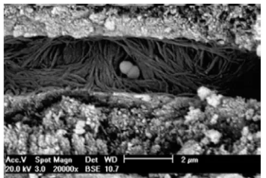
Nair et al. 2005 observed microbial organizations in the root canal system which very often give rise to a complex network embedded in a matrix the so called biofilm communities’ adherent to the root canal walls, isthmuses and ramifications accordingly to Ricucci & Siqueira 2010a. Costerton et al. 1999 that bacteria organized in this way are more resistant to host defence mechanisms and disinfectants than planktonic bacteria and, therefore, are reported to be the most common cause of persistent inflammation, underpinning again the concept, argued by Siqueira & Rôças 2008 that the eradication of bacteria from the root canal system is critical to the endodontic treatment of teeth with apical periodontitis.
Despite conflicting technologies the principles for root canal treatment laid at the beginning of the last century - Hall 1928 cited by Ng et al. 2011- remain consistent with contemporary quality guidelines approved by Endodontic Societies in Europe and North America (Ng et al. 2011). The final objective of root canal treatment is prevention (in the case of pulp inflammation) or resolution (in case of pulp infection) of periapical disease, by eradication of bacteria and their sources of nutrient supply from the root canal system (Reit et al. 1999, Trope & Debelian 2008). That’s to say elimination of the source of infection in an effort to obtain conditions to promote the cure and health of the root and the tissues around it. Accordingly, endodontic infections are treated by intracanal procedures such as chemomechanical preparation (instrumentation with files with copious antibacterial irrigants) in an excellent isolation field supplemented or not by an interappointment intracanal medication, followed by filling and ended with the definitive restoration.
Although sterilization seems practicable, the fact that we are dealing with a complex anatomy renders the available clinical resources frequently unable to free all canal space from all microorganisms, as was first described by Byström & Sundqvist 1983. A more realistic goal of reducing the bacterial populations to a level below the clinically relevant threshold (the level necessary to induce or sustain disease) has been accepted by many clinicians and researchers in an almost dogmatic manner (Ricucci & Siqueira 2010a). The persisting bacteria can
31
survive in treated canals and are able to induce or sustain periradicular tissue inflammation and longstanding endodontic infections (Siqueira & Rôças 2008, Nair 2004): microorganisms, like Enterococcus faecalis and Candida albicans, are true “persisters” since they seem to have a natural ability to survive more harsh environments and stressed conditions (Chavez 2007). Later, Siqueira & Rôcas 2009a affirmed that some members may occupy critical niches within a complex microbial community and, therefore, are potentially important in maintaining the stability and virulence of the microbial community.
For many years, the major technique available to researchers to identify bacteria was the culturing of microorganisms and identification of sampled species by their phenotypic traits. Undoubtedly, knowledge of endodontic infections, based on data involving Microbiologic Root Canal sampling (MRS), has increased significantly during the last 30 years (Siqueira & Rôças 2008), but several questions still await elucidation.
Sampling is important to determine the composition of the endodontic microflora, because in accordance with Gomes et al. 1994 this may be related to the various clinical presentations and symptoms or stages of development of an endodontic infection as well as its responses to different treatments.
In spite of the adoption of molecular approaches with the promising emergence of new data about endodontic microbiology, we are still currently faced with an old controversy: to perceive the usefulness of the MRS over the endodontic’s performance.
1.1. Mollander’s concept: reasons to perform MRS
MRS was an early recommendation for clinical routine use, but did not become widespread among general dental practitioners as noticed by Molander et al. 1996a. By contrast, it has been extensively used in scientific evaluations of antimicrobial intracanal treatment strategies and for the characterization of endodontic microbiology (Reit et al. 1999).
Mollander et al. 1996a reminded that the concept that MRS should form an essential part of endodontic treatment strategy was first suggested by Onderdonk in 1901 and also that Coolidge in 1919 proposed it to be part of the clinical routine. However, it was not until 30 years later that the technology received widespread recognition, mainly through the public applications of Appleton (1932) and Grossman (1938).
32
In Sweden, for instance, after the influential work of Engström (1964) and Möller (1966), the technology became compulsory for undergraduate endodontic education in all four Swedish Dental Schools, accordingly to Molander et al. 1996a.
The American Board of Endodontics made an attempt to evaluate the attitude of their members toward microbiologic assay of root canals prior to obturation. Responses to a questionnaire indicated that the Culture was currently being used along with the endodontist's clinical evaluation of the tooth and not necessarily alone as the final mandatory for obturation of the root canal(s). Culturing was also recommended as a check on one's aseptic technique and, in cases which do not respond to routine endodontic treatment, for identification of the microorganism and antibiotic-sensitivity testing (Lane & Grossman 1971).
However, despite these efforts to disseminate the “new idea”, the methodology is complex and its diagnostic accuracy has been questioned early on by Buchbinder 1971, Sims 1973 and Reit et al.1999.
Nowadays, there is another reason to persist with MRS in the clinical protocol. In fact, we must keep alert to the increased resistance of oral microflora to antibiotics probably associated with an antibiotic overprescription in daily dental practise, often without sufficient rationale for choosing a particular drug. Thus, any effort to study the microbial composition and the susceptibilities of endodontic pathogens will, most likely, facilitate the choice of appropriate clinical protocols or, in occasional cases, of an antibiotic as adjunctive to the clinical treatment of the infection.
1.2. How to correctly perform sampling
1.2.1. Inclusion and exclusion criteria
Sampling includes only one tooth with complete root formation per person and only one root canal per tooth. If the tooth is multi-rooted, either the largest one or the one with periradicular radiolucency is sampled to confine the microbial assessment to a single ecological environment.
Authors like Molander et al. 1998, Shatorn et al. 2007a and Rôças & Siqueira 2011a suggest that, generally, patients must be excluded in endodontic microflora’s analysis in cases of:
33
• systemic debilitating diseases such as diabetes mellitus, liver disease, chronic infections, rheumatoid arthritis or any other systemic disease that compromises the immune system;
• use of systemic steroids or chemotherapeutic agents;
• requirement of prophylactic antibiotic before dental treatment; • active chronic or aggressive marginal periodontitis;
• pregnancy at the time of initial treatment.
Besides the single-rooted, before the sample collection and according to the scope’s analysis, the following features must be checked for each patient: response to sensitivity tests, tenderness to percussion, presence of a sinus tract or swelling, depth of periodontal pocket, history of previous antibiotic medication and root canal treatment.
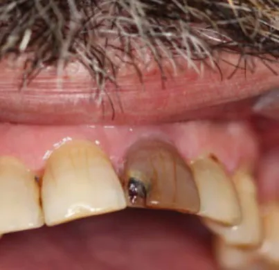
For those investigations on primary endodontic infections, the criteria comprises inclusion of asymptomatic necrotic pulps (neither spontaneous pain nor response to pulpal tests or sinus tract, although tenderness to percussion may be present) with or without radiographic evidence of periapical lesions. Patients who received antibiotic therapy in the last 3 months or exhibited periodontitis (presence of periodontal pockets deeper than 4 mm) must be excluded, due to putative influences on the final results. Also excluded are badly broken teeth (with extensive caries at the time of endodontic treatment) that could not be suitably isolated from the gingiva and saliva by the application of rubber dam: a straight probe is mandatorily suggested by Gomes et al. 2008 and by Rôças & Siqueira 2011a to investigate for pulp space exposure and, if necessary, restorations ought to be replaced before root canal treatment is initiated.
In investigations about treatment failure, root-fillings should end within 5mm of the radiographic apex (Ollander et al. 1998). Furthermore, Endodontic European Society in 2006 recommend that the recall period must be at least 1 year for the symptomatic cases and 4 years for the asymptomatic ones, because no case with residual radiolucency can be assessed as failure before a 4-year observation period, unless the lesion increases in size or signs and symptoms of infection arise. This selection must include teeth with apical periodiontitis diagnoses on the basis of strict clinical and radiographic criteria: in agreement with Sjögren et al. 1997 a diagnosed apical radiolucency is measured horizontally and vertically with a ruler to the nearest millimetre and its size is determined as the mean value of the two measurements.
34
1.2.2. When to sample?
Root canal treatment alters root canal microflora profiles in both quantity (bacterial amount) and quality (bacterial composition). Accordingly to Ito et al. 2005, these differences could be due to the drastic environmental changes in root canals brought about by both mechanical cleaning with files and topical application of drugs.
As a result, to assess those differences, studies of the bacteria occurring in the root canal system involve several basic conditions:
• Pre-instrumentation samples – collected immediately after the execution of the access cavity;
• Post-instrumentation samples – collected immediately after the completion of chemomechanical procedures;
• Post-medication samples – collected immediately after the removal of interappointment dressings;
• Post-obturation samples – collected from root canal-treated with associated periapical periodontitis lesion at a given time, months to years after the initial treatment.
Basically, MRS is a passive way to obtain a sample of the pulpal space at varying moments before, during or after pulpar treatment. Aseptic techniques must be used throughout the endodontic sample acquisition with special care being taken in the control of leakage between the rubber dam and the tooth.
The sampling and bacterial evaluation methodology is invariant, regardless of the sampling time (before, during or after root canal treatment).
Independently of the sampling technique, samples should be processed in the laboratory no later than four hours post collection as suggested by Alsunaien et al. 2010.
1.2.3. How to Sample
In order to obtain MRS one can use charcoaled paper points to absorb the fluid of the root canals or small files and swabs or aspiration in acute cases when soft tissues are affected.
35
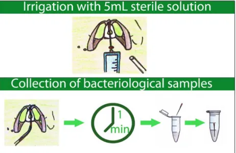
1.2.3.1. Collection of
pre and post-instrumentation and
post-medication samples in vivo
Studies investigating bacteria remaining in the root canals after chemomechanical procedures or intracanal medication potentially allow identification of species with the potential to influence treatment outcome (Siqueira & Rôças 2008).
Once the access cavity is created using sterile burs, the canals to be sampled cannot be dry. One can achieve humidification by irrigation with 5mL of sterile saline solution. This is crucial if an adequate collection of microorganisms with paper cones is to be achieved. If the canal is wet, all fluid inside the pulpal space should be absorbed, using as many paper points as necessary.
The most widely used technique in root canal sampling involves the insertion of 3 or 4 sterile paper points ISO 25 or 30 (Figure 1.1). The paper points should be inserted 1mm shorter than the estimated radiographic length. Each cone must stay inside the root canal for 60 seconds with pumping movements to generate a suspension with bacteria of the main pulpal area. Collection of the soaked paper points should be performed without any contact with potential external contaminators. The paper points should immediately be placed in a sterile microtube with 2mL of Reduced Transport Fluid (RTF) as it offers protection against oxidation, fact demonstrated by Spiegel et al. 1979 and Byström & Sundqvist 1983. This procedure is repeated for each cone.
Figure 1.1. Collection of Bacteriological Samples. Cavity is first irrigated with 5mL of sterile saline solution. Then the cone is inserted and left on cavity for 1 minute. Cones are then immediately immersed in sterile microtube with 2ml of Reduced Transport Fluid (RTF) which protects any anaerobe in sample from oxygen induced damage.
36
1.2.3.2. Collection of exsudates samples
If it is a study of exudates, the lesion can be aspirated via a sterile 16 gauge needle to syringe after disinfection of the oral mucosa with 2% chlorohexidine gel and before surgical drainage. If aspiration is unsuccessful (no pus being collected) sampling can be conducted by swabbing the lesion after incision and discharge of the pus. The microbial pus specimens sampled are then immediately placed into a test tube containing RTF. Use of the aspiration technique may help prevent sample contamination with residual oral flora.
1.2.3.3. Collection of Post-obturation samples
Sunde et al. 2002 reported that the high percentage of endodontic treatment failure in teeth with periapical lesions have been related to circumstances of microbial origin. In fact, refractory cases and post-operative pain (interappointment flare-ups) are often related to an ongoing overgrowth of anaerobic bacteria in the periapical area (Sunde et al. 2002). Thus, it may be helpful to identify treatment resistant bacteria, and this can only be achieved by laboratory studies of post-obturation samples.
It is obviously critical that aseptic techniques are strictly followed throughout endodontic sampling. If there is a post, it is desirable to remove it by ultrasonic vibration, a method less invasive than the use of burs.
After removal of coronal restoration and localization of the root canal orifice, the filling material is removed, either manually (with type K and/or H files) or with an appropriate mechanical system under irrigation with sterile physiological saline solution, as avoidance of chemical solvents minimizes disruption of the bacterial milieu (Sathorn et al. 2007a). The retrieved material can be transferred to microtubes containing TE buffer (10mmol/L tris-HCl, 1mmol/L EDTA, pH 7.6) according to Rôças et al. 2010 if Nucleic Acid (NA) studies are the only aim, or RTF, if samples are to be anaerobically cultured irrespective of the performance of NA studies. Radiographs are an easy way to verify that all filling material had been removed. Pulp space is humidified prior to the collection of the sample with paper points inserted to a level approximately 1mm short of the root apex, based on diagnostic radiographs. This material must be aseptically transferred to the tubes containing the above mentioned solutions. No irrigant is used till the initial sampling is complete.
Preceding sampling, some investigators like to establish canal patency with a file ISO 15 in order to produce minimal instrumentation, running the risk of removing
37
some target material. If this is the case, sample should also include the file, but it should have its head removed to minimize potential external contamination.
1.2.3.4. Investigation on Apical Periodontitis of Treated Root-Canals
Apical lesions assessed after apicetomy
The protocol explained by Subramanian & Mickel 2009 initiates with disinfection of the surgical site and that should be obtained by a one-minute-long oral rinse with 0.12% chlorhexidine digluconate followed by swabbing the surgical area with the same solution. A full-thickness mucoperiosteal flap is then reflected, using both a submarginal or intrasulcular incision after local anesthesia, and the root end is accessed with a surgical bur cooled with sterile water. The periradicular tissue is removed by curettage and stored in tubes containing 1 mL sterile water. Two to three millimetres of root ends are resected after curettage and similarly stored. All samples collected are stored at -20˚C until processed in the lab.
A portion of the periradicular tissue sample is sent for histopathological examination.
Its desirable than in randomly selected cases, immediately after flap reflection, periosteal tissue samples are collected from areas adjacent to the surgical sites using curettes and paper points to test for bacterial contamination of the surgical site (Subramanian & Mickel 2009).
Apical lesions assessed in extracted teeth
Without delay, after extraction, each tooth is profusely rinsed with sterile saline solution and a ISO 15 sterile scalpel is used to remove all attached soft tissue, including the apical periodontitis lesion, from the root. Cleaning of the external root surfaces is made with 3% hydrogen peroxide and disinfected with 2.5% sodium hypochlorite; the latter is inactivated by sterile 5% sodium thiosulphate. The solutions are scrubbed onto the root surfaces by using sterile cotton applicators. After disinfection, a sterility control sample is obtained from the external root surfaces using an ISO 80 sterile paper point dampened with TE buffer or RFT. After decoronation with a diamond disc under saline cooling, the root can be cut into two halves horizontally (coronal and apical) with the use of another diamond disc. Apical segments are transferred to tubes containing 1 mL TE buffer and immediately frozen at -20°C (Rôças et al. 2010, Özok et al. 2012).
38
1.3. Problems during collection
1.3.1. Exchange of samples
As with any clinical sample, collection tubes must be correctly labelled with the pre-established designation of each sample, desirably at the very beginning of the appointment, and always before any collection procedure.
1.3.2. Maintenance of paper-point shape throughout sampling
As explained above, once wet, the root canal microbiological content is collected with paper points. These present the problem of not maintaining the original shape as they become impregnated with solution. A further difficulty arises when dealing with narrow root canals, as the paper points become very difficult to handle properly, making sampling of the apical third a difficult accomplishment. The last paper point is the most important because it will absorb the liquid from the most apical peripheral areas of the apical region (Dahlén 2009). This is especially true if the preceding paper cones became easily soaked and, therefore, hardly collected valid samples of those root canal region.
Wide canals do not usually present major challenges. In these canals, even a 30 paper point will easily fit into the termini of the pulp space. Thus, samplings in post-instrumented root canals do not usually present any kind of problems to insert and collect paper points. This is clearly not the situation for thin root canals before debridement. To overcome this problem, one can use sterile small files (ISO 008, 010, 015), but these will create a different dilemma: if the files are conducted by hand, the head must be cut off before insertion into the microtube in order to prevent introduction of external contaminants. This can be accomplished with a sterilized orthodontic plier. Alternatively, files can already have no cable and be individually sterilized. Handling of these files should be performed with sterile tweezers, although it complicates travelling through the root canal anatomy.
1.4. The Question of False Negatives/Positives
The major issue of any test is its validity: does it measure what it claims? (Reit et al. 1999).
The strict conditions under which MRS must be performed (as described above) emphasizes how potentially error-prone it can be (Sathorn et al. 2007a). Indeed, false positive and false negative results may adversely affect the performance of
39
MRS. However, despite these risk, adopters comfortable with the clinical protocols most often appeared to produce valid samples (Molander et al. 1996b); that’s to say that irrespective of the detection method used, the sampling method resulted in an appropriate collection of the microorganisms present in that particular root canal.
1.4.1. False Negatives
1.4.1.1. Inaccessible Areas
False negative results can occur if there are bacteria located in inaccessible areas for MRS as alerted by Heling & Shapira 1979, Wu et al. 2006 and Siqueira 2008. In practice, these bacteria can repopulate root canals after the first MRS showed a negative result.
In histological observations of Ricucci & Siqueira 2010b, bacteria have been found in inaccessible inter-canal isthmuses, dentinal tubules, irregularities and accessory canals or even in some untouched areas of the main canal often in the form of biofilms. Low-level ultrasonic agitation has been used by Nguyen et al. 2002 in microbiological research to segregate clumped bacteria organized in biofilms without injuring cells and a similar approach could probably be applied in root canal sampling. Its ability to dislodge bacteria from inaccessible locations especially deep within dentinal tubules is however unknown (Sathorn et al. 2007a).
In vitro studies as the one already mentioned of Rôças et al. 2010 have shown the
usefulness of cryogenically ground samples. In this technique all tooth is destroyed allowing recovery not only of the pulp space microorganisms, but also of the anatomically hidden ones. Samples are cryogenically pulverized with the use of a freezer mill. The powdered root segments are frozen at -80˚C in 5mL UV-treated RNA stabilization reagent (RNAlater Qiagen, Hilden, Germany). Accordingly to Alves et al. 2009, this procedure can also be useful if the intention is to compare the microbiota between the coronal and apical part of the root canal system.
1.4.1.2. The legacy of Drugs
At the end of the chemomechanical preparation, and after the use of intracanal dressings, it is mandatory to do MRS only after neutralization of the chemicals used. This is because both classes of chemicals cause bacterial latency (Dahlén
40
2009) and thus prevent bacterial growth (Sathorn et al. 2007a). This fact justifies the usage of inhibitors such as 5% sodium thiosulphate for halogen-containing antiseptics (iodine or chlorine) or the combination of 3% Tween 80 and 0.3% L-α-lecithin (L-α-phosphatidylcholine) when chlorhexidine was the chosen therapeutic. Furthermore, if nucleic acid testing is to be used in lab procedures, alcohol could be avoided and be substituted by 5% sodium thiosulphate, as the former increases the likelihood of free nucleic acid precipitation at the time of specimen collection.
1.4.1.3. Sample Transport and storage
Special concern must be taken regarding the transport medium as it not only needs to keep the viability of all microorganisms, but also be bacteriostatic in the sense that no cell division should take place (Dahlén 2009). Moreover, desirably it ought to inactivate therapeutic substances used in clinical endodontic procedures, which otherwise prevent bacterial cells from growing in the laboratory media as well as contain reducing substances (as cysteine) to keep the medium from being oxygenized (Dahlén 2009).
1.4.1.4. Procedures in the Laboratory
The overwhelming majority of isolates from infected root canals were found to be anaerobic bacteria, having Sato et al. 2012 suggested that the environment in root canals is mostly anaerobic and therefore supports their growth.
It is well-know that Culture has important limitations including low sensitivity, time-consuming and misidentification as consequence of inability to grow many oral species under laboratory artificial conditions (Siqueira & Rôças 2009b). This is especially true as we may be dealing with anaerobe fastidious bacteria that have stringent environment and nutritional requirements as noticed by Wade 2002 and by Sathorn et al. 2007a. Since these methods depend heavily on the viability of the Culture and on phenotype based species identification, the results may be far from in vivo reality.
In the lab, the lack of appropriate culture media for the bacteria in the sample may also result in a false negative result. This is especially important, since as noted above, the endodontic infection is usually polymicrobial in nature, forcing multiple selective growth broads to be used.
41
Finally, the false-negative samples are especially difficult to avoid when taking samples at revision of a previously root-filled tooth. They may be in a stressed situation, after mechanical removal of gutta-percha and sealer, which may not allow in vitro growth (Siqueira 2008).
1.4.2. False Positives
False positive results are usually the result of sample contamination. Thus, to accurately perform MRS, special care should be taken to remove any source of contamination material from the handling area.
Indeed, it seems that false-positive results, as long as endodontic sampling relies on evidence-based principles, are well controlled. Generally they are limited to contaminants of the operative field. Facultative anaerobic species like polysaccharide-producing Streptococci (Streptococci mutans, Streptococci
sanguis, Streptococci oralis and Streptococci salivarius), Corynebacterium spp., Neisseria spp. and Haemophilus spp are oral bacteria that are empirically known
not to establish in the anaerobic and non-saccharolytic environment of the root canal and, thereby, strongly indicate leakage (Dahlén 2009). Equally, Micrococci, coagulase-negative Staphylococci, spore-forming bacteria (Bacillus spp.) and enteric rods are most likely contaminants from the surroundings by careless handling of the samples in the office or lab (Dahlén 2009).
1.4.2.1. Proper Control of External Contaminants
1.4.2.1.1. Use of Sterilized Material
Prior to the procedure, all plaque debris and caries should be removed and existing restorations should be checked. Also, the procedure field should be prepared, first by mouth rinsing with chlorhexidine solution, and then by the use of a sterilized rubber dam tightly adjusted to the cervical part of the crown. Furthermore, only sterilized clinical material should be used. Despite all these precautionary measures, the endodontic field might not be entirely sterile or completely immune from saliva leakage and air contamination. As a result, false positives can still occur, although the above procedures certainly keep these to a minimum.
The performance of the pre-endodontic restoration (if necessary) is well proved: defined as a restoration main made with glass ionomer or composite, before the beginning of an endodontic treatment. Jensen et al. 2007 pointed out that it is decisive in providing better conditions to the application of the rubber dam, thus
42
increasing its efficiency, enhances the irrigants action inside the root canal space as it reduces “extrusion” to adjacent areas and promotes the stability of the temporary restoration between visits.
1.4.2.1.2. Decontamination protocol of the operative field
Also critical in the exclusion of contaminants is a decontamination protocol for the entire clinical field (clamp, rubber dam and tooth surface) after it is fully applied. This decontamination commonly uses sterile tweezers and cotton rolls or pellets impregnated first with 3% hydrogen peroxide (one should wait till bubbling is finished), followed by 3% sodium hypochlorite or 10% iodine tincture. Only after this first disinfection, can the root canal be accessed via the opening of an access cavity, removal either of temporary fillings, caries, existing restorations or, if it is the target, root fillings. Subsequently, a novel disinfection (performed as above described) is made to guarantee the absence of contaminants at the operative field. At the end of this, a disinfection drug inactivation step with 5% sodium thiosulphate fluid must be performed using the same procedure. This inactivation is crucial to avoid false-negative samples, due to viable but non-cultivable bacteria (that’s to say bacteriostatically affected by iodine or sodium hypochlorite) (Dahlén 2009).
After the inactivation step, a swab impregnated in sterile physiologic serum is scrubbed into the operative field and external tooth surface, specially the cavo-superficial angle, and immediately transferred to a transport medium in order to check the sterility: if culture positive results are observed, all samples collected must be regarded as contaminated and excluded from the study.
1.4.2.2. Proper management of each paper point
Extreme care must be taken when handling the paper cones: any contact with any external surface of the root canal space (even the access cavity, especially at the cavo-superficial angle) dramatically increases the chance for false positive results. Normally, the paper points are inside a paper package that has an appropriate site to be open: this must be performed using both hands and taking extremely caution to avoid touching the sterilized coins. That means that when pulling the back part of box it is recommendable not to till it completely apart, so that the paper points stay protected from contamination injuries during sampling collection.
43
It is also crucial to touch only one cone at each time, leaving the untouched ones inside the paper package suitable for a valid sample.
1.4.2.3. Intracanal Dressing
Another issue that can also give rise to false-positive results is the removal of intracanal dressing before sampling: the remnants left in the canals can be equally collected and become part of the sample, altering the true results of the clinical protocol. For example, the residual calcium hydroxide may affect the viability of bacteria on the paper point.
On the other hand, bacteria can re-enter the pulp space between appointments through coronal leakage of the temporary restoration and/or marginal deficiency, cracks and exposed dentinal tubules as demonstrated by Fors et al. 1986. In those cases, despite the efficiency of endodontic procedures, cultures will be positive since the hermetic seal was not achieved.
1.5. Processing of samples
1.5.1. Culture
Traditionally, infections of the oral cavity have been studied by classical microbiological methods as no real alternatives were available (Nair 2007, Dahlén 2009, Rôças & Siqueira 2011a).
Briefly, the microorganism’s Culture starts with sample dilutions in Phosphate Buffered Saline (PBS) of the transport medium. Carlsson & Sundqvist 1980 suggested inoculating them into appropriate enriched medium under conditions that prevent oxygen diffusion so that toxic intermediates of oxygen do not accumulate and interfere with viability of anaerobic bacteria. Plates are then aerobically, microerobically and anaerobically incubated for a period of time (long enough to allow even slowly growing species to form colonies; not less than 2 weeks in the case of strict anaerobic bacteria). Gomes et al. 1994 and Sunde et al. 2002 affirmed that the use of all these conditions is important as former results indicate that 60–70% of the bacterial isolates are found to be either strict anaerobes or microerophiles and Zielke et al. 1976 observed that an aerobic culturing technique alone is not sufficient to reflect the microbiologic status of the root canal system.
After detection of bacterial growth, the procedure includes the isolation of the representative Colony-Forming Units (CFUs) in order to obtain pure cultures. After