Integrated Master in Bioengineering
Antimicrobial activity of selected phytochemicals
against Escherichia coli and Staphylococcus aureus
cells and biofilms
Dissertation for Master Degree in Bioengineering – Specialization in Biological Engineering
Joana Isabel Carvalho Monte
Supervisor: Manuel José Vieira Simões (PhD)
The present thesis was developed for the obtention of Master degree in Bioengineering, in the Faculty of Engineering of University of Porto.
The work was carried out at LEPAE during 6 months. The main objective of the thesis was the evaluation of the efficacy of phytochemicals against Escherichia coli and Staphylococcus aureus planktonic cells and biofilms.
The judge that approved the present document was composed by three elements: Luís de Melo (Cathedratic Professor), Maria da Conceição Fernandes (PhD), Manuel Simões (PhD).
The author
____________________________________________________ (Joana Isabel Carvalho Monte)
The supervisor
____________________________________________________ (Manuel Simões)
“Try to learn something about everything,
and everything about something.”
Acknowlegments
Em primeiro lugar, gostaria de agradecer ao meu orientador, o Professor Doutor Manuel Simões, por toda a dedicação, disponibilidade e empenho ao longo da duração deste projeto.
Gostaria também de agradecer à Ana Abreu, pela incansável ajuda e auxílio na realização deste trabalho. Estou também grata à Anabela e à Joana pelo apoio que me deram nalguns dos ensaios realizados no laboratório.
Quero agradecer ainda a todos do laboratório E007, Carla, Luciana, Ritas, Catarina, Paula, Joanas, Madalena e Renato pela disponibilidade e simpatia.
A todos os meus amigos, em especial às minhas colegas de laboratório Inês, Helena e Carolina pelos ótimos momentos proporcionados e pela sua companhia. Quero agradecer ainda ao João Manuel, ao Francisco, à Catarina e ao João Paulo, por estarem incondicionalmente do meu lado durante os últimos cinco anos. Um especial obrigada ao Nelson pela companhia, apoio e gargalhadas que me proporcionou. À Ana e à Cató por serem as minhas amigas, companheiras e confidentes e por sempre estarem do meu lado.
Finalmente, de modo muito especial, um obrigada à minha família, mãe, pai, Helena e Marta, pelo incentivo, paciência e apoio incondicional durante a realização deste projeto.
Abstract
Antimicrobial resistance is one of the biggest problems facing global public health. The effectiveness of antimicrobial drugs has been lost due to the evolution of pathogen resistance. Plants are considered the greatest source to obtain new antimicrobials. They produce secondary metabolites, phytochemicals, which protect the plant against pathogens.
The aim of this study was to assess the antimicrobial activity of four phytochemicals - 7-hydroxycoumarin (7-HC), indole-3-carbinol (I3C), salycilic acid (SA) and saponin (SP) – against Escherichia coli and Staphylococcus aureus and also understand their ability to control biofilm formation.
Several experiments were carried out in order to: i) test the ability of phytochemicals to control planktonic bacteria growth through the measurement of minimal inhibitory concentration (MIC) and the minimal bactericidal concentration (MBC); ii) evaluate the phytochemicals action in the control of biofilms; iii) understand aspects of the phytochemicals mode of action against the bacteria.
Results have shown that MIC values were higher for E. coli than for S. aureus. The 7-HC and I3C were the most effective, with MICs of 200 and 400 µg/mL for S.
aureus, respectively, and 800 µg/mL against E. coli. Regarding MBC, 1600 and 5000
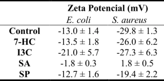
µg/mL were obtained for I3C and SA, respectively. It was also observed that 7-HC and SP has no significative effect in surface charge of E. coli; in contrast, I3C and SA make the membrane more and less negative, respectively. S. aureus surface charge was changed in contact with SA and SP. It was observed that phytochemical concentration did not affect the biofilm removal for both bacteria. E. coli biofilms are more susceptible to phytochemicals comparing to S. aureus biofilms.
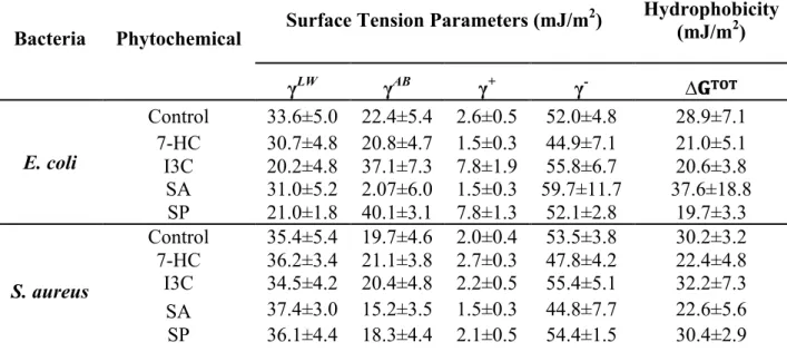
SA and SP promoted the increase and decrease of hydrophilic properties of E.
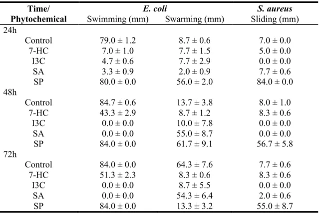
coli, respectively. S. aureus became less hydrophilic in contact with 7-HC and SA. E. coli showed the highest motility and also an increasing in swimming and swarming
motility over time. Motility was mostly affected when I3C was added. Swimming and sliding motilities were completely inhibited and swarming motility was not affected by I3C. The quorum-sensing results indicated that inhibition of violacein production was detectable with 7-HC, I3C and SA, with halos ranging from 5 to 19 mm. I3C was also the most effective phytochemical. The increasing of concentration resulted in an
increasing of pigment inhibition zone. The OMPs expression in E. coli was not affect after the exposure to phytochemicals. Dual combinations between antibiotics and I3C produced synergictic effects against S. aureus resistant strains.
This study suggests that 7-HC and I3C are the most important phytochemicals against E. coli and S. aureus. Both phytochemicals affected the motility and QS activity, which means that they can play an important role in the interference of cell-cell interactions and in biofilm control.
Resumo
A resistência antimicrobiana é um dos maiores problemas enfrentados pela saúde pública global. A eficácia dos agentes antimicrobianos é cada vez mais inferior devido à evolução de mecanismos de resistência a infecções e microrganismos patogénicos. As plantas são consideradas a melhor fonte para a obtenção de novos agentes antimicrobianos. Estas produzem metabolitos secundários, os fitoquímicos, que fazem parte de mecanismos de proteção da planta contra agentes invasores e patogénicos.
O principal objetivo foi a avaliação da actividade antimicrobiana de 4 fitoquímicos – 7-hidroxicumarina (7-HC), indol-3-carbinol (I3C), ácido salicílico (SA) e saponina (SP) – nas estirpes Escherichia coli e Staphylococcus aureus e também compreender a sua capacidade de controlo na formação de biofilmes.
Vários estudos foram realizados de modo a: i) testar a capacidade dos fitoquímicos no controlo do crescimento bacteriano de células planctónicas através da deteção da concentração minima inhibitória (CMI) e concentração mínima bacteriana (CMB); ii) avaliar a ação dos fitoquímicos no controlo de biofilmes; iii) entender os aspetos dos fitoquímicos no modo de ação contra as bactérias.
Os resultados mostraram que os valores de CMI são mais elevados para E. coli do que para S. aureus. A 7-HC e I3C foram os mais eficazes, obtendo-se valores de CIM de 200 e 400 µg/mL para S. aureus, respectivamente, e 800 µg/mL para E. coli. Relativamente a CMB, obtiveram-se valores de 1600 e 5000 µg/mL para I3C e SA, respectivamente. Foi também observado que 7-HC e SP nao tiveram efeito significativo na carga da membrana da E. coli; ao contrário de I3C e SA que tornaram a carga da membrana mais e menos negativa, respectivamente. A carga da membrana de S. aureus foi alterada em contato com SA e SP. Foi verificado que a concentração de fitoquímico nao influencia a remoção de biofilmes para ambas as bactérias. Os biofilmes de E. coli são mais susceptíveis aos fitoquímicos comparativamente aos biofilmes de S. aureus.
O SA e a SP promoveram o aumento e diminuição das propriedades hidrofílicas da E. coli, respectivamente. S. aureus tornou-se menos hidrofílico em contato com 7-HC e SA. E. coli apresentou a mobilidade mais elevada e também um aumento na mobilidade swimming e swarming ao longo do tempo. A mobilidade foi
maioritariamente afetada pela adição de I3C. A mobilidade swimming e sliding foi completamente inibida e swarming foi afetada após a adição de I3C. Os resultados de quorum-sensing indicaram que a inibição da produção de violaceína foi detetada com 7-HC, I3C e SA, com halos compreendidos entre 5 e 19 mm. I3C foi também o fitoquímico mais eficaz. O aumento da concentração resultou num aumento da zona de inibição de pigmento. A expressão das OMPs na E. coli não foi afetada após a exposição aos fitoquímicos. A combinação dupla entre os antibióticos e I3C produziu efeitos sinérgicos contra estirpes resistentes de S. aureus.
Este estudo sugere que 7-HC e I3C são os fitoquímicos mais promissores contra E. coli e S. aureus. Ambos os fitoquímicos afetam a mobilidade e a actividade de quorum-sensing, o que significa que apresentam um papel determinante na interferência de interações célula-célula e no controlo de biofilmes.
Content List
Acknowlegments ... i
Abstract ... iii
Resumo ... v
Content List ... vii
Figures List ... ix
Tables List ... xi
Glossary ... xiii
Chapter 1 ... 1
Work Outline ... 1
1.1. Background and Project Presentation ... 1
1.2 Main objectives ... 2
1.3 Thesis Organization ... 3
Chapter 2 ... 5
Literature Review ... 5
2.1 Antimicrobial resistance and phytochemicals ... 5
2.2. A new therapy against resistance – Phytotherapy ... 7
2.3. Phytochemicals and their classes ... 9
2.4. Mode of Action of Phytochemicals ... 12
2.5. Biofilms and phytochemicals: ... 12
Chapter 3 ... 17
Activity of Selected Phytochemical Products as Antimicrobials and in Biofilm Control ... 17
3.1 Introduction ... 17
3.2 Matherial and methods ... 19
3.2.1. Bacterial Strains ... 19
3.2.2. Phytochemicals ... 19
3.2.3. Determination of Minimum Inhibitory Concentration ... 19
3.2.4. Determination of Minimum Bactericidal Concentration ... 20
3.2.5. Biofilm formation and control in sterile 96-‐well polystyrene microtiter plates ... 20
3.2.6. Biofilm analysis ... 21
3.2.6.1 Crystal Violet method ... 21
3.2.6.2 Resazurin Method ... 21
3.2.7. Classification of biofilm producer bacteria ... 22
3.2.8. Statistical analysis ... 22
3.3 Results and Discussion ... 23
3.3.1. Antimicrobial activity of phytochemicals and biofilm control potential ... 23
Chapter 4 ... 29
Aspects Underlying the Antibacterial and Biofilm Control Action of Phytochemicals ... 29
4.1 Introduction ... 29
4.2 Materials and methods ... 32
4.2.1. Bacterial Strains ... 32
4.2.3. Determination of Zeta Potential ... 32
4.2.4. Physico-‐chemical charecterization of bacterial surface ... 33
4.2.5. Free energy of adhesion ... 34
4.2.6. Motility ... 34
4.2.7. Detection of quorum-‐sensing inhibition ... 35
4.2.8. Outer Membrane Proteins ... 36
4.2.8.1. Extraction ... 36
4.2.8.2. OMP Analysis ... 36
4.2.9. Antibiotic-‐Phytochemical Dual Combinations Assay – Efflux Pumps Inhibition ... 37
4.2.9.1. Classification of dual combinations ... 37
4.2.10. Statistical analysis ... 38
4.3 Results and Discussion ... 39
4.3.1. Surface characterization of cells in presence of phytochemicals ... 39
4.3.2. Analysis of E. coli and S. aureus free energy of adhesion ... 41
4.3.3. Motility assays ... 43
4.3.4. Quorum-‐sensing assays ... 45
4.3.5. Characterization of cell membranes ... 48
4.3.5.1. Outer Membrane Proteins ... 48
4.3.5.1. Efflux pumps ... 50
Chapter 5 ... 53
Concluding remarks and perspectives for further research ... 53
5.1. Conclusions ... 53
5.2. Perspectives for further research ... 55
References ... 57
Appendix ... I A. Experimental data ... I
Figures List
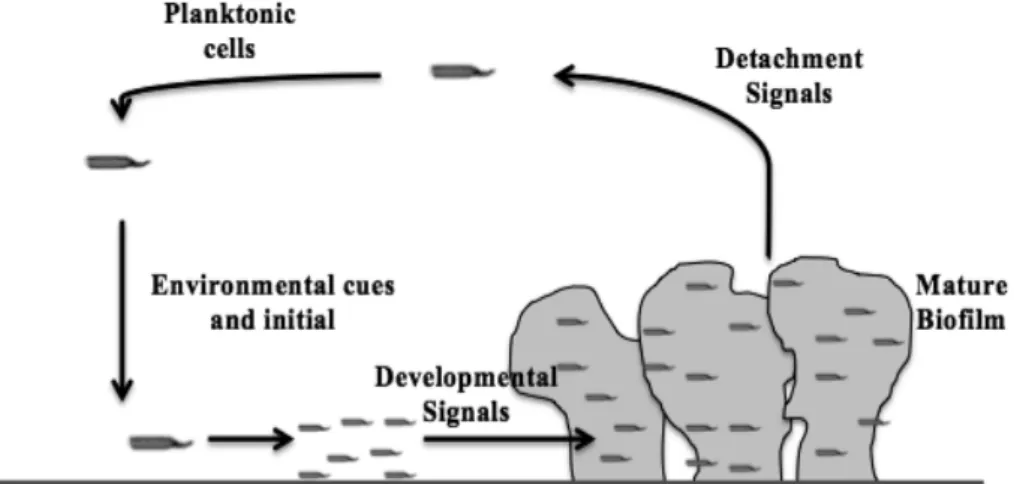
Figure 1. Traditional targets of antimicrobial compounds……….…...……7 Figure 2. Model of biofilm development. Planktonic cells contact with surface and
cells resulting in the formation of microcolonies. Cells in the biofilm can return to a planktonic lifestyle to complete the cycle of biofilm development…..……14
Figure 3. OD570 nm as a measure of biofilm mass (a) and fluorescence (λex: 570 nm and λem: 590 nm) results as a measure of biofilm viability (b) for E. coli and S.
aureus. The mean ± standard deviation values resultant from three independent
experiments are depitecd………...………...25
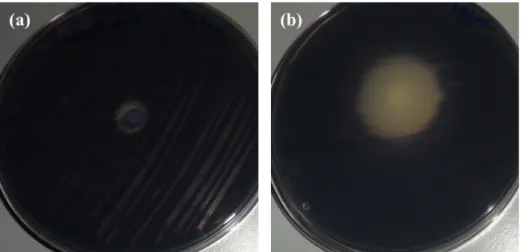
Figure 4. Examples of results obtained with quorum-sensing assay: (a) no bioactivity
(SA at 5000 µg/mL); (b) antibacterial and anti-QS halos are observed (I3C at 5000 µg/mL) with addition of phytochemicals………46
Figure 5. OMPs profiles of E. coli. The profile of molecular weight standards (1),
control (2), 7-HC (3), I3C (4), SA (5), SP (6) are presented………...…...…49
Tables List
Table 1. MIC for E. coli and S. aureus. ... 23 Table 2. MBC values for E. coli and S. aureus. ... 24 Table 3. Percentages of biofilm removal and inactivation by the selected
phytochemicals against E. coli and S. aureus. ... 26
Table 4. Zeta potential (mV) results of suspensions of E. coli and S. aureus in contact
with phytochemicals at the MIC. ... 39
Table 5. Hydrophobicity (∆𝐆𝐓𝐎𝐓), and apolar (γLW) and polar (γAB) components of the surface tension of untreated and treated cells. The means ± SDs are illustrated. 40
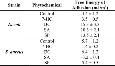
Table 6. Free energy of adhesion (∆𝐆𝐓𝐎𝐓bws) of bacterial cells to polystyrene, treated and untreated with phytochemicals. ... 42
Table 7. Motility results for bacteria with and with phytochemicals. The drop
baseline was 6mm which was subtracted from the results presented. ... 44
Table 9. Antimicrobial activity of antibiotics. The means (mm) ± standard deviation
for at least three replicates are illustrated. ... 51
Table 10. Classification of the effect of dual combinations of phytochemicals and
antibiotics. ... 52
Table A.1. Physico-chemical characterization of polystyrene (PS). ... I
Glossary
Indexes and parameters: AB – Lewis acid-base component B – Bacteria
B - α-bromonaphtalene F – Formamide
LW – Lifshitz-van der Waals component S – Bubstratum
TOT – Total W – Water
sws- Between teo entities od a given surface when immersed in water
bws – Between one bacteria and a substratum that are immersed or dissolved in water bwb – Between two bacterial surfaces, when immersed in water
bw – Between bacteria and substratum sw – Between substratum and bacteria TOT – Total
∆G – Free energy of interaction (mJ/m2) γ - Surface free energy (mJ/m2)
θ - Contact angle (°)
+ - Electron acceptor paramenter of Lewis acid-base component - - Electron donor parameter of Lewis acid-base component
Abreviations: AHL - N-acyl-homoserines AI – Autoinducers
CIP – Ciprofloxacin CV – Crystal Violet
DMSO – Dimethyl sulfoxide
EPS – Extracellular polymeric substances ERY - Erythromycin
I3C – Indole-3-carbinol
MBC – Minimum Bactericidal Concentration MDR – Multi-drug resistant
OD – Optical Density
ODC – Cut-off optical density OMPs – Outer membrane proteins p – Statistical significance level PS – Polystyrene
QS – Quorum-sensing
QSI – Quorum-sensing inhibition RMAs – Resistance-modifying agents SA – Salycilic Acid
SDS-PAGE – Sodium dodecyl sulfate polyacrylamide gel electrophoresis SP – Saponin
SPSS - Statistical Package for the Social Sciences TET - Tetracycline
WHO – World Health Organization 7-HC – 7-hidroxycoumarin
C
hapter 1
Work Outline
1.1. Background and Project Presentation
Since the discovery of the first antibiotic, penicillin, the employment of any novel antibiotic has been followed by the appearance of bacterial resistance to that antibiotic in as little time as a few years. Antibiotics have the ability to kill bacteria or inhibit their growth.
Resistance to antibiotics is one of the biggest problems that global public health is facing. Antimicrobial resistance is a natural consequence of the adaption of pathogens to the exposure to antimicrobials used in medicine, food, crop production and to disinfectants in farms and households. Resistant organisms cause infections that are more difficult to treat and more expensive; some strains have become resistant to all available antimicrobial agents (Byarugaba, 2004). Resistant infections affect treatment costs, disease spread and duration of illness (Okeke et al., 2005)
In order to find novel antimicrobial agents with new modes of action, plants have been explored as sources for the identification of new and effective antibacterials. An endless number of plant species have been reported to act against several bacteria in vitro, and many medicinal plants produce secondary metabolites (phytochemicals) capable of inhibiting the growth of a wide range of microorganisms including fungi, yeasts and bacteria. Phytochemicals have been studied for the treatment of microbial infections since 1990, due to the increasing inefficacy of conventional antibiotics (Simões et al., 2009).
1.2 Main objectives
The main aim of this work was to assess the antimicrobial efficacy of selected phytochemicals against Escherichia coli and Staphylococcus aureus planktonic cells and also to evaluate them on biofilm control.
In the present study, four different phytochemicals – 7-hydroxycoumarin (7-HC), indole-3-carbinol (I3C), salicylic acid (SA) and saponin (SP) – were tested against E. coli and S. aureus in both planktonic and sessile states. The strains tested are considered the most clinical significant bacteria due to their capacity to resist against several antibiotics (Simões et al., 2008; Xu et al., 2006).
To evaluate the antimicrobial activity of the several phytochemicals two experiments were performed to determine the minimum inhibitory concentration (MIC) and minimum bactericidal concentration (MBC). The biofilm control was also performed to understand the efficacy of phytochemicals to remove biomass and inactivate biofilm cells. The biofilm control assay was studied in 24 h aged biofilms after 1 h in contact with the phytochemicals. The biomass removal and metabolic inactivation were calculated through the optical density (OD) and fluorescence measurements.
Several aspects of planktonic cells were evaluated to understand the mode of action of the selected phytochemicals. The surface charge of bacteria was studied through the measurement of Zeta potential and the hydrophobicity of cells was also assessed. To evaluate the potential activity of the phytochemicals to prevent E. coli and S. aureus adhesion to polystyrene (PS), the prediction of theoretical adhesion
through the measurement of contact angles was performed. The phytochemicals were also studied on the ability to interfere with bacterial motility and quorum-sensing (QS), two microbial aspects involved in biofilm formation.
The OMPs expression of E. coli was studied in contact with phytochemicals through a sodium dodecyl sulfate polyacrylamide gel electrophoresis (SDS-PAGE) in order to detect the possible expression of resistance proteins. Regarding S. aureus resistant strains, dual combinations of phytochemicals and antibiotics – tetracycline (TET), ciprofloxacin (CIP) and erythromycin (ERY) - were tested to understand their ability to act in efflux pumps inhibition.
1.3 Thesis Organization
In chapter 1, the context, motivations and main goals for the development of this thesis are explained. This chapter is also a guideline to the overall work, composed by 5 chapters.
Chapter 2 includes the literature review about the main subjects of this work. In this chapter, the main problems associated with the appearance of bacterial resistance are highlighted. Plant products, especially secondary metabolites, are introduced as one of the solution for the antimicrobial resistance. The mode of action of phytochemicals is also developed in this chapter. Finally, it is reported the problem of the higher resistance associated to bacterial growing in biofilms and quorum-sensing inhibition (QSI) is presented as one possible solution for the prevention of biofilm formation.
In the third chapter are described the results of the activity of phytochemicals as antimicrobial agents against E. coli and S. aureus. The MIC and MBC of the selected phytochemicals are presented. In this chapter are also studied the phytochemicals in the control of biofilm, showing the biofilm removal and metabolic inactivation for each one of the phytochemicals at the MIC and 5 × MIC.
Chapter 4 describes the study of surface charge and hydrophobicity characteristics of E. coli and S. aureus when exposed to phytochemicals. The influences of phytochemicals in motility and QS are also presented in this chapter. To finalize the chapter, OMPs expression of E. coli are studied when exposed to
phytochemicals; and dual combination of phytochemicals and antibiotics are described in order to study the antimicrobial activity and their synergistic effects in S.
aureus efflux pump inhibition.
Finally, in chapter 5 the main conclusions of the work are referred and some recomendations for future research are given.
C
hapter
2
Literature Review
2.1 Antimicrobial resistance and phytochemicals
There is a continuous search for new drugs and antibiotics in order to heal the main infectious diseases. However, the microorganisms have become resistant to most of the antibiotics. The microorganisms are successful when facing adverse conditions, because they seem to sense and respond to the external environment and modulate gene expression accordingly. Antimicrobial resistance is one of the biggest problems threatening global public health (Byarugaba, 2004; Okeke et al., 2005). This problem is a natural consequence of the adaption of infectious agents to antimicrobials used in several areas, including medicine, food animals, crop production and disinfectants in farms, hospitals and households (Bloomfield, 2002; McEwen and Fedorka-Cray, 2002; Vidaver, 2002; Wise and Soulsby, 2002). Resistance allows microorganisms to survive in the presence of toxic conditions. The effectiveness of many antimicrobial drugs has been lost due to the evolution of pathogen resistance. Many of the microorganisms are no longer susceptible to most of the existing antibiotics and therapeutic agents (Byarugaba, 2004). Bacteria generally
acquire drug resistance in a multitude of ways (Saleem et al., 2010). The acquisition can be done by de novo mutation or acquisition of resistance genes from other microorganisms and passed on during replication (Fajardo et al., 2008; McManus, 1997; Sefton, 2002; Smith and Lewin, 1993). Resistance genes are able to act in different ways enabling bacterium to: produce the enzymes that inactivate the antimicrobial agent; modify the target site; produce an alternative metabolic pathway that inhibits the antimicrobial agent; express efflux mechanisms, preventing the antimicrobials to reach intracellular targets (Spratt, 1994; Webber and Piddock, 2003; Woodford and Ellington, 2007).
There are several factors influencing the efficacy of antimicrobial agents. Examples include the use of an inefficient product, in other words, of an antimicrobial product that presents an incomplete spectrum of activity; the application of antimicrobial agents at sublethal concentrations, which can allow the adaptation of microorganisms to these new conditions; or an insufficient contact time between the antimicrobial product and microorganisms (Bessems, 1998; Heinzel, 1998; Russel 2003).
Beyond all the environmental factors affecting the resistance explained before, there are cellular mechanisms influencing this process. The Gram-negative bacteria are less susceptible to antimicrobial agents than Gram-positive bacteria, because they present a thick cell wall, more difficult to entry, and an outer membrane. The waxy envelope presented in mycobacteria inhibits the uptake of antimicrobial agents, so they are even more resistant (McDonnell and Russell, 1999). Efflux is another process related with the increasing of resistance. Through efflux pumps, Gram-negative bacteria pump out the antimicrobial, detergents, organic solvents and disinfectant agents, contributing to the resistance (Beumer et al., 2000; Cloete, 2003; Kumar and Schweizer, 2005).
The resistant microorganisms can cause infections that are more difficult to treat, and it is necessary drugs that are more expensive and toxic and also less available (Howard et al., 2001). The effective microbial therapy is usually delayed because of the acquisition of resistance from microorganisms (Ibrahim et al., 2000; Kollef, et al., 1999; Lautenbach et al., 2001). Bacteria have shown resistance with increasing trends. Consequently, the rate of discovery of new antimicrobial agents has decreased since the 1970s (Byarugaba, 2004). The traditional antibiotics have been recognized because they are able to kill bacteria or inhibit their growth, through
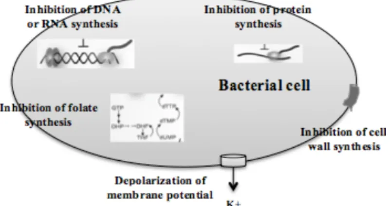
inhibition of bacterial functions, such as: cell wall synthesis, DNA replication, RNA transcription and protein synthesis, which are essential for cell growth (Figure 1) (Clatworthy et al., 2007). In order to treat infections caused by antimicrobial-resistant organisms, it is necessary a more effective therapy (Cosgrove and Carmeli, 2003).
The methods to evaluate antimicrobial activity are essential for the selection of a new drug. However, there are several factors that can affect the antimicrobial susceptibility test: pH, moisture, effects of thymidine or thimine, effects of variation in divalent cations and growth conditions (Lalitha, 2004). Magnesium and calcium are divalent cations that affect the results of aminoglycosides and tetracycline. Finally, the components needed for growth should be fulfilled (Lalitha, 2004). The antimicrobial susceptibility tests are divided into 3 principles: diffusion, dilution and diffusion and dilution. The most popular methods are: the Kirby-Bauer, E-Test, agar dilution, Stokes, microdilution and macrodilution (Anvisa, 2012; Lalitha, 2004; Othman et al., 2011).
2.2. A new therapy against resistance – Phytotherapy
Natural new antimicrobial can be present in several products used by humankind. For example, aromatic plants and spices, used as flavour enhancers, have already been demonstrated to have significant antimicrobial activity (Christaki et al., 2012). Also, antimicrobials from vegetal origin can be obtained from essential oils, seeds, flowers, fruits and roots. These kind of antimicrobial systems have been used
centuries ago, in the preservation of food. Plants produce a wide range of organic compounds. However, the secondary metabolites present special interest because of their importance as pharmaceuticals, venoms, fragrances, industrial materials and cosmetics (Amaral and Silva, 2003; Mendes, 2007). The discovery of all the potential of plants brings a great interest in phytotherapy.
The World Health Organization (WHO) defines medicinal plants those that present traditional use as therapeutic compounds (Severiano et al., 2010). According to the WHO, medicinal plants would be the greatest source to obtain a wide range of drugs with antimicrobial properties (Nascimento et al., 2000). Consequently, interest in medicinal plants has increased in recent years. Indeed, plants were reported to be used by about three quarter of the world population (Rao et al., 2004). The story of medicinal plants says that they are part of the humankind evolution and were the first therapeutic sources used. However, the use of medicinal plants, phytotherapy, had major impact in the beginning of XIX century. There are 250.000-500.000 plant species, however just a small percentage has been investigated phytochemically (Mahesh and Satish, 2008).
It is interesting to realize that most of the medicines used nowadays were identified in plants, and some of the most important pharmaceutical drugs are natural drugs and linked directly to natural product research (Bighetti et al., 2005). Also, most of the commercial drugs were used before in crude form in traditional and folk medicine, suggesting their potential biological activity (Ciocan and Bara, 2007). Also, plants present numerous constituents and their extracts can show synergistic effects between the different active principles due to the presence of classes of compounds or different structures contributing for the same activity (Maciel et al., 2002). The plants can prevent cardiovascular diseases, carcinogenesis, inflammation, atherosclerosis, and others health problems (Albano and Miguel, 2001).
Some plants have potential as remedies for diseases. Examples include the use of bear-berry (Arctostaphylos uvaursi) and cranberry juice (Vaccinium macrocarpon) to treat urinary tract infections, essential oils of tea tree (Melaleuca alternifolia) as active ingredients in many topical formulations to treat cutaneous infections and
Hydrastis canadensis and Echinacea species for tuberculosis infections (Abreu et al.,
2012).
Regarding the plants, they can be divided in several families, for example Asteraceae, Lamiaceae, Apeaceae, Euphorbiaceae, Rutaceae and Fabaceae. Regarding
to these families, some of them are more incident in Portugal. Lamiaceae is one of the families found in our country and frequently found in Mediterranean region, Middle East and tropical mountains (Severiano et al., 2010). Currently, there are 3.500 species of this family (Severiano et al., 2010). Lamiaceae are composed of herbs, shrubs and trees. Plants belonging to this family can be applied in medicine, cosmetics and food industry (Mendes, 2007). Asteraceae is another plant family present in Portugal and includes around 23.000 species and 1.500 genus. They can be found in temperate and subtropical climates. Herbs, subshrubs and vines belong to this family (Severiano et al., 2010). Finally, Apiaceae is another family present in Portugal and is composed for 3.000 species and 300 genus (Moreno-Dorado et al., 2000). The plants that belong to this family can be found in tropical or temperate regions (Judd et al., 2002). Such as Lamiaceae, plants of this family are commonly used in medicine and food industry.
2.3. Phytochemicals and their classes
The increasing of antimicrobial resistance has led to the study of plants products for searching new antimicrobials (Clardy et al., 2006). The chemical diversity and structure are the main causes for the success of phytochemicals (Gibbons, 2004). The phytochemicals have become interesting since the 1990’s because of the increasing dissemination of bacterial resistance mechanisms worldwide, due to the excessive and inappropriate use of antimicrobials.
Plant products are divided into two classes: phytoanticipins and phytoalexins. Molecules that are present in an inactive form (example: glucosides) belong to the first group; the second group is composed by molecules whose levels increase in response to microbial attack or is generated in response to a specific infection (Tegos et al., 2002). Phytoalexins are a large group of chemically diverse molecules, including: simple phenylpropanoids derivatives, alkaloids, glycosteroids, flavonoids, isoflavonoids, sulfur products, terpenes and polyketides (Hammerschmidt, 1999). The same molecule can be a phytoalexin or a phytoanticipin in different organs of the same plant. Examples of phytoanticipin are terprenoids, quinones and tannins (Abreu et al., 2012).
There are several phytochemical classes with antimicrobial properties; however, the medical community does not recognize them as therapeutics agents. This is mainly explained because the majority of phytochemicals have weak spectra of activities (Tegos et al., 2002) and the concentrations required are too high to be clinically significant (Aeschlimann et al., 1999; Schmitz et al., 1998). The major problem for the identification of antimicrobial agents from plants is the variability in the extraction methods and antibacterial tests used (Simões et al., 2009).
Beyond the antimicrobial properties, phytochemicals are able to present antiviral (Jassim and Naji, 2003; Muhtar et al., 2008; Naithani et al., 2008), antiparasitic (Atawodi and Alafiatayo, 2007; Chan-Bacab and Peña-Rodríguez, 2001; Sriram et al., 2004) and antifungal (Morel et al., 2002; Rahman and Moon, 2007; Treyvaud Amiguet et al., 2006) activities and also exert cytotoxic activity against tumor cells (Rimando and Suh, 2008; Suffredini et al., 2006; Udenigwe et al., 2008).
The groups of secondary metabolites produced by plant include: terpenoids, phenolics, alkaloids, essential oils constituents, lectins polypeptides and polyacetylenes (Mendes, 2007; Simões et al., 2009). The secondary compounds are responsible for defending against external aggressions and the main subclasses are: simple phenols and phenolic acids, quinines, flavones, flavonoids, tannins, coumarins, and others (Simões et al., 2009). Usually, the plants belonging to one family produce the same type of compounds. Normally, alkaloids are produced from the Apocinaceae and Solanaceae families; flavonoids are produced from Rutaceae plants; and terpenoids are produced from Asteraceae and Lamaceae families (Amaral and Silva, 2003). According to the classes that substances belong, they can produce different effects. Flavonoids present anti-inflammatory action, protecting the blood vessels, and are hypotensive and sedative; digestion action is guaranteed by antraquinones; bronchodilator action is made by coumarins; bactericidal action is achieved with tannins and essential oils (Severiano et al., 2010); and essential oils presents, apart from the others already mentioned, sedative, stimulating, analgesic and expectorant properties (Souza et al., 2011). Antimicrobial properties are expressend by most of plants that are composed by: tannins, terpenoids, glycosides, alkaloids, saponnins, flavonoids, polyphenols and coumarins (Das et al., 2010; Hill, 1952; Neelima et al., 2011; Padmini et al., 2010).
The phytochemical compounds are mostly secondary metabolites of plants and deposit in specific parts of the plant (Ciocan and Bara, 2007). Regarding phytochemicals, the main classes with health benefits are present below.
Phenolics and polyphenols form one of the simplest groups of bioactive phytochemicals, consisting of a single substituted phenolic ring (Das et al., 2010). This group seems to be toxic to microorganisms because of the site(s) and number of hydroxyl groups present on the phenolic ring (Scalbert et al., 1991; Urs et al., 1975). Increasing the hydroxylation results in the increasing of toxicity (Ciocan and Bara, 2007). Quinones are composed by an aromatic ring with two ketones substitutions (Das et al., 2010). These molecules react with nucleophilic amino acids in proteins, causing the inactivation of the protein or loss of function (Ciocan and Bara, 2007). Flavones, flavonoids and flavonols belong to a group of molecules with phenolic structures containing one carbonyl group (Ciocan and Bara, 2007; Das et al., 2010). They are active against a wide range of microorganisms, probably because they can react with extracellular and soluble proteins and also react with cell walls (Bennet and Wallsgrove, 1994; Ciocan and Bara, 2007). Tannins are another group that can be divided into two groups: hydrolysable and condensed. The first is based on gallic acid; the second group is composed by numerous condensed tannins derived from flavonoid monomers. The antimicrobial mode of action is related with their ability to inactivate microbial adhesin, enzymes and cell envelope transport proteins (Ciocan and Bara, 2007). Coumarins are derivatives of cinnamic acid and comprise a large class of phenolic substances (Hoult and Payát, 1996; Thuong et al., 2009). These compounds are the simplest members of the group of oxygen heterocyclic, also known as 1,2-benzopyrene, consisting of fused benzene and α-pyrone ring (Hoult and Payát, 1996). More than 1300 coumarins have been studied from natural sources and several properties are related with them, such as: antimicrobial, anti-inflammatory, antioxidant, anticoagulation, antiestrogenic and sedative (Hoult and Payát, 2012; Mello, 2009; Paramjeet et al., 2012). Essential oils and terpenoids are compounds based on an isoprene structure. They occur as diterpenes, triterpenes and tetraterpenes (C20, C30, C40), they are called terpenoids when contain other elements, such as oxygen. These compounds are active against bacteria, fungi, viruses and protozoa (Ciocan and Bara, 2007; Das et al., 2010).
The alkaloids are heterocyclic nitrogenous compounds that present analgesic, antispasmodic and bactericidal action (Ciocan and Bara, 2007; Das et al., 2010; Stary, 1996).
2.4. Mode of Action of Phytochemicals
Bacterial growth can be inhibited by phytochemicals through several mechanisms. These plant products can act on various biochemical targets on the bacterial cells. The mode of action of phytochemicals is not completely understood, neither the phytochemical antibacterial specificity (Simões et al., 2009).
Some experiments have been done to study the mode of action of several phytochemicals. The chemical structure and properties influence the site of action of phytochemicals. The mechanism of action of essential oils against bacteria involves membrane disruption through the lipophilic structure (Griffin et al., 1999; Mendoza et al., 1997). Alkaloids, such as berberine and piperine, interact with bacterial cytoplasmic membrane, intercalate with DNA or inhibit efflux pumps in S. aureus (Khan et al., 2006). Phenols act by interruption of energy production due to enzyme inhibition by the oxidized products, which react with sulfhydyl groups or non-specific interaction with proteins (Mason and Wasserman, 1987). In the case of flavonoids, they inhibit the synthesis of nucleic acids of Gram-negative and Gram-positive bacteria (Cushnie and Lamb, 2005; Mori et al., 1987). Other authors, shown that glycoside saponins are able to induce pore-like structures that change the membrane permeability; they can also interfere with energy metabolism (Mandal et al., 2005; Melzig et al., 2001; Sinha Babu et al., 1997).
2.5. Biofilms and phytochemicals:
Natural products have been isolated from plants for usage in biodeterioration control. Biodeterioration is the chemical and physical alteration resulting from
biological activity. The microorganisms associated to biodeterioration growth as biofilms that adhere to substrates. The treatment for this problem involved the use of biocides, however these are chemical agents and most of them are cytotoxic. So, natural compounds from plants with biocidal activity have emerged, and they are promising alternative for the control of biodeterioration without negative impacts of the environment (Guiamet et al., 2006). The chemical biocides are toxic and difficult to degrade, being persistent in the environment, causing chemical contaminations and the spread of resistance. In contrast, natural biocides, extracted from plants, are biodegradable and environmental friendly (Guiamet and Saravia, 2005). The phytochemicals can act as control agents on the bacterial biofilm formation and development (Simões et al., 2009).
Guiamet and co-workers (2006) performed a study with Cichorium intybus,
Arctium lappa and Centaurea cyanus from Asteraceae family to test their ability to be
used as antimicrobial agents against different microorganisms associated with biodeterioration. These plants showed moderate activity against two species of
Pseudomonas and no activity against Bacillus cereus. Rosmarinus officinalis L.
extracts act as an antimicrobial agent against S. aureus. Allium sativum produce allicin, which is one of the most effective antimicrobial products isolated from garlic (Abreu et al., 2012).
Biofilm formation is a feature closely related to pathogenicity (Ren et al., 2005). A biofilm is formed by planktonic bacteria that adhere to a surface and initiate the development of sessile microcolonies surrounded by an extracellular matrix (Otto, 2009) (Figure 2). Bacteria form complex surface-attached communities, also called biofilms (Hentzer and Givskov, 2003). Biofilms develop structures that are morphologically and physiologically differentiated from free-living bacteria (Davies et al., 1998).
The process of biofilm formation includes several steps: preconditioning of the adhesion surface; planktonic cells are transferred from the bulk liquid to the surface; adsorption of cells at the surface; desorption of reversible adsorbed cells; the bacterial cells are adsorbed irreversibly at the surface; transport of substrates to the biofilm; substrate metabolism by the biofilm cells and transport of products out of the biofilm; finally, biofilm is removed by detachment or sloughing (Simões, 2005). The formation of biofilm is dependent of several parameters. Regarding to the surfaces, the attachment is easier on rough, hydrophobic and coated surfaces (Donlan, 2002;
Pereira, 2001). Parameters like flow velocity, water temperature and nutrient concentration also influence the biofilm attachment (Pereira, 2001; Vieira, 1995).
Biofilms are an example of physiological modifications and they also increase the tolerance to antimicrobial therapies and to the host immune response (Hentzer and Givskov, 2003; Simões et al., 2009). Most of the bacterial infections detected in human body involve the formation of biofilms. The biofilm mode of growth permits an increased bacterial survival in hostile conditions, such as in the presence of antibiotics and disinfectants (Hentzer and Givskov, 2003; Trentin et al., 2011). There is an increasing interest in preventing, controlling and eradicating biofilms. Biofilms by bacterial cells are thought to be regulated by autoinducer molecules; in a process called quorum-sensing (Ren et al., 2002). The impairment of bacterial adhesion and biofilm formation by a pathway that does not affect the bacterial death is an important feature of the new concept in antivirulence therapies (Trentin et al., 2011). This alternative should maintain the cells in a planktonic state, switching off the virulence expression and attenuating the pathogen, making the microorganisms more susceptible to antimicrobial agents and immune system (Macedo and Abraham, 2009; Martin et al., 2008).
Bacteria in biofilms present a reduced susceptibility to antimicrobial agents caused by a variety of factors, such as: nutrient depletion within the biofilm, reduced access to cells in the biofilm, production of degradative enzymes and neutralizing Figure 2. Model of biofilm development. Planktonic cells contact with surface resulting in the
formation of microcolonies. Cells in the biofilm can return to a planktonic lifestyle to complete the cycle of biofilm development.
chemicals, between others (Brown and Gilbert, 1993). Biofilms have been reported to be 100-1000 more protectors to bacteria than populations of planktonic cells (Gilbert et al., 2002; Mah and O’Toole, 2001; Stewart and Costerton, 2001). The main difference between planktonic cells and biofilms is the presence of a polysaccharide matrix, delaying the diffusion of antimicrobials into the biofilm (Brooun et al., 2000).
Traditional treatment of infectious diseases is related with compounds that inhibit the growth of bacteria. But, it has been observed that they develop resistance to antimicrobial compounds. So, quorum-sensing seems to be the next opportunity to improve bacterial infection. Quorum-sensing influences bacterial biofilm growth and biofilm development that is related with cell-cell interactions (Simões et al., 2009). Quorum-sensing inhibitory compounds are the new line of antimicrobial agents and can be applied in several areas: medicine, agriculture and aquaculture (Hentzer and Givskov, 2003).
Several biotechnology companies have already tried to develope some strategies to interrupt the bacterial quorum-sensing, such as: inhibition of N-acyl-homoserines (AHL) signal recognition, signal dissemination and signal reception (Hentzer and Givskov, 2003). By interfering with cells communication, it is possible to interfere also in the resistance of biofilms and their ability to form resistant structures, causing cell dispersion. Quorum-sensing systems are involved in a wide range of microbial activities: extracellular enzyme biosynthesis, biofilm development, antibiotic biosynthesis, biosurfactant production, extracellular polymeric substances (EPS) synthesis and production of extracellular virulence factors (Chatterjee et al., 1995; Davies et al., 1998; Daniels et al., 2004; Fux et al., 2005; McGowan et al., 1995; Passador et al., 1993; Pearson et al., 1995; von Bodman and Farrand, 1995).
Autoinducers (AI) are molecules to perceive the size of bacterial population and AHL are the major AI molecules. Several Gram-negative bacteria use AHL signals to coordinate the behaviour of cells in a population. An important achievement was the discovery of molecules produced by plants that mimic AHL signals, affecting quorum-sensing behavior. Several plants of medicinal use demonstrated potential to inhibit quorum-sensing (Hentzer and Givskov, 2003; Waters and Bassler, 2005).
Studying the grapefruit and its furanocoumarins as inhibitors of biofilm formation, it has been shown that dihydroxybergamottin and bergamottin exhibit strong inhibition of both AI-1 and AI-2 activities even at concentrations as low as 1
µg/mL (Girennavar et al., 2008). Furanocoumarins are able to interfere with cell-cell signalling and also inhibit biofilm formation. However, the mechanisms of action are not completely understood (Girennavar et al., 2008). Delissea pulchra, an Australian macroalga, produces halogenated furanones, which are inhibitors of AHL, inhibiting bacterial quorum-sensing and biofilm formation (McLean et al., 2004). Auraptene and lacinartin, two compounds belonging to coumarin family, are promising natural compounds that can be used to prevent and treat periodontal diseases. They were also evaluated on the growth, biofilm formation/desorption, and adherence to human oral epithelial cells of Porphyromonas gingivalis. Lacinartin was able to inhibit biofilm formation and to cause desorption of a pre-formed biofilm of P. gingivalis. This suggests that coumarins may contribute to reducing tissue destruction (Marquis et al., 2012). Some information about the structure-activity relationship of coumarins showed that the group on position 5 and position 2’/3’ of the isoamylene chain can affect the antifouling activities against both Balanus albicostatus and Bugula neritina (Wang et al., 2013).
Therefore, phytochemicals are not only important for the antimicrobial response and the substitution of antibiotics because of bacterial resistance, but also for the control of biofilms formation. Natural products are important sources of bioactive compounds and the medicinal plants used in folk medicine can facilitate the search of new agents.
C
hapter
3
Activity of Selected Phytochemical Products as
Antimicrobials and in Biofilm Control
3.1 Introduction
Microbiologists have learned to assess antibiotic effects in vivo by evaluating the MIC and MBC in vitro. These methods assess the influence of antibiotics against planktonic microorganisms in the exponencial phase of growth and predict the efficacy of antibiotcs against bacteria in infections (Ceri et al., 1999; Fux et al., 2005).
Staphylococcus aureus and Escherichia coli are two human pathogens that can
cause a variety of infections. The main charactheristic of these infections is the formation of biofilms (Beenken et al., 2004).
In order to prevent biofilm formation, several studies have been performed to find antimicrobial agents that affect the viability of bacteria in biofilms. Natural products from plants have been shown to influence microbial biofilm formation (Rasooli et al., 2008). Plants are an important source of disinfection compounds as they produce a wide range of phytochemicals with antimicrobial properties, most of them against microorganisms, insects, nematodes and other plants (Abreu et el., 2013a). Phytochemicals are able to inhibit peptidoglycan synthesis, damage microbial
membrane structures, modify bacterial membrane surface hydrophobicity and also modulate quorum-sensing (Rasooli et al., 2008). These processes can also inhibit or affect biofilm formation.
To reach high killing rates and to avoid resistance and adaption, disinfectants are used at very high concentrations relative to their MICs (Abreu et al., 2013a). So, after the detection of MIC and MBC for the selected phytochemicals, they were tested at MIC and 5×MIC. The effects of phytochemicals on E. coli and S. aureus biofilms were evaluated through biomass prevention and metabolic inactivation. The quantification of biofilm removal takes into account both live and dead cells assessed by the Crystal Violet method (CV). CV is a dye wich binds to negatively charged surfaces in the extracellular matrix (Extremina et al., 2011; Peeters et al., 2008). Resazurin is used to quantify the viability of cells, based on the live cells. This compound is a blue redox indicator and it reduces to pink by contact with viable bacteria in the biofilm (Extremina et al., 2011).
Pathogens are increasing their capacity to survive after contact with antimicrobials and disinfectants. Many of the existing antibiotics are ineffective due to their extensive and inappropriate use (Abreu et al., 2013a). Plants are important sources for the development of antimicrobials and strategies to control growth and biofilm formation (Abreu et al., 2013a).
The purpose of this study was to assess the antimicrobial efficacy of selected phytochemicals (7-HC, I3C, SA and SP) against E. coli and S. aureus planktonic cells. Moreover, the effects of these phytochemicals were assessed on biofilm control.
3.2 Matherial and methods
3.2.1. Bacterial Strains
The bacteria used in this study were obtained from the Spanish Type Culture Collection (CECT): the Gram-negative bacterium Escherichia coli (CECT 434) and the Gram-positive bacterium Staphylococcus aureus (CECT 976). The bacteria were distributed over the surface of Plate Count Agar (PCA – Merck) and incubated for 24 h at 30 ± 3 ºC.
3.2.2. Phytochemicals
The phytochemicals used were: 7-hidroxicoumarin (7-HC), indol-3-carbinol (I3C), salicylic acid (SA) and saponin (SP). These compounds were obtained from Sigma-Aldrich (Portugal) and prepared in dimethyl sulfoxide (DMSO, Sigma).
3.2.3. Determination of Minimum Inhibitory Concentration
The MIC is considered the lowest concentration of an antimicrobial that will maintain or reduce the growth of a microorganism after 24 h incubation (Andrews, 2001). MIC of phytochemicals was determined by microdilution method in sterile 96-wells microtiter plates (McBain et al., 2004). The cell suspensions of S. aureus and E.
coli were obtained by overnight cell cultures in Mueller-Hinton broth (MHB) (Fluka,
Portugal) and were adjusted to a OD620nm at 0.1±0.02 (corresponding to approximately 1×10⁶ cells/mL) in the spectrophotometer (VWR V-1200). The suspension cells were added to sterile 96-well polystyrene microtiter plates (Orange Scientific) with different phytochemicals in several concentrations (25, 50, 100, 200, 400, 800, 1600, 3200 µg/mL) in a final volume of 200 µL. The volume of 7-HC, I3C, SA and SP added to each well was 10 µL. DMSO was used as a negative controls. No antimicrobial activity was detected by DMSO at this concentration (data not shown). After 24 h at 30 °C, the MIC of each sample was determined by measuring the optical density in the spectrophotometer (620 nm) (SpectraMax M2E, Molecular devices). MIC corresponds to the concentration in which the final OD is inferior or equal to the initial OD.
3.2.4. Determination of Minimum Bactericidal Concentration
The MBC is defined as the lowest concentration of antimicrobial that will prevent the growth of an organism after subculture on to antibiotic-free media (Andrews, 2001). MBC of phytochemicals was determined by the drop method. After measuring the MIC, the wells corresponding to the phytochemicals concentrations equal and above the MIC were added (10 µL) to PCA plates. The drops were drained along the plate. After 24 h at 30 ºC, the plates were analysed and the MBC of each phytochemical corresponding to the concentration which inhibited the growth of the bacteria.
3.2.5. Biofilm formation and control in sterile 96-well polystyrene microtiter plates
Biofilms were developed according to the modified microtiter plate test proposed by Stepanović et al. (2000). For both bacteria, at least 6 wells of a 96-well polysytrene microtiter plate were filled with 200 µL of overnight batch cultures in MHB (OD620nm= 0.04 ± 0.02). The plates were incubated overnight at 30 ºC and 150 rpm. The negative control wells were also placed on the plates, being sterile water and medium. Plates were incubated for 24 h at 30 ºC and agitated at 150 rpm. In order to test the effects of phytochemicals in several steps of the process there are various treatments that can be applied. However, in this case, the treatment applied consisted in incubating overnight the strains without phytochemicals. The biofilms were formed in microtiter plates for 24 h, and subsequently, were incubated with phytochemicals for 1 h.
After biofilm development, the content of wells was removed and the wells were washed three times with 200 µL of NaCl (8.5 g/L) to remove reversibly adherent bacteria. The phytochemicals were added to the wells at the MIC and 5 × MIC. The microtiter plates were incubated for 1 hour. The remaining attached bacteria were analysed by using crystal violet and resazurin.
3.2.6. Biofilm analysis
3.2.6.1 Crystal Violet method
Before phytochemicals application, the inoculum in the walls was removed and the wells were washed with 200 µL of sterile water. Later, 250 µL of ethanol were loaded for 15 minute to promote biofilm fixation. The supernatant was removed and the plates were air-dried. Subsequently, 200 µL of CV solution (Gram color staining set for microscopy, Merck) was added for 10 minutes to stain the fixed bacteria. After washing in water, the plates were dried and finally, the wells were loaded with 200 µL of aceditic acid 33% (v/v) (Merck) to release and dissolve the stain. To analyse the biofilm, the OD of the solutions was measured at 570 nm using a microtiter plates reader (SpectraMax M2E, Molecular Devices). After obtaining the values of absorbance, the percentage of biomass removal is obtained according equation 1:
% 𝐵𝑖𝑜𝑚𝑎𝑠𝑠 𝑟𝑒𝑚𝑜𝑣𝑎𝑙 = !" !"#$%"&!"#!!" !!!"#$!!"#$%&!"#
!" !"#$%"&!"# ×100 (1)
where OD control570 represents the optical density of the control at 570 nm, and OD phytochemicals570 is the optical density of the phytochemical.
3.2.6.2 Resazurin Method
In this method, a commercially available resazurin solution (Sigm) was used. The plates were loaded with 190 µL of sterile MH medium and 10 µL of resazurin solution. After 20 minutes of incubation at room temperature, fluorescence (λex: 570
nm and λem: 590 nm) was measured using the microtiter plates reader. After measuring
the fluorescence, it is possible to calculate the percentage of metabolic inactivation:
% 𝑀𝑒𝑡𝑎𝑏𝑜𝑙𝑖𝑐 𝑖𝑛𝑎𝑐𝑡𝑖𝑣𝑎𝑡𝑖𝑜𝑛 = !"#$ !"#$%"&!!"#$ !!!"#$!!"#$%&
!"#$!"#$%"& ×100 (2)
where FLUOcontrol represents the fluorescence intensity of biofilms not exposed to phytochemicals and FLUOphytochemical represents the fluorescence intensity value for biofilms exposed to phytochemicals.
3.2.7. Classification of biofilm producer bacteria
According to Stepanović et al. (2000), the bacterial strains can be classified using the following groups:
§ Non-producer (-): OD ≤ ODC;
§ Weak producer (+): ODC < OD ≤ 2xODC;
§ Moderate producer (++): 2xODC < OD ≤ 4xODC; § Strong producer (+++): 4xODC < OD.
where ODC represents the cut-off OD for the microtiter plates test as three standard deviations above the mean OD of the negative control. The negative control wells contained broth only.
3.2.8. Statistical analysis
The data was analyzed using One-Way Anova and the statistical program SPSS 21.0 (Statistical Package for the Social Sciences). The results were presented as the means ± standard deviation. Significance level for the differences was set at p<0.05 and the calculations were based on confidence level equal or higher than 95%.
3.3 Results and Discussion
3.3.1. Antimicrobial activity of phytochemicals and biofilm control potential
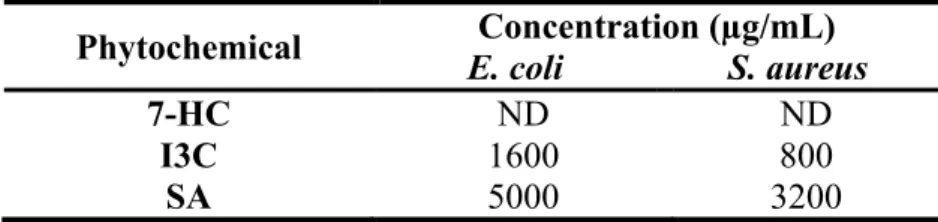
The MIC of 7-OH, I3C, SA and SP were measured for both bacteria (E. coli and S. aureus). The MIC value was considered to be the lowest concentration of phytochemical able to totally inhibit microbial growth. Table 1 shows the results obtained.
Table 1. MIC for E. coli and S. aureus.
Strains Phytochemicals (µg/mL)
7-HC I3C SA SP
E. coli 800 800 3200 ND
S. aureus 200 400 1600 ND
ND – Not detectable
The values of MIC for E. coli are higher than those for S. aureus. This result can be explained because E. coli is a Gram-negative bacterium and it is less susceptible to antimicrobials than Gram-positive bacteria. The Gram-negative bacteria tend to be more resistant to lipophilic and amphiphilic inhibitors than those Gram-positive, including dyes, detergents, free fatty acids, antibiotics and chemotherapeutics agents (Nikaido, 1996). In other study, antibiotics from natural origin showed activity against Gram-positive bacteria, but more than 90% of them have no useful effect against E. coli (Vaara, 1993). This increased resistance of Gram-negative bacteria can be attributed to the presence of the outer membrane. The porin channels slow down the penetration of small hydrophilic solutes and the low fluidity of the lipopolysaccharide layer decreases the rate of transmembrane diffusion of lipophilic solutes (Nikaido and Vaara, 1985; Plésiat and Nikaido, 1992).
Regarding to the different phytochemicals tested, SP was the unique compound that had no detectable MIC for concentrations lower than 3200 µg/mL. The 7-HC and I3C were the most effective compounds against both bacteria, since they presented the lowest values of MIC.
The literature is full of reports describing natural products and extract with antimicrobial activity with MIC values over 1000 µg/mL, which has little relevance for clinical application (Gibbons, 2004).
After the determination of MIC values for each phytochemical, MBC values were measured, using several concentrations sub and above-MIC. Table 2 presents the values of MBC for both bacteria. Regarding SP, the MBC was not possible to be quantified due to the absence of a MIC value.
Table 2. MBC values for E. coli and S. aureus.
ND – Not detectable
Similarly to the results of MIC, the values of MBC were higher for E. coli than for S. aureus. MBC represents the lowest concentration of antimicrobial product necessary to kill a bacterium; so Gram-negative need a higher concentration of antimicrobial, because these bacteria are more resistant (Nikaido, 1996). Regarding to 7-HC, the MBC was not defined for both bacteria, until the maximum value tested (10000 µg/mL). I3C seems to be the most effective phytochemical against both bacteria.
After testing the effects of the selected phytochemicals against the E. coli and
S. aureus strains, the effects of the same phytochemicals on their biofilms were tested.
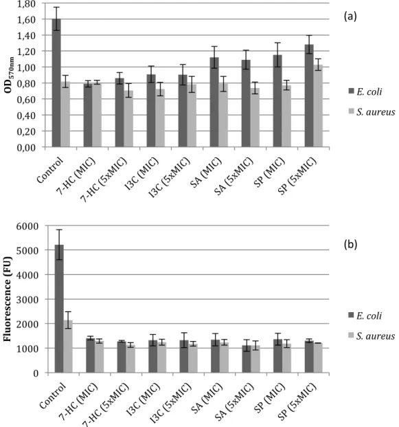
Biofilm formation was performed in sterile 96-well polystyrene microtiter plates. In this case, the bacteria were incubated overnight without phytochemicals. Biofilms were formed for 24 hours and after that, biofilms were incubated with phytochemicals at the MIC and 5 × MIC for 1 hour. According this procedure, it would be possible to conclude about the efficacy of phytochemicals in control (removal and inactivation) of biofilms. Figure 3 shows the results obtained from this assay.
Phytochemical E. coli S. aureus Concentration (µg/mL)
7-HC ND ND
I3C 1600 800
Figure 3. OD570 nm as a measure of biofilm mass (a) and fluorescence (λex: 570 nm and λem:
590 nm) results as a measure of biofilm viability (b) for E. coli and S. aureus. The mean ± standard deviation values resultant from three independent experiments
are depicted.
According to the classification proposed by Stepanović et al. (2000), both strains are weak biofilm producers (+).
In the Table 3 are presented the percentages of biofilm removal and inactivation with the selected phytochemicals at different concentrations. The ability of phytochemicals to control 24 h aged biofilms was analysed, based on their effects on biomass and metabolic activity.
0 1000 2000 3000 4000 5000 6000 Fl u or esc en ce ( FU ) E. coli S. aureus 0,00 0,20 0,40 0,60 0,80 1,00 1,20 1,40 1,60 1,80 OD 570n m E. coli S. aureus (a) (b)
Table 3. Percentages of biofilm removal and inactivation by the selected phytochemicals against E. coli and S. aureus.
The results show the capacity of phytochemicals to remove/inactivate the biofilms of E. coli and S. aureus. The biomass removal is dependent on the phytochemical and on its concentration. Comparing the values obtained, it is concluded that the phytochemical concentration (MIC and 5 × MIC) did not influence the removal of the biofilm (p>0.05), for both bacteria. Probably, the cells are able to adapt to the phytochemicals and with a higher concentration, the effect is not so noticed. Other possibility is the occurrence of changes in the biofilm phenotype (Cerca et al., 2006). However, the type of bacteria influences the values of biofilm removal. The same phytochemical, at the same concentration, presents a different behaviour in Gram-negative and Gram-positive bacteria (p<0.05). The percentages of biomass removal were always higher for E. coli than S. aureus with all the phytochemicals and concentrations performed. Total biofilm removal was not achieved with any of the selected phytochemicals. The highest reduction in biomass was found for E. coli with 7-HC. All the phytochemicals were more active in removing E. coli biofilms than S. aureus biofilms.
In terms of metabolic activity, the phytochemicals promoted higher reduction for E. coli biofilms than S. aureus biofilms. The inactivation percentages refer to the quantity of cells not viable present in the biofilm. The selected phytochemicals induced a higher quantity of cells not viable in biofilms of E. coli comparing to those of S. aureus. The biofilms of E. coli treated with phytochemicals at different concentrations did not show significative differences in the percentage of viable cells (p>0.05). Probably, the same effect explained before, for the percentage of removal, occurred in this case and the cells are able to adapt or the biofilm phenotype is
Phytochemicals
E. coli S. aureus
% Biomass
removal % Metabolic inactivation % Biomass removal % Metabolic inactivation
7-OH (MIC) 51.03±2.53 73.01±1.48 9.63±2.30 39.79±4.15 7-OH (5 × MIC) 46.67±4.47 75.50±0.70 1.68±0.32 47.30±4.59 I3C (MIC) 43.63±6.47 74.67±4.45 0.14±0.04 42.03±5.61 I3C (5 × MIC) 43.98±7.95 74.68±5.78 4.02±0.09 44.99±4.68 SA (MIC) 33.36±5.15 74.24±4.86 0.00±0.00 42.21±5.34 SA (5 × MIC) 34.19±6.52 78.75±4.57 0.00±0.00 48.26±8.55 SP (MIC) 31.80±6.92 73.86±4.62 0.00±0.00 44.71±7.65 SP (5 × MIC) 23.86±5.80 75.09±1.37 0.00±0.00 43.54±0.29