Universidade de Aveiro 2013
Departamento de Biologia
Noémia Rita Alves
Fernandes
Fragmentos de tRNA com capacidade de
silenciamento em peixe zebra
Assessing silencing ability of tRNA derived
fragments in zebrafish
DECLARAÇÃO
Declaro que este relatório é integralmente da minha autoria, estando devidamente referenciadas as fontes e obras consultadas, bem como identificadas de modo claro as citações dessas obras. Não contém, por isso, qualquer tipo de plágio quer de textos publicados, qualquer que seja o meio dessa publicação, incluindo meios eletrónicos, quer de trabalhos académicos.
Universidade de Aveiro 2013
Departamento de Biologia
Noémia Rita Alves
Fernandes
Fragmentos de tRNA com capacidade de
silenciamento em peixe zebra
Assessing silencing ability of tRNA derived
fragments in zebrafish
Dissertação apresentada à Universidade de Aveiro para cumprimento dos requisitos necessários à obtenção do grau de Mestre em Biologia Molecular e Celular, realizada sob a orientação científica da Doutora Ana Raquel Santos Calhôa Mano Soares, Investigadora de Pós-doutoramento do Departamento de Biologia da Universidade de Aveiro
Apoio financeiro da FCT e do FSE no âmbito do III Quadro Comunitário de Apoio
o júri
presidente Prof. Doutora Maria de Lourdes Gomes Pereira
Professora Associada com agregação da Universidade de Aveiro
arguente Doutora Joana Carvalho
Investigadora de Pós-doutoramento no Institute of Molecular Pathology and Immunology of the University of Porto (IPATIMUP)
orientadora Doutora Ana Raquel Soares
agradecimentos A realização desta dissertação apenas foi possível devido ao contributo de várias pessoas a quem não posso deixar de expressar o meu sincero reconhecimento e gratidão:
À Doutora Ana Soares, minha orientadora, pela competência científica, acompanhamento e disponibilidade reveladas ao longo deste ano de trabalho, assim como pelas críticas, correções e sugestões relevantes feitas durante a orientação.
Ao Professor Doutor Manuel Santos pela oportunidade de desenvolver este trabalho no seu laboratório, pelo seu entusiasmo contagiante por esta área científica e por todos os conhecimentos e conselhos partilhados.
A todos os colegas de laboratório por toda a ajuda disponibilizada, em especial à Violeta pela partilha dos seus conhecimentos sobre a manutenção e cuidados com os peixe-zebra.
Ao Tomás, pela compreensão, paciência e carinho que sempre me dedicou e por me fazer acreditar em mim e nas minhas capacidades nos momentos mais difíceis.
A todos os meus amigos, em especial à Inês, à Corine e à Raquel pela amizade e carinho incondicionais, pelo apoio, o incentivo e a coragem e por estarem presentes nos momentos bons e menos bons.
À minha família, em especial aos meus pais pela oportunidade, pelo incentivo e pelo carinho e compreensão imprescindíveis ao longo de todo este tempo que sempre demonstraram. E ao meu irmão Pedro que com a sua personalidade peculiar consegue animar-me em qualquer momento.
Por fim quero agradecer à Universidade de Aveiro, em especial ao Departamento de Biologia pela disponibilização das instalações e de todas as condições necessárias à realização deste projeto.
palavras-chave Pequenos RNAs não-codificantes, Fragmentos de tRNA, Capacidade de silenciamento, peixe zebra
resumo Desde a sua descoberta que os pequenos RNAs não codificantes (pRNAnc) têm sido implicados em diversos mecanismos de regulação em organismos eucarióticos, nomeadamente no silenciamento génico pós-transcricional, na regulação da cromatina e no desenvolvimento das células germinativas. Apesar dos microRNAs (miRNAs) serem a classe de pRNAnc mais estudada, o desenvolvimento de novas tecnologias de sequenciação permitiu a descoberta de novas classes de pRNAnc.
Recentemente foram identificados novos pRNAnc que derivam de tRNAs (fragmentos de tRNA) cujas vias de produção e funções ainda não estão bem definidas. Nos últimos anos, o laboratório de RNA da Universidade de Aveiro identificou fragmentos derivados de tRNAs (tRFs) em peixe zebra. A abundância de dois destes fragmentos - tRF_3 e tRF_4, que derivam da porção 5' de tRNAs maduros – e o facto de ambos serem conservados em vertebrados, processados pela Dicer e apresentarem alguma capacidade de associação com proteínas Argonautas, sugere que estas moléculas representam uma nova classe de RNAs reguladores que evoluíram cedo nos vertebrados e que podem estar envolvidos em mecanismos de regulação pós-transcricional de forma análoga aos miRNAs.
Tendo esta informação em consideração, nesta tese estudou-se a capacidade de silenciamento tanto do tRF_3 como do tRF_4. Para isso foram realizadas experiências com um sistema repórter duplo que permite a análise da regulação do gene alvo por moléculas endógenas. Este ensaio revelou que ambos os fragmentos têm capacidade de silenciamento, embora apenas o tRF_4 tenha uma região cuja destabilização inibe a sua capacidade de silenciamento, de forma semelhante aos miRNAs. Foi também realizada a previsão computacional de genes alvo para o tRF_4, sendo que estes se relacionam com o padrão de expressão do tRF_4. Esta tese demonstra que os tRFs em peixe zebra possuem capacidade de silenciamento e estudos futuros são necessários para determinar experimentalmente os seus alvos moleculares.
keywords Small non-coding RNAs, tRNA-derived fragments, Silencing ability, Zebrafish
abstract Since their discovery, small non-coding RNAs (sncRNAs) have been implicated in several eukaryotic regulatory mechanisms, such as post-transcriptional gene silencing, chromatin regulation and germline development. Although miRNAs constitute the most studied class of sncRNAs, the advent of high-throughput sequencing technologies allowed the discovery of new classes of sncRNA molecules.
Recently, novel sncRNAs derived from tRNAs (tRNA-derived fragments) have been identified, whose biogenesis and functions are not yet well defined. In the last years the RNA laboratory of the Aveiro University has also identified tRNA derived fragments (tRFs) in zebrafish. The abundance of two of these tRFs, namely tRF_3 and tRF_4, which derive from the tRNA 5’-portion – and the fact that both are conserved in vertebrates, processed by Dicer and exhibit some ability to associate with Argonaut proteins, suggest that these fragments represent a novel class of regulatory RNAs that have evolved early in vertebrates and may be involved in ancient mechanisms of genome regulation.
Taking this information into account, in this thesis the silencing ability of both tRF_3 and tRF_4 was studied. In order to do that, experiments were performed with a dual reporter system, which allows for the analysis of target regulation by the endogenous molecule. This assay revealed that both fragments have silencing ability, although only tRF_4 has a region whose destabilization inhibits its silencing ability, similarly to miRNAs. Gene target computational predictions for tRF_4 were also performed. The results obtained are related with tRF_4 expression pattern. This thesis shows that tRFs in zebrafish have silencing ability and further studies are required in order to determine, experimentally, their molecular targets.
Contents
List of abbreviations ... I List of Figures ... III List of Tables ... V
1. State of the art ... 1
1.1 The RNA ... 3
1.2 The RNA interference ... 4
1.3 A novel class of small RNAs derived from tRNAs ... 5
1.3.1 The tRNA ... 5
1.3.2 The tRNA fragments ... 7
1.3.2.1 tRNA fragments biogenesis ... 8
1.3.2.2 Biological functions ... 12
1.4 Zebrafish and gene silencing ... 15
1.4.1 sncRNAs in Zebrafish ... 17
1.5 Objectives ... 18
2. Materials and Methods ... 19
2.1. Zebrafish development and tissue tRF profiling ... 21
2.2 Reporter Assay ... 21
2.2.1 Primer Annealing ... 22
2.2.2 Ligation ... 22
2.2.3 Bacterial Transformation ... 23
2.2.4 Plasmid Extraction and Linearization ... 23
2.2.5 Zebrafish maintenance ... 24
2.2.6 Zebrafish Embryos Microinjection ... 24
2.3 Gene Target Prediction ... 24
2.3.1 Duplex Injections ... 24
2.3.2 Alcian Blue Assay ... 25
2.3.3 RNA extraction ... 25
2.3.4 cDNA preparation ... 26
2.3.5 sec23b and tub1a RT-qPCR ... 26
3. Results ... 27
3.2 tRF_3 and tRF_4 have Silencing Ability ... 31
3.3 Gene Target Prediction... 35
3.3.1 Alcian blue staining ... 38
4. Discussion ... 43
5. Conclusion ... 49
I List of abbreviations
aaRS - Aminoacyl-tRNA synthetases Ago - Argonaute
AMP - Adenosine monophosphate ATP - Adenosine tri-phosphate bp - Base pair
cDNA - Complementary DNA
DFRS - Dual fluorescent reporter sensor
DGCR8 - DiGeorge syndrome critical region gene 8 DNA - Deoxyribonucleic acid
dpf - Days post fertilization
dsRBD - Double-stranded RNA-binding eIF4A - Eukaryotic initiation factor-4A eIF4E - Eukaryotic initiation factor-4E eIF4G - Eukaryotic initiation factor-4G FW - Forward
GFP - Green fluorescent protein hpf – Hours post fertilization hrs - Hours
lincRNA - Long intergenic non-coding RNAs min - Minutes
miRISC - miRNA-induced silencing complex miRNA - microRNA
MO - Morpholino
mpf - Months post fertilization mRFP - Red fluorescent protein mRNA - messenger RNA ncRNA - non-coding RNA ng – Nanograms
nt - Nucleotides
PCR - Polimerase chain reaction piRNA - Piwi-interacting RNA
II
Piwi - P-element induced wimpy testis pL - Picoliter
rasiRNA - Repeat associated small interfering RNA Rev - Reverse
RISC - RNA-induced silencing complex RNA - ribonucleic acid
RNAi – interference RNA rpm - rotations per minute rRNA - ribosomal RNA
RT-PCR - reverse transcription-polymerase chain reaction scaRNA - Small Cajal body-specific RNAs
sec - seconds
siRNA - small interfering RNA snoRNA – small nucleolar RNA snRNA - small nuclear RNA
TALENS - Transcription Activator-Like Effector Nucleases TILLING - Targeting Induced Local Lesions in Genome tiRNA - tRNA-derived stress-induced fragments
TRBP - HIV-1 TAR RNA binding protein tRNA - transfer RNA
tRNAf/tRF – tRNA-derived fragments UTR - un-translated region
YB-1 - Y box binding protein 1 ZFN - Zinc Finger Nucleases µg - microgram
µL - microliter µM - micromolar
III List of Figures
Figure 1. The RNA content of a cell Figure 2. tRNA synthesis pathway Figure 3. tRNA aminoacylation Figure 4. tRFs classes
Figure 5. tRNA halves biogenesis Figure 6. miRNA Biogenesis Figure 7. Small tRFs biogenesis
Figure 8. 24hpf zebrafish embryo and organ formation
Figure 9. Transient assays for unveil miRNA functions in Zebrafish Figure 10. Primers inserted in DFRS plasmid
Figure 11. Plasmid construction
Figure 12 . tRF sequences and their possible mature tRNAs Figure 13. tRF_3 abundance increases during development Figure 14. tRF_3 is tissue specific
Figure 15. tRF_4 expression increases during development Figure 16. tRF_4 expression is tissue specific
Figure 17. tRf_3 has silencing ability Figure 18. tRf_4 has silencing ability Figure 19. tRF_3 do not have a seed region
Figure 20. tRF_4 5’-portion is crucial for silencing
Figure 21. Protein processing in endoplasmic reticulum in Danio rerio
Figure 22. 5 day-post-fertilization zebrafish larvae skeleton stained for cartilage with
Alcian Blue
Figure 23. tRF_3 over-expression does not affect cartilage development Figure 24. tRF_4 over-expression affects cartilage development in zebrafish Figure 25. sec23b does not alter its expression in the presence of tRF_4.
V List of Tables
1
3 1. State of the art
1.1 The RNA
The RNA was first extracted in 1960, by James Darnell and colleagues (Scherrer, 2003), and since then, many RNA molecules have been described. The RNA is a single-stranded nucleotide chain, which makes it more flexible and capable of making a wide variety of complex 3D molecular shapes. Each nucleotide is comprised of a phosphate group and a ribose sugar which is bond to one of the four bases, adenine (A), cytosine (C), guanine (G) and uracil (U). Also, like some proteins, RNA can have catalytic functions, known as ribozymes (Griffiths, 2004; Pierce, 2010).
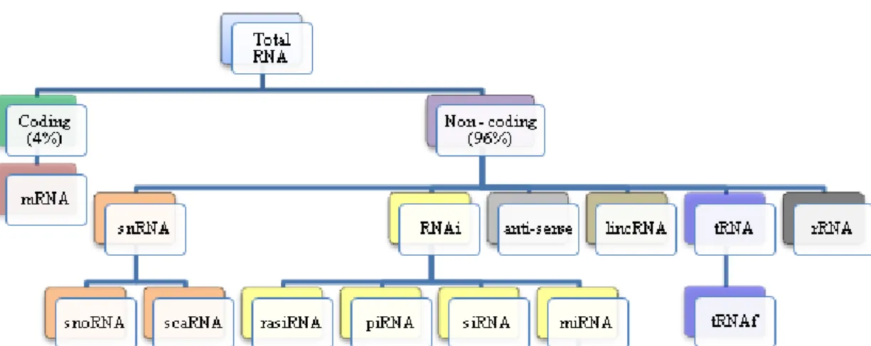
RNA molecules are divided into several classes according to their function, size and location in the cell. There are two main types of RNA, namely coding and non-coding RNA. Messenger RNA (mRNAs) constitutes the group of coding RNAs. These molecules are translated into proteins and their size varies with the size of the gene being transcribed. Despite the fact that the mRNA represents 60% of the total transcription of the cell it rarely makes up more than 4% of the total RNA due to its degradation soon after synthesis, being a short-lived molecule (Brown, 2002; Liu, et al., 2006; Arraiano and Fialho, 2007). On the other hand, thenon-coding RNAs (ncRNAs) do not encode proteins (Liu, et al., 2006) and account for the remaining 96% of the total RNA (Brown, 2002). The most abundant non-coding RNA is ribosomal RNA (rRNA), which makes up 80% of the total cell RNA. These molecules are important components of the ribosomes, the structures on which protein synthesis takes place, and are found in the cytoplasm (Brown, 2002). The other 16% of RNA contains the transfer RNA (tRNA) and small non-coding RNAs (Brown, 2002). Transfer RNA length ranges from 70 to 90nt and can be found in the cytoplasm and in mitochondria (Elliott and Ladomery, 2011) and plays an important role in protein synthesis, as it carries the correct aminoacid to the ribosome on which translation occurs. Small non-coding RNAs include small nuclear RNA (snRNA), interference RNA (RNAi), long intergenic non-coding RNAs (lincRNA) and anti-sense RNA (Figure 1) (Brown, 2002; Smith, 2009). These RNAs play key roles in several cellular processes and have regulatory functions, being involved in several biological processes, namely development, differentiation, immune response, infection and cancer (Arraiano and Fialho, 2007).
4 1.2 The RNA interference
The RNA interference (RNAi) was one of the most remarkable discoveries of the last 20 years. For the first time, it was clear that RNA molecules not only have important roles in translation, but also in regulation of gene expression in almost all eukaryotic organisms (Nilsen, 2007). The RNAi includes any post-transcriptional gene regulation event in which a small sized single-stranded RNA (20 to 30nt) forms a RNA-induced silencing complex (RISC) with an Argonaute protein (Ago), regulating, therefore, the expression of a cognate mRNA either by repressing translation or degradation. In this complex, the role of the small RNA is to guide it to the proper targets by complementar base-paring interactions, while the Ago proteins function as effector molecules (Nilsen, 2007; Kelly and Hurlstone, 2011; Kang, 2011). Some Ago proteins have catalytic activity and can specifically cleave the mRNA, but others confer the RISC complex the ability to induce DNA methylation, chromatin structure alteration and repression of mRNA translation (Nilsen, 2007).
There are several types of small non-coding RNAs, namely repeated-associated small-interfering RNA (rasiRNA), piwi-interacting RNA (piRNA), small interfering RNA (siRNA) and microRNA (miRNA), being the last two the most studied classes (Lee, et al., 2009) (Figure 1). All of the above have gene expression regulatory functions. Both rasiRNAs and piRNAs are thought to be involved in germline silencing of repeat transcripts, in Drosophila and mammals, possibly by chromatin modification (Matera, et
al., 2007 and Elliott and Ladomery, 2011). piRNAs are also responsible for cleaving RNAs
made by transposable elements and for regulating the embryonic development and
Figure 1. The RNA content of a cell (Adapted from Smith, 2009). In a human cell 4% of the RNA
(mRNA) encodes proteins. The other 96% contribute for the translation process like rRNA and tRNA or have regulatory functions like RNAi.
5
telomere protections (Huang, et al., 2013). siRNAs cleave RNAs derived from viruses, retroelements and repeat sequences. They also regulate long-term gene expression by directing cytosine methylation through RNA directed DNA methylation (Matera, et al., 2007 and Huang, et al., 2013). miRNAs, which are the most well-known class, regulate fundamental biological processes like proliferation, metabolism, embryogenesis, aging, and cell death, by targeting and silencing a huge number of human mRNA sequences (Elliott and Ladomery, 2011 and Huang, et al., 2013).
The advances in high throughput sequencing technologies allowed the discovery and characterization of an increasing number of small ncRNAs and prevented many to be discarded and considered as merely transcriptional “noise”. These small ncRNAs have turned out to be a hotspot in the life sciences, as it is becoming more and more evident that they have key regulatory functions (Huang, et al., 2013). The new sequencing technologies have, among others, unveiled a novel class of non-coding RNAs derived from tRNAs, the tRNA derived fragments (Lee, et al., 2009).
1.3 A novel class of small RNAs derived from tRNAs 1.3.1 The tRNA
The transfer RNA (tRNA) is the bridge that links the genetic code and the polypeptide sequence, in other words it is the molecular adaptor that associates the mRNA codon to an amino acid (Elliott and Ladomery, 2011). This molecule is usually 70 to 90 nt long and its secondary structure is described as a cloverleaf as it folds into four base paired stems. These structures are called the D loop, T loop, anticodon loop and the accept stem. The anticodon loop contains the anticodon, which recognizes its correspondent codon in the mRNA. The acceptor stem consists in the base paired between the 5’ and the 3’ ends, having the 3’ end a CCA residue which is the location for the amino acid binding. The tRNA has an extensive base modification which allows its tertiary L-shape structure (Elliott and Ladomery, 2011; Sobala and Hutvagner, 2011).
6
In summary, the synthesis of the transfer RNA comprises three steps: cleavage/trimming, modification and aminoacylation (Figure 2).
Transfer RNA from eukaryotic organisms is synthesized by RNA polymerase III (Alberts,
et al., 2008). The tRNA precursor (pre-tRNA)
chain is usually synthesized with additional nucleotides in one or both ends, the 5´ leader and the 3´ trailer. These extra sequences are then removed by the endonuclease RNase P, which recognizes the global L-shape tRNA structure and can specifically hydrolyze the phosphodiester bond, liberating the 5´-end of the molecule leaving a 5´-phosphate group (Krebs, et al., 2011; Elliott and Ladomery, 2011). The 3´-end is first cleaved in the middle by RNase E and RNase III and then trimmed by RNase II, RNAse BN, RNase PH and RNaseZ. This last enzyme cleaves the phosphodiester bond leaving a free 3´-OH end for the CCA residues addition by tRNA
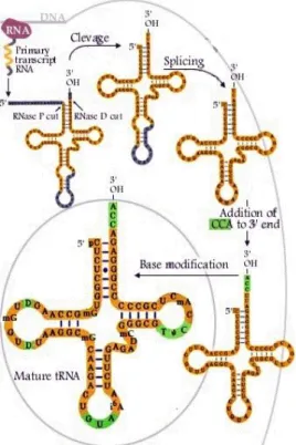
nucleotidyltransferase (Elliott and Ladomery, 2011). After, the tRNA suffers modifications in specific bases. The modifications are performed by specific tRNA-modifying enzymes. Usually 15 to 20% of the tRNA nucleotides are modified. These modifications are important for the tertiary structure formation and, when occurring in the anticodon, it creates a phenomenon known as wobble, in which a particular anticodon can recognize more than one codon (Krebs, et al., 2011; Elliott and Ladomery, 2011). Once all these steps have taken place, the tRNA can be charged by the aminoacyl-tRNA synthetases in the 3’-CCA terminal (Figure 3) (Krebs, et al., 2011).
Figure 2. tRNA synthesis pathway
(Adapted from (Nobel Media AB, 2013). tRNA genes transcription by RNA-Pol III produce a pre-tRNA which contains 5' and 3' extensions. RNase P and RNAse Z cleave 5' and 3' end respectively. Then tRNA nucleotidyltransferases create modifications in specific nucleotides and aminoacyl-tRNA synthetases add a CCA triplet at the 3´end.
7
Figure 3. tRNA aminoacylation (Maxwell, 2012). First, a specific aminoacid and an ATP molecule
bind to the aminoacyl-tRNA synthetases (aaRS). The aminoacid is activated by binding to the AMP and a phosphate is released. Then, the correct tRNA binds to the synthetase and the aminoacid is covalently attached to it, releasing the AMP. Finally the charged tRNA is released.
1.3.2 The tRNA fragments
In 1971, it was observed that, when infected by bateriofage T4, E. coli would produce specific leucine tRNA fragments, one with 48 nt and the other one with 39 nt corresponding to the 5’ end and to the 3’ end, respectively (Yudelevich, 1971).
Until recently, little importance was given to this finding, as it was thought that these molecules were random degradation products of tRNA, and, therefore, junk. Using protocols usually applied to miRNA profiling, the reads coming from tRNAs often constitute a minor fraction of the total library (1,5% to 10%) and were therefore, ignored from a deeper analysis as sequencing artefacts or degradation products of the mature tRNAs. However, comparative analysis between new and existing sequencing data revealed that tRNA fragments are conserved across distantly related species and correspond to a particular domain of tRNAs, drawing researcher’s attention for their possible cellular importance. Also, several deep sequencing, genetic screens, transcriptional, and molecular analyses studies showed that these small RNAs, are associated with several biological processes, such as development and stress response (Martens-Uzunova, et al., 2013).
These newly identified tRNA fragments seem to have different sizes and origins and can be specific to a tissue or a developmental stage (Cole, et al., 2009; Haussecker, et
al., 2010; Saikia, et al., 2012).
Since tRNA fragments were discovered, their nomenclature has been highly inconsistent. Several researchers have been using names like tRNA halves, tRNA-derived
8
Figure 4. tRFs classes (Garcia-Silva, et al., 2012).
3’U tRF is represented in yellow and derives from the immature tRNA. Green and blue segments correspond to 5’tRF and 3’CCA tRF respectively and derived from the mature tRNA.
halves, tRNA-derived RNA fragments (tRNAf), tRNA-derived stress-induced fragments (tiRNAs) or tRNA-derived small RNAs to refer to similar entities (Martens-Uzunova, et
al., 2013). Recently, two different nomenclatures have been proposed, one based on the
tRNA fragments size and another on the part of the tRNA or pre-tRNA from which the tRNA fragments are derived (Sobala and Hutvagner, 2011).
Therefore, tRNAfs can be divided into two major groups: the tRNA halves, which include tRNA fragments that range from 30 to 35nt and are produced in response to stress conditions, by cleavage near the anticodon;
and the small tRNA fragments (tRFs) (Figure 4). This group contains tRFs with approximately 20nt and can be divided in three sub-classes: 5’tRFs, 3’CCA tRFs and 3’U tRFs. The first class includes all the tRFs derived from the 5’ end of the tRNA and cleaved near its D-loop. The second class consists in all tRFs derived from the 3’ end and cleaved near the tRNA T-loop. The third class of tRFs is composed by those derived from the 3’ end of the pre-tRNA.
Most of these tRFs end in a series of U residues, generated by RNA polymerase III (Sobala and Hutvagner, 2011; Martens-Uzunova, et al., 2013).
1.3.2.1 tRNA fragments biogenesis
The tRNA halves, or the tRNA fragments-derived from cleavage within the anticodon, have been associated with response to stress conditions (Thompson and Parker, 2009; Fu, et al., 2009; Wang, et al., 2012). As stated before, tRNAs are a key component in the translation machinery, however they also have roles in cell proliferation and stress response. For example, during amino acid starvation, tRNA uncharged can act as signaling molecules and activate Gcn2 kinase, which instead phosphorylates eIF2α (Thompson and Parker, 2009; Saikia, et al., 2012). Stress-induced tRNA cleavage occurs through specific enzymes secreted from stressed cells such as Rny1 in yeast and angiogenin in mammals (Figure 5) (Fu, et al., 2009; Saikia, et al., 2012). Both of these ribonucleases belong to families, RNase T2 and RNase A, respectively. These enzyme families have little substract
9
specificity, pointing to possible cleavage of other RNAs during stress. Wang, et al. noticed that the nucleotides before the major cleavage were Gs and Cs, which reduces the randomness theory (Wang,
et al., 2012). Rny1 and angiogenin are
secreted ribonucleases. Rny1 is secreted in the vacuole and released to the cytoplasm during oxidative stress, where
it can access the cytosolic RNAs (Thompson and Parker, 2009). Angiogenin is most located in the nucleus or the nucleolus, but is also found in the cytoplasm, bound to its inhibitor RNH1. Stress conditions promote the dissociation of angiogenin from RHN1 or the ribonuclease release from the nucleus (Thompson and Parker, 2009).
tRNA cleavage may inhibit specific steps of translation, leading to its global decrease or produce tRNA halves that can target specific mRNAs, which in conjunction with tRNA processing enzymes or Argonaute proteins, inhibit their translation or leads to their degradation (Thompson and Parker, 2009).
Small tRNA fragments have been often compared with miRNAs. Their features, namely size and association with Ago proteins and even part of their biogenesis are very similar to those of miRNAs (Pederson, 2010).
Very briefly miRNAs, which in general are present in the genome as clusters, are processed from precursor transcripts (pri-miRNAs), which are mostly transcribed by RNA polymerase II from independent genes (Ghildiyal and Zamore, 2009; Carthew and Sontheimer, 2009; Krol, et al., 2010). The pri-miRNA usually has a long imperfect stem and, like most mRNAs, has a 5’-cap structure and a polyadenylated tail (Hussain, 2012). After being transcribed, the pri-miRNA is cleaved in the nucleus by the Microprocessor Drosha-DGCR8 (Pasha in Drosophila and C. elegans) into a 60-70 nt precursor hairpin, the pre-miRNA, which contains in its 3’ end 2 nt overhang and a 5’-phosphate group (Ghildiyal and Zamore, 2009; Krol, et al., 2010). Then a nuclear export protein, Exportin-5 in a RanGTP-dependent manner, exports the pre-miRNA through the nuclear pore (Ghildiyal and Zamore, 2009).
Figure 5. tRNA halves biogenesis (Garcia-Silva, et al.,
10
Once in the cytoplasm, Dicer and its dsRBD assistant protein TRBP (or Loqs in flies) cleave the pre-miRNA. This connection to Dicer, can only be possible because, unlike Drosha, it possesses a Paz domain that interacts with the 2 nt overhanging in 3’UTR, left by Drosha (Ghildiyal and Zamore, 2009; Carthew and Sontheimer, 2009). Dicer cleaves off the loop and originates an approximately 20 nt double stranded RNA, a miRNA/miRNA* duplex, with two nucleotides overhanging at each 3’ end. (Ghildiyal and Zamore, 2009; Winter, et al., 2009; Krol, et al., 2010). Dicer and TRBP then dissociates from the duplex and an helicase unwinds and separates both strands (Winter, et al., 2009). Usualy only one strand of the miRNA/miRNA* is incorporated into a miRNA-induced silencing complex (miRISC) being the other strand degraded. The criteria for choosing one strand over the other is probably thermodynamic (Ghildiyal and Zamore, 2009; Carthew and Sontheimer, 2009; Krol, et al., 2010) (Figure 6). miRISC then targets the homologous transcrips by base paring interactions and silences the target mRNA. The crucial feature for target is excellent complementarity between the 3’ end of the target mRNA and the miRNA 5’ end, also called “seed” region. This “seed” region starts at position 2 in the guide sequence, up to the 6th or 8th nucleotide. The complementarity between this two regions must be perfect in order to have an effective silencing by the miRNA (Elliott and Ladomery, 2011).
Figure 6. miRNA Biogenesis (Winter, et al., 2009). RNA polymerase II transcribes miRNA genes,
generating a primary miRNA transcript with hairpin structures. Microprocessor Drosha-DGCR8 then cleaves the pri-miRNA and the resulting hairpin, pre-miRNA, is exported from the nucleus to the cytoplasm by Exportin-5-Ran-GTP. Once in the cytoplasm Dicer-TRBP cleaves the the pre-miRNA into miRNA duplexes. The duplexes are unwond and the functional strand as well as Dicer are loded into the RNA-induced silencing complex (miRISC). The mature miRNA can then guide the RISC complex to the 3’UTR of a mRNA causing its cleavage or translation repression or deadenilation.
11
The tRFs are originated from tRNA gene transcripts which may be cleaved by Dicer, ELAC2 or RNase Z, giving rise to 5’tRFs, 3’CCA tRFs and 3’U tRFs. 5’tRFs biogenesis can be Dicer-dependent (Cole, et al., 2009) in mammals or Dicer-independent as it was reported in Schizosaccharomyces pombe (Buhler, et al., 2008). The differences in their size (19nt and 23nt, respectively), support the idea that there must be different mechanisms/enzymes for each organisms (Cole, et al., 2009; Sobala and Hutvagner, 2011). Computational studies also point that some 5’tRFs can actually acquire an hairpin structure, like the one observed for miRNAs, which may point towards a similar mechanism of action or function (Cole, et al., 2009 and unpublished data). 3’CCA tRFs are cleaved by dicer after CCA addition to the tRNA. In fact, it was reported that 5’tRFs studied in Tetrahymena carry a 5’ monophosphate, which is typical of Dicer cleavage and differs from the 5-hydroxyls described on longer half-tRNAs generated by starvation-induced cleavage (Couvillion, et al., 2010). Moreover, Lee et al., found that the preferential cleavage site for these fragments was located in AU sites in prostate cancer cells (Lee, et al., 2009). The 3’U tRFs are generated from pre-tRNA, which still did not lost the polyuridine tract. These fragments are cleaved by RNase Z (Sobala and Hutvagner, 2011) or, ELAC2, a cytoplasm enzyme (Lee et al., 2009). In both studies 3’U tRFs are only found in the cytoplasm, leading to speculate that these transcripts may be cleaved in the nucleus and rapidly exported to the cytoplasm (Haussecker, et al., 2010).
After cleavage both tRNA halves and tRFs are thought to bind to Ago proteins. Until now investigators have found that these molecules associate more efficiently with Ago 3 and Ago4 (Haussecker, et al., 2010). In mammals it is known that only Ago2 has cleavage activity and Ago1, 3 and 4 do not have this activity, therefore their function has not been totally elucidated yet. It was also seen that, in Tetrayemena, 3’CCA tRFs associate with a PIWI protein, Twi12, which does not have cleavage activity (Couvillion,
et al., 2010).
Although both miRNAs and tRFs are generated by Dicer and associate with Ago proteins, they differ from each other in several aspects. Mammalian miRNAs are not methylated in the 3’ terminal ribose. Yet, Cole et al., saw that tRFs-derived from tRNAGln
have 3’ terminal modification. Still they cannot tell if the modification occurs after or prior Dicer processing. Also, miRNAs form a stable complex, when incorporated with Argonautes, so when Dicer is knocked down there is no significant change in miRNA steady-state level.
12
However, when knocking down Dicer, Cole et al., noticed smaller fractions of tRFs than in the minimal RISC (AGO2 and tRF), which suggests that either the tRFs associate inefficiently with Ago proteins, or they do not associate with the proteins at all, or the tRFs are incorporated into non-Argonaute complexes, which have a faster turnover rate. This inefficiency in RISC incorporation can be due to the 3’ terminal modification (Figure 7) (Cole, et al., 2009).
1.3.2.2 Biological functions
The tRNA derived fragments have long been regarded as random byproducts of tRNA biogenesis or degradation. Yet, the discovery of the small tRFs, which biogenesis is dependent on Dicer automatically raised the question of whether or not these fragments have a biological function (Cole, et al., 2009; Gebetsberger, et al., 2012). Several lines of work suggested that tRFs are indeed novel biological entities. First of all, tRFs biogenesis seems to be regulated and the tRFs have approximately the same size distribution. In fact the cleavage sites showed to be conserved and specific enzymes are required to cleave the
Figure 7. Small tRFs biogenesis (Sobala and Hutvagner, 2011) 3’U tRFs can be generated from
pre-tRNA by RNase Z, either in the cytoplasm or in the nucleus. After processed by RNase P and Z and added the CCA-terminal, the tRNA can be permaturely exported to the cytoplasm and processed by Dicer. Mature aminoacylated tRNAs may enter the small RNA pathway via Dicer, which produces the 5’tRfs and the 3’ CCA tRFs. White triangle - RNAse Z cleavage site; Black triangle - RNase P cleavage site; Red arrows - premature nuclear export.
13
tRNA. (Lee, et al., 2009; Cole, et al., 2009; Sobala and Hutvagner, 2011; Martens-Uzunova, et al., 2013). Furthermore the results show that tRFs are produced after intron excision and even after 5’-CCA-3’ addition (Martens-Uzunova, et al., 2013). Also tRNA halves biogenesis seems to be also controlled by RNA methylation and induced by specific stress (Martens-Uzunova, et al., 2013) or cell proliferation (Wang, et al., 2012). Secondly, until now only tRFs corresponding to the 5’end and the 3’end of the tRNA were reported, and no tRF matching the anticodon loop was stated in mammals (Sobala and Hutvagner, 2011). Pederson, stated that this feature might be selected by evolution, and as the anticodon is optimally configured for base-paring, it did not have any selective advantage on a regulation path being, therefore, excluded from the sequence (Pederson, 2010). Third, when performing deep sequencing it is clear that the sequencing number of different tRFs does not correlate to those of the tRNA gene copies or with tRNA gene expression or codon usage distribution. Moreover, when RNA degradation increases there is not an increase in the fragments abundance (Lee, et al., 2009; Sobala and Hutvagner, 2011; Martens-Uzunova, et al., 2013). At last, there has been evidence that tRFs can associate with Ago proteins and some can actually have trans-silencing activity (Lee, et al., 2009; Sobala and Hutvagner, 2011).
These evidences indicate that the tRNA-derived fragments can be novel biological entities. Although, their roles are not totally clarified yet, there are already several lines suggesting that tRNA-derived fragments participate in important biological processes like stress response and gene expression regulation.
Haussecker, et al., focused their work on tRFs (3’U and 3’CCA) ability to associate with Ago proteins and their propensity to have an effect on luciferase reporters. They found that both 3’U and 3’CCA bind more effectively to Ago 3 and Ago4 than with Ago1 and Ago2 and that 3’CCA and that the latter has a moderate effect on the reporter while 3’U does not (Haussecker, et al., 2010).
Lee, et al, also studied 3’U but focus their attention on their possible function. The authors studied in particular tRF-1001, and by knocking down this tRF with a siRNA, they saw a decrease in cell proliferation (Lee, et al., 2009).
Using HeLa cells, Cole et al. conjecture that tRNAs may compete with pre-miRNA for Dicer cleavage leading to an unbalanced miRNA homeostasis. An increase in tRNA levels leads to cell transformation maybe because Dicer would be forced to accept them as
14
a substrate and thus altering miRNA homeostasis and function. They also verified that 5’tRFs do not associate with Ago2, but Ago1. Ago1 has been implicated in transcription gene silencing by miRNAs and siRNAs that target promoter regions and promotes heterochromatin formation. This suggests that tRFs may also be involved in transcriptional gene silencing pathway (Cole, et al., 2009). In another study, using deep sequencing and computational comparison, it was showed that there are 3’CCA tRFs that do not undergo Dicer processing, but can Associate with Ago2 and guide down regulation in vitro (Li, et
al., 2012). Wang et al. also demonstrated that a 5’tRF derived tRNAglu (CTC) has trans-silencing activity. This tRF is located in the cytoplasm and the mechanism by which silencing is carried out is different from miRNA/siRNA pathway, as the binding region to the target RNA is positioned in the 3’end and not in the 5’end (Wang, et al., 2012).
Recently, several works from Paul Anderson group have shown that tiRNAs (tRNA-derived stress-induced fragments) can inhibit translation. They have demonstrated that Angiogenin is responsible for producing tiRNAs in stress conditions and U2OS cells transfection with 5´-tiRNAs could induce phospho-eIF2α-independent translational arrest, and promote stress granules assembly. The tiRNAs efficiency was higher when possessing a 5’-monophosphate in the 5´-end. Ivanov et al, found that tiRNAs can efficiently inhibit translation of uncapped mRNAs. 5’-tiRNAs can directly or indirectly bind eIF4G, eIF4A, or to the complex eIF4G/ eIF4A and inhibit translation. It can also bind to the eIF4F, favouring eIF4E:4E-BP complex formation over eIF4F. These tiRNAs also interact with YB-1, whose function is to regulate transcription and translation. The cold shock domain of this protein can displace eIF4E from m7G cap and the carboxyl terminus interferes with eIF4G binding. This suggests that tiRNAs and YB-1 cooperate to eIF4G/A binding and consequently translation initiation and that this protein is require for stress granule assembly by the tiRNAs (Yamasaki, et al., 2009; Emara, et al., 2010; Ivanov, et al., 2011). Sobala and Hutvagner, also found that 5’tRFs can inhibit protein translation in human cells, by interfering with the elongation process. Using HeLa cells, they studied a highly-express 5’tRF (Gln) and find that it can inhibit reporter genes translation in vitro and in vivo. Unlike miRNA/siRNA, they noticed that this tRF did not need complementary sites, instead it recognizes a “GG” dinucleotide sequence, which appears to be conserved in 5’tRFs at position 17-8 or 18-19, depending on the parent tRNA. They observed that the 5’tRF cellular fraction can associate actively with polysomes, interfering with the
15
elongation process or with other important translation processes like tRNA-charge specificity (Sobala and Hutvagner, 2013).
Using 454 DNA pyrosequencing technologies to sncRNA discovery in zebrafish, the laboratory of RNA in the university of Aveiro unveiled 11 tRFs, which align with the tRNAs in the 3’end (6tRFs), the 5’ end (4tRFs) and the pre-tRNA (1tRF) (unpublished data). Some of these tRFs showed to be more abundant in adult tissues like muscle, fins and skin, than in developmental stages. Three of the tRFs identified are homologous to other tRFs identified in human cells or tissues. tRF_3 is similar to tRF-5002 found in prostate cancer cell lines and also to another tRNAglu fragment in human fetus hepatic tissue and it was recently described in human cells after a RSV infection. tRNAf_2 was also found in human fetus hepatic tissue and derives from tRNAval, tRNAf_11 is identical to cand15 and derives from tRNAarg. These results show that these tRFs are likely conserved in vertebrates and therefore they must have a biological function. In vitro biogenesis assays performed with two of the most expressed tRNA derived fragments found in zebrafish - tRF_3 and tRF_4, showed that they were cleaved by Dicer and may fold into a similar structure to that of pre-miRNAs (unpublished data). Also, both tRF_3 and tRF_4 have an uridine at the 5’ end, which is a miRNA characteristic and bind, although weakly, to Ago1 (unpublished data), suggesting that these fragments can act like miRNAs and be involved in gene silencing.
1.4 Zebrafish and gene silencing
Danio rerio, also known as zebrafish, is a small size tropical water fish, which
inhabits Asia. For the past 20 years this organism has become one of the favourite in vivo model systems for studying developmental processes and human diseases (Santoriello and Zon, 2012; Kardash, 2013). Recently sequencing studies unveiled that zebrafish has 26,000 protein coding genes and by direct comparison to the human genome it was possible to conclude that 71,4% of the human genes have at least one zebrafish orthologue. On the other hand, 69% of zebrafish genes have at least one orthologue in humans (Santoriello and Zon, 2012; Chitramutu, 2013; Howe, et al., 2013; Kardash, 2013). Also, it was possible to see that 82% (2,601 out of 3,176) of the genes related to human diseases can be related to a zebrafish orthologue (Howe, et al., 2013). Besides the great genetic similarity, zebrafish has many advantages for which it is considered an attractive model in research.
16
Figure 8. 24hpf zebrafish embryo
and organ formation. (Weigta, et al., 2010)H – Head; E - Eye; S/O - sacculi/otoliths;T – Tail; TT – Tail Tip
Zebrafish has a high fecundity and a single female can lay 200-300 eggs. The embryos develop outside the body and are transparent, which allows following organogenesis. The development is rapid and the major organs are formed within 24hours after fertilization (Figure 8). Furthermore, its raise and maintenance is easy and inexpensive when compared to other vertebrate models (Santoriello and Zon, 2012; Chitramutu, 2013).
At first, zebrafish was used in forward genetics, in which chemical and insertional mutation, namely the DNA transposon system known as Sleeping Beauty, has
led to the identification of the causative genes for a given phenotype, by positional cloning or through candidate gene approach (Santoriello and Zon, 2012; Chitramutu, 2013). Recently, reverse genetic techniques have been developed and successfully applied to zebrafish. These techniques include antisense morpholino (MO) oligonucleotide mediated gene knockdown technology, Targeting Induced Local Lesions In Genome (TILLING), Zinc Finger Nucleases (ZFN), Transcription Activator-Like Effector Nucleases (TALENs), Tol2 mediated Transgenesis, GAL4-UAS System, Tol2-mediated Gal4/UAS, Cre/Lox system and a tamoxifen-inducible Cre/lox method (Chitramuthu, 2013).
Microinjection is the most frequently used method when working with zebrafish embryos. This is an efficient and rapid vehicle for adding either mRNA, morpholino or RNA duplex into hundreds of embryos per hour (Figure 9) (Rosen, et al., 2009, Fjose and Zhao, 2010). Microinjection of fertilized eggs, easily obtained by mating adult fish, provides the opportunity to study directly their functions throughout zebrafish development. Injection of chemically synthesized miRNA mimics allows determining the effect of an over-expression of a specific miRNA during development and, in some cases, to detect variations in predicted mRNA targets. Also the validation of these targets and their binding sites can be achieved by co-injection of a reporter gene, which consists in mRNA of GFP and the miRNA binding site in its 3’UTR (Fjose and Zhao, 2010).
17 1.4.1 sncRNAs in Zebrafish
According to miRBase, 346 microRNAs have already been unveiled in zebrafish (miRBase, 2013). Recently, Wei et al, using Next-Generation sequencing technology unveiled the expression pattern of 198 known miRNAs within 122 different miRNA families in zebrafish embryos. The same study showed that 225 of the 56,311 reads derived mostly from 3’ and 5’ ends of mature tRNAs. These fragments were different and had different cleavage-sites from those detected in angiogenin-dependent fragments found in stressed cells. 3’ tRNA reads size did not fluctuate much (18nt), unlike the 5’ which were distributed more widely and lead to the conclusion that they might me under different selection mechanisms or have different functions. Also, it was possible to notice that tRFs abundance increases dramatically from 24hpf, being more abundant those derived from the 5’end (Wei, et al., 2012).
As stated before, 454 DNA pyrosequencing technologies applied to sncRNA discovery in zebrafish by our laboratory unveiled 11 tRFs in this organism. From those, tRF_3 and tRF_4 were chosen to study in this thesis, as they were the most expressed ones. tRF_3 is a ~30nt fragment which derives from the 5’end of a mature tRNA through Dicer cleavage. This fragment is conserved in zebrafish, humans and mouse and appears to bind to Ago1. Preliminary studies also indicate that its expression is tissue dependent. tRF_4 is a smaller fragment, with ~19nt. This is not a “common” tRF, as it does not start at the 5’end of the mature tRNA, but near its D-loop. Its biogenesis is Dicer dependent and it can also bind to Ago1 protein. tRF_4 expression varies according to the tissue and in some tissues its
Figure 9. Transient assays for unveil miRNA
functions in Zebrafish (Adapted from: Fjose and Zhao, 2010). A) Gain-or-loss of function experiments can be performed by injection of miRNA or miRNA inhibitors in embryos. The effects can be analyzed by assessing the phenotype or the expression levels of the mRNA. (B–D) Illustrates the effects on the mRNA levels by injection of either a mature miRNA duplex or MOs. B) miRNA duplex injection leads to a decreased level of the target mRNA and therefore the protein encoded by it. C) MOs injection blocks miRNA from binding its target site, leading to an increase in protein level. D) Mature miRNA and GFP reporter mRNA co-injections can be used to validate predicted binding sites of a specific mRNAs.
18
expression is higher than its corresponding tRNA. As the tRF_3, tRF_4 is conserved in zebrafish, humans and mouse.
Zebrafish has been used to a widely range of molecular studies. Although, until now,studies on the function of tRNA-derived fragments are scarsed.
1.5 Objectives
The aim of this work was to elucidate the function of two tRFs - tRF_3 and tRF_4 - previously described in zebrafish, by our lab. The main objective was to test if these tRNA-derived fragments have silencing ability, similarly to miRNAs and investigate putative targets that can be silenced by them.
19
21 2. Materials and Methods
2.1. Zebrafish development and tissue tRF profiling
tRF profiling in zebrafish was performed by Northern blot. Northern blot is a technique that allows detecting specific RNAs by using a radioactive hybridization probe. For profiling of tRNA fragments during development samples from 24hpf, 48hpf, 72hpf, 10dpf, 1mpf and 2mpf were used. For zebrafish tissue profiling samples from gut, muscle, eyes, brain, fins, bones, skin and gills were used. After RNA extraction, 30µg of total RNA were fractionated on a denaturing polyacrylamide gel (10%). The RNA was transferred to an Hybond-N membrane (GE Healthcare) for 35min in a semidry transfer system followed by UV-crosslinked. Complementary antisense oligonucleotides to tRF_3 and tRF_4 were used as hybridation probes after being labeled with [32P]-ATP and T4 polynucleotide kinase (Takara). Membrane pre-hybridization lasted for 4hrs in hybridization buffer (5× Denhardt's solution, 1% SDS and 6,6x SSPE) ate 64ºC (tRF_3) and 40ºC (tRF_4). The probes were added and incubated with the membranes over night at the previous temperatures. The membranes were washed twice with washing solution (2xSSPE and 0,1%SDS) at room temperature and twice at 57ºC for 3min each and then exposed to a phosphor screen (Biorad®) overnight followed by scanning with a Molecular Imager® FX (Biorad), equipped with Quantity One FX software.
The probes used were the following:
tRF_3: 5’-GCCGAATCCTAACCACTAGACCACCAGGGA-3’ tRF_4: 5’-GCGAGAATCATACCCCTA-3’
2.2 Reporter Assay
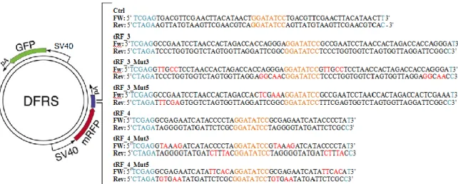
In this work a double reporter system, provided by Huttner’s lab, was used in order to verify the tRFs silencing ability (Tonelli, et al, 2006). The DFRS plasmid (pDSV2-EGFP-mRFP) has two fluorescent proteins, GFP (green fluorescent protein) and RFP (red fluorescent protein), which are controlled by the samepromoter. GFP identifies the tissues expressing the plasmid and the RFP, which contains a 3'UTR cassette complementary to the tRF of interest, functions as a silencing sensor. Two different assays were performed. First, a total complementary sequence to the tRF_3 and tRF_4 was used in order to see whether they have silencing ability or not. Then, mutations were made in specific locations in order to study the tRFs functional domains.
22 2.2.1 Primer Annealing
In order to have an efficient ligation of the insert (tRF and its complementar sequence) to the DFRS plasmid, the primers must be phosphorilated. This is accomplished by using T4 PNK enzyme. A mix with 3µL of the sense and anti-sense oligo at 100µM, 3 µL of 10x PNK buffer, 2µL of 10µM ATP, 2 µL of T4 PNK and 17µL of miliQ H2O was
prepared, for a total of 30µL of solution. The mix was then incubated at 37ºC for 90min. Then 4µL NaCl 0,5M was added, incubated at 95ºC for 2min and cool to the room temperature (~2hrs). The mix was then stored at 4ºC until use.The primers used were the following:
Forward and reverse primers both from tRF_3 and tRF_4 were ordered from Sigma and the tRF_3_Mut3, tRF_3_Mut5, tRF_4_Mut3, tRF4_Mut5 were ordered from Eurofins.
2.2.2 Ligation
The ligation of the insert to the plasmid was performed following the protocol described below. Briefly, three mixes with different ratios of plasmid (concentration 10ng/µL) and insert (concentration 10ng/µL) were prepared: 1:0, 1:3 and 1:5, respectively. 2µL of T4 DNA ligase 5x buffer and 1µL of T4 DNA ligase were added to each mix (ligation reaction) in a total volume of 20 µL. All ligation reactions were incubated at 16ºC overnight (Figure 11).
Ctrl
FW: 5’ TCGAGTGACGTTCGAACTTACATAACTGGATATCCTGACGTTCGAACTTACATAACTT 3'
Rev: 5’CTAGAAGTTATGTAAGTTCGAACGTCAGGATATCCAGTTATGTAAGTTCGAACGTCAC - 3'
tRF_3
Fw: 5’TCGAGGCCGAATCCTAACCACTAGACCACCAGGGAGGATATCCGCCGAATCCTAACCACTAGACCACCAGGGAT3’
Rev: 5’CTAGATCCCTGGTGGTCTAGTGGTTAGGATTCGGCGGATATCCTCCCTGGTGGTCTAGTGGTTAGGATTCGGCC3’
tRF_3_Mut3
Fw: 5’TCGAGGTTGCCTCCTAACCACTAGACCACCAGGGAGGATATCCGTTGCCTCCTAACCACTAGACCACCAGGGAT3’
Rev: 5’CTAGATCCCTGGTGGTCTAGTGGTTAGGAGGCAACGGATATCCTCCCTGGTGGTCTAGTGGTTAGGAGGCAACC3’
tRF_3_Mut5
Fw: 5’TCGAGGCCGAATCCTAACCACTAGACCACTCGAAAGGATATCCGCCGAATCCTAACCACTAGACCACTCGAAAT3’
Rev: 5’CTAGATTTCGAGTGGTCTAGTGGTTAGGATTCGGCGGATATCCTTTCGAGTGGTCTAGTGGTTAGGATTCGGCC3’
tRF_4
FW: 5’TCGAGGCGAGAATCATACCCCTAGGATATCCGCGAGAATCATACCCCTAT3’
Rev: 5’CTAGATAGGGGTATGATTCTCGCGGATATCCTAGGGGTATGATTCTCGCC3’
tRF_4_Mut3
FW: 5’TCGAGGTAAAGATCATACCCCTAGGATATCCGTAAAGATCATACCCCTAT3’
Rev: 5’CTAGATAGGGGTATGATCTTTACGGATATCCTAGGGGTATGATCTTTACC3
tRF_4_Mut5
FW: 5’TCGAGGCGAGAATCATATTCACAGGATATCCGCGAGAATCATATTCACAT3’
Rev: 5’CTAGATGTGAATATGATTCTCGCGGATATCCTGTGAATATGATTCTCGCC3’
Figure 10. Primers inserted in DFRS plasmid. The primers are composed by two sequences (black)
separated by EcoRV cleavage site (orange). Restriction sites are shown in blue and mutated nucleotides in red.
23 2.2.3 Bacterial Transformation
Transformation is the process by which cells uptake a foreign DNA present in its surroundings. In this case the cell is E.coli and the DNA is the DFRS plasmid. 20 µL of plasmid DNA was added to 200 µL competent cells, incubated on ice for 30 min, heat shocked at 42°C for 90 sec, placed on ice and to it, 800 µL SOC media was added. The cultures were then covered and incubated with shaking at 37 °C for 1 hour. Positive transformants were selected by plating on LB agar medium containing ampicilin and growing overnight at 37 °C.
2.2.4 Plasmid Extraction and Linearization
Plasmid extraction was performed with GeneJET™ Plasmid Miniprep Kit from Fermentas, following the manufacturers’ protocol and quantified by nanodrop. The plasmid was sequenced by stabvida.
For a most effective expression in zebrafish, the plasmid has to be injected in its linear form. The linearization was performed using 2µL of 10x FD buffer, 2µL of plasmid DNA (up to 12µg), 1µL of FastDigest BglII and miliQ H2O up to a final volume of 20 µL.
The mix was then incubated for 40 minutes at 37ºC. The final digestion product was cleared using QIAquick® Nucleotide Removal Kit from QIAGEN, following the manufactures’ protocol. Briefly, 100 µL of PNI buffer was added and the mixture was applied to the QIAquick spin column followed by centrifugation (6000rpm, 1min). 750 μL of PE buffer was added and the mixture was centrifuge first at 6000rpm, 1min and then
Figure 11. Plasmid construction. The DFRS plasmid contains GFP (green) and a mRFP (red) sequences.
Both are controlled by similar promoters (SV40). mRFP 3’UTR, where all the sequences were inserted, is shown in blue. PA is the poliadenilation site.
24
13,000 rpm, 1min to remove residual ethanol residues from PE buffer. 30 μL of MiliQ water were applied to the centre of the QIAquick membrane and after 2 min the tubes were centrifuged (13rpm, 1min).
2.2.5 Zebrafish maintenance
In this study wild-type AB zebrafish embryos were used. The embryos were collected and kept at 28ºC under standard laboratory conditions. Wild-type AB zebrafish were maintained in CESAM laboratory zebrafish facility in Aveiro University. The study was approved by the DGV (Direção-Geral de Veterinária).
2.2.6 Zebrafish Embryos Microinjection
One cell zebrafish eggs were microinjected with a solution containing 20ng/µL of the reporter plasmid. The solution was made with Phenol red and 0,9M KCl.
One-cell stage eggs were injected with approximately 1000pL of the solution. Only wild type AB zebrafish strain was used in this study and the embryos were collected and kept at 28ºC in the CESAM’s zebrafish facility of Aveiro University.
They were analyzed at 24hpf by fluorescent microscopy.
2.3 Gene Target Prediction
3’UTRs from zebrafish mRNAs were acquired from Biomart (http://www.biomart.org) and blasted against tRF_4 sequence. Those which showed perfect match between nucleotides 2 and 7 (seed region) and no more than 5 mismatches were confirmed thermodynamically by RNAhybrid and considered for further analysis.
2.3.1 Duplex Injections
tRF_3 and tRF_4 RNA duplexes, a duplex containing a scrambled sequence were obtained as siRNAs, the first two from IDT and the last one from SIGMA:
tRF_3 duplex: sense: 5’-UCCCUGGUGGUCUAGUGGUUAGGAUUCGGC-3’ antisense: 5’- GCCGAAUCCUAACCACUAGACCACCAGGGA-3’ tRF_4 duplex: sense 5’- UAGGGGUAUGAUUCUCGC-3’ antisense 5’- GCGAGAAUCAUACCCCUA-3’ Scrambled duplex:
25
sense 5’-GUGUAACACGUCUAUACGCCCA-3’;
antisense 5’-GGCGUAUAGACGUGUUACAC [dT][dT]-3’;
The siRNAs were injected into one-cell fertilized embryos at 5 μM concentration in TE Buffer (pH=7,5). Approximately 1000pL of the RNA duplex were injected.
2.3.2 Alcian Blue Assay
Alcian blue assay stains cartilage and bones allowing its visualization and analysis. Five days post fertilization (dpf) zebrafish embryos were fixed for 5hrs in 4% (p/v) phosphate-buffered formaldehyde and maintained in methanol 100% at -20ºC until use. The embryos were washed 2 times in phosphate-buffered saline with 0.1% Tween-20 (PBT). Then they were bleached in 30% hydrogen peroxide until the eyes became sufficiently translucent (~2hrs). Next the embryos were rinsed twice with 1 ml of PBT and transferred into 0,5ml of an Alcian blue solution (1% concentrated hydrochloric acid, 70% ethanol, 0.1% Alcian blue), overnight. The specimens were rinsed 3 times with 1 ml acidic ethanol (5% concentrated hydrochloric acid, 70% ethanol, HCl-EtOH) and incubated for 20 min in 1ml of the same solution. Re-hydration was performed in 1ml HCl-EtOH/H2Od
series: 75% / 25% HCl-EtOH/ H2Od; 50/50 HCl-EtOH/ H2Od; 25/75 HCl-EtOH/ H2Od and
100% H2Od. Finally the H2Od was removed and the embryos were stored in 1ml KOH. The
embryos were visualized in a dissecting microscope (Nikon SMZ1500).
2.3.3 RNA extraction
The RNA was extracted using TRIZOL® Reagent, from approximately 100 injected embryos (24hpf), and following the manufacturer’s protocol. For cDNA preparation the samples were treated with DNAse I (Invitrogen), according to the protocol provided by Invitrogen, with minor changes. Briefly, 500ng of total RNA were mixed with 1µL DNase I and DNaseI buffer incubated for 40 min at 37ºC. 1µL of EDTA is added in order to inhibit the reaction and the mix is incubated for 10 min at 65ºC. Two RNA extractions were performed, for the first 100 µL phenol:chlorophorm:iso-amyl was used followed by centrifugation (12,000rpm for 15minutues at 4ºC ), for the second 1volume of chlorophorm was added to the supernatant and the samples were centrifuge (12,000rpm for 15minutues at 4ºC). The supernatant was removed, the pellet washed with 500µL of 75% ethanol and centrifuged again at 7,500rpm for 5minutes at 4ºC. The pellet was dried, ressuspended in 30µL of miliQ H2O and quantified by nanodrop.
26 2.3.4 cDNA preparation
cDNA is DNA obtained from mRNA template in a reaction catalysed by the enzymes reverse transcriptase. This technique can be combined with PCR (RT-PCR) allowing detection of gene expression.
cDNA was prepared using SuperScript®II RT (Invitrogen) from 500ng of total RNA. In the first step 1µL of Oligo d(T) 12-18, 1µL of dNTPs mix (10µM) and H2O up to
a total volume of 12µL were added. The mix was incubated at 65ºC for 5 minutes and then cooled down for another 5 minutes. Next, 4µL of 5xFirst Strand buffer, 2µL of 0,1 µM DTT and 1µL of RNase out were added. The solution was mixed and incubated at 42ºC for 2 minutes. One uL of SSII RT was added and the samples were incubated for 15 minutes at 70ºC. Then 0,5µL of RNase H was added and after incubated for 20 minutes at 37ºC.
2.3.5 sec23b and tub1a RT-qPCR
In order to quantify sec23b transcripts a quantitative real time PCR was performed. qPCR was carried out by using the synthesized cDNA and SYBR® Green detection reagent, Universal qPCR Primer provided in the kit, and the forward primer for sec23b and tub1a (the endogenous control) detection (the same used for RT-PCR). It was performed in the ABI Prism 7500 Real time PCR System (Applied Biosystems) and the reactions were incubated in a 96-well optical plate. The cycling began with template denaturation at 95ºC for 2min, followed by 40 cycles at 95ºC for 30 sec and 60ºC for 1 min. The samples and the no-template controls were carried out in triplicate and run at the same time.
The primers used were the following:
Sec23b_fw–5’-TGGTGGGATCGGCCATGTG-3’; Tub1a_fw–5’-GGCATCAACTACCAGCCTCC-3’;
27
29 3. Results
3.1 tRF_3 and tRF_4 profiling
The tRF_3 is a 30nt non-coding RNA derived from the 5’end of a mature tRNA
(Figure 12). Previous work from our lab has shown that this fragment requires Dicer for its biogenesis and that its expression is conserved in humans, mouse and in zebrafish. The tRF_4 derives from the 5’ end of a mature tRNA and it is 19nt long (Figure 12). This fragment is rather peculiar as it does starts near the D-loop and not in the 5’end. Its biogenesis is also dependent on Dicer and it derives from tRNAs that can acquire pre-miRNA-like conformations. Given all these evidences and the similarities between this fragment and miRNA/siRNA molecules, it is possible to assume that tRF_4 is not a random product of tRNA degradation and may have analogous biological functions in the cell.
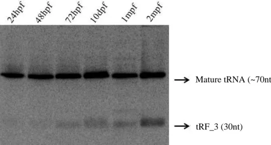
Despite of being a tRNA halve, previous results shown that the tRF_3 expression is not related to stress conditions in zebrafish and it is different from tissue to tissue. In this study the expression of this fragment was analysed both during zebrafish development and in its different tissues in adulthood. It was possible to see that its expression increases during its development (Figure 13), being more prominent in adult tissues such as gut, skin, bone and fins (Figure 14).
tRF_3
tRF_4
Figure 12. tRF sequences and their possible mature tRNAs. tRFs are shown in orange and the mature
30
Like the tRF_3, tRF_4 has also shown to be differently expressed from tissue to tissue. Its profiling revealed that it increases during development, especially after 10 days post fertilization (Figure 15) and it is more abundant in certain tissues, namely eyes, gills, bone, gut and skin (Figure 16). It was also possible to see that in these tissues, the abundance of the fragment is higher than the mature tRNA, which confirmed the previous results obtained in the lab (Figure 16).
Figure 14.tRF_3 is tissue specific. tRF_3 tissue profiling was obtained by northern blot using RNA samples from different tissues from adult fish.
Mature tRNA (~70nt) tRF_3 (30nt)
Figure 13. tRF_3 abundance increases during development. tRF_3 development profiling was performed
by northern blot, using RNA samples from different stages of development (24hpf, 48hpf, 72hpf, 10dpf. 1mpf and 2mpf).
tRF_3 (30nt)
31 3.2 tRF_3 and tRF_4 have Silencing Ability
In order to study the silencing ability of the highly expressed tRF_3 and tRF_4 in zebrafish, a dual-fluorescense GFP-Reporter/mRFP-Sensor plasmid (Dual fluorescence reporter system – DFRS) was injected in one cell-stage zebrafish embryos. The zebrafish was chosen as a model to this work, as the fragments in this study had already been reported in this organism and are conserved among vertebrates (unpublished data). The DFRS plasmid contains both GFP and mRFP under the control of identical constitutive promoters, the SV40 promoter. As the sensor-based strategy relies on the silencing of a transcript, the GFP-reporter is used to identify the cells that are actually expressing the plasmid, and the mRFP-sensor contains a 3’untranslated region with a tandem cassette, which is complementary to the tRF of interest (Tonelli, et al, 2006). Therefore, only the embryos expressing GFP, and hence the DFRS plasmid were analysed and the expression
Figure 15. tRF_4 expression increases during development. Development profiling of tRF_4 was
performed using RNA samples from different stages of development (24hpf, 48hpf, 72hpf, 10dpf. 1mpf and 2mpf), by northern blot.
Mature tRNA (~70nt)
tRF_4 (19nt)
Figure 16.tRF_4 expression is tissue specific. tRF_4 profiling was obtained by northern blot using RNA samples from different tissues from adult fish.
Mature tRNA (~70nt)
32
of the injected plasmid was visible in the zebrafish embryo muscle. For each condition three types of pictures were taken, one showing only GFP signal, another showing RFP signal and a third containing both GFP and RFP signals (Merge).
Using the DRFS plasmid, with the reverse complementary sequence to the tRFs inserted in the mRFP 3’UTR, it was evident that tRF_3 and tRF_4 have silencing ability, as there was a decrease in the expression of mRFP signal when compared with the control. For both tRF_3 and tRf_4, the majority of the embryos injected with the plasmid containing the GFP and the RFP, it was visible that the RFP signal was absent, although the GFP was still detectable (Figures 17 and 18 respectively). This means that the plasmid was being expressed in the zebrafish embryo muscles cells, but due to their ability for silencing by establishing a bound with a complementary sequence, the RFP signal was not detected. However, it was also noticed that the silencing ability for the tRF_4 fragment is stronger than in tRF_3. This may be due to the higher expression of tRF_4 compared to tRF_3 in these tissues (Figure 16). The tRF_3 and tRF_4 have, therefore, ability to recognize a complementary sequence as a target site and suppress its expression.
Figure 17. tRF_3 has silencing ability. A, B, C – Embryo injected with 20ng/µl of DFRS control plasmid, showing
GFP, mRFP and merge signal respectively. D, E, F - Embryo injected with 20ng/µl of DFRS plasmid containing the reverse complementary sequence to the tRF_3, showing GFP, mRFP and merge signal respectively.
tR F_ 3 ( 2 0 n g /µl) GFP mRFP Merge Co n tr o l (2 0 n g /µl) A B C D E F * * * *