The impact of HSF on endometrium
HONGCHU BAO1, GUICHAN WANG2, XIN HUANG1, MEIMEI WANG1, XINRONG WANG1, CUIFANG HAO1*
1Department of Reproductive Medicine, The Affiliated Yantai Yuhuangding Hospital of Qingdao University, Yantai, Shandong, China 2Department of Gynecology and Obstetrics, The Affiliated Yantai Yuhuangding Hospital of Qingdao University, Yantai, Shandong, China
S
UMMARYStudy conducted at the Department of Reproductive Medicine, The Affiliated Yantai Yuhuangding Hospital of Qingdao University, Yantai, Shandong, China
Article received: 5/11/2017
Accepted for publication: 6/16/2017
*Correspondence:
Department of Reproductive Medicine, The Affiliated Yantai Yuhuangding Hospital of Qingdao University Address: No 20, Yudong Road
Zhifu District, Yantai, Shandong – China Postal code: 264000 [email protected] http://dx.doi.org/10.1590/1806-9282.63.12.1069
Objective: We conducted the research in order to explore the impact of hydrosalpinx
fluid (HSF) on endometrium.
Method: HSF group: 261 patients with HSF scheduled to undergo laparoscopic
surgery 3 to 7 days after menstruation in our center. Hysteroscopy would also be performed in order to observe the endometrial morphology during the surgery. Sixty (60) patients would be randomly selected for endometrial biopsy in order to detect the inflammatory cytokines TNF-α and IL-2 mRNA. Non-HSF group: 210 patients with no evidence of HSF due to chronic salpingitis or pelvic adhesion. IVF-ET treatment was performed after eliminating the factor of male infertility and hysteroscopy was conducted before the treatment. Fifty (50) patients underwent endometrial biopsy in order to detect TNF-α and IL-2 mRNA.
Results: Hysteroscopy was performed in 261 patients with HSF and 210 patients
without HSF. The incidence rate of endometritis manifestation among these two groups of patients was 37.2% (97/261) and 20.5% (43/210), respectively. The incidence rate of endometritis in the patients with HSF is significantly higher than in the patients without HSF (p<0.05). Sixty (60) patients from the HSF group and 50 patients from the non-HSF group were regrouped according to inflammatory and normal manifestation after the endometrial biopsy. There were 49 patients in the inflammatory manifestation group and 61 patients in the normal manifestation group. RT-PCR technology was adopted to detect the expression of inflammatory cytokines TNF-α and IL-2 mRNA in endometrial tissue. The level of TNF-α mRNA expression in endometrial tissues with inflammatory manifestation was higher than in normal endometrium (76.75±11.95 vs. 23.45±9.75, p<0.01). There are significant differences between them. The level of IL-2 mRNA expression in endometrial tissues with inflammatory manifestation was higher than that found in normal endometrium (80.56±13.35 vs. 35.12±8.35, p<0.01). There are significant differences between them.
Conclusion: Chronic endometritis is related to HSF and may therefore affect
endometrial receptivity.
Keywords: HSF, endometrium, immunohistochemistry, polymerase chain reaction.
I
NTRODUCTIONEndometrial receptivity is an important factor to deter-mine the success of pregnancy and hydrosalpinx fluid (HSF) may have a bad effect on endometrial receptivity through various mechanisms. Using immunohistochem-ical examination on patients with HSF after endometrial biopsy, Mayer et al.1 found that endometrial integrin α
υβ3 of the patients with HSF during implantation window
re-sponse, integrin, matrix metalloproteinases, endometrial blood flow, leukemia inhibitory factor are moderately af-fected.2-4 Our study explores the connection between HSF patients and chronic endometritis (CE) and verifies wheth-er HSF would influence endometrial receptivity by inducing CE. We observed the general performance of endometrium in patients with HSF, first through hysteroscopy and then collecting endometrial samples from different patients for the detection of TNF-α and IL-2 mRNA in order to assess whether HSF has negative effects on the endometrium.
M
ETHODStudy object
Four hundred seventy-one (471) patients undergoing infertility treatment in the Reproductive Medicine Centre due to salpingitis from July 2010 to June 2014 were cho-sen. Their average age was 31.5 years and infertility lasted from 2 to 9 years.
Inclusion criteria
• (1) The female partner of infertile couples needing treatment due to salpingitis.
• (2) Age range from 25 to 35 years old.
• (3) Normal menstrual cycle lasting from 25 to 35 days with a variation of 3 days.
• (4) Weight range from 45 to 70 kg and body mass in-dex between 18 and 25 kg/m2.
Exclusion criteria
• (1) Infertility due to unknown reasons other than salpingitis.
• (2) Routine examination of male semen abnormal more than twice.
• (3) Female partner with one of the following endo-crine abnormalities: polycystic ovary or polycystic ovary syndrome, hyperprolactinemia.
• (4) Female partner presenting abnormal cervical cy-tological examination: HPV infection or atypical cell hyperplasia or more severe lesions indicated by TCT.
• (5) Female partner’s baseline endocrine levels (mens-truation D2~D5) FSH and/or LH > 10 IU/mL.
• (6) Acute inflammation of the genital tract.
• (7) Organic lesions present including submucosal ute-rine fibroids, endometrial polyps, uteute-rine adhesions.
Diagnostic criteria
HSF group: HSF was indicated by hysterosalpingography (HSG). Non-HSF group: no HSF found after HSG that would be caused by chronic tubal inflammation or pel-vic adhesion.
Experiment group
HSF group: 261 patients with HSF scheduled to undergo laparoscopic surgery 3 to 7 days after menstruation in our center. Hysteroscopy would also be performed in order to observe the endometrial morphology during the surgery. Sixty (60) patients would be randomly selected for endometrial biopsy in order to detect the inflamma-tory cytokines TNF-α and IL-2 mRNA.
Non-HSF group: 210 patients with no evidence of HSF due to chronic salpingitis or pelvic adhesion. IVF-ET treatment was performed after eliminating the factor of male infertility and hysteroscopy was conducted before the treatment. Fifty (50) patients underwent endometrial biopsy in order to detect TNF-α and IL-2 mRNA.
Experiment methods
Methods and procedures for endometrial examination with hysteroscopy
• (1) Evaluation time: the best period is the early to the middle stage of endometrial proliferation, i.e., 3 to 7 days after menstruation.
• (2) Anesthesia: Hysteroscopy can be performed in outpatient setting without anesthesia and few pa-tients need intravenous or general anesthesia. In the case of inpatients who prepare to undergo combined laparoscopic surgery, general anesthesia was used.
• (3) The objective lens of hysteroscope is placed into the cervical canal slowly under direct vision after rou-tine disinfection and speculum placement. At the same time, physiological saline is applied for cervical canal expansion and uterine distention, the disten-ding pressure is 100-120 mmHg.
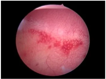
• (4) Endometrial morphology observation: Normal uterine cavity was covered by newly formed smooth endometrium, yellowish and reddish, with few blood capillary and open glandular ducts (Figure 1). Endo-metritis patients often show endometrial hyperemia, edema and exudation, even necrosis. In this study, the biopsy would be performed only for patients with normal endometrial appearance and inflammatory manifestations like endometrial hyperemia, edema etc. Other lesions such as endometrial polyp, submu-cosal myoma of uterus etc. were excluded from our study. Diagnostic criteria for CE under hysteroscope can refer to the description of Zolghadri et al.:5 en-dometrial hyperemia presents a crimson red or fire red color and the subepithelial vascular network is significantly dense and thickened (Figure 2).
sam-ple was then rinsed by physiological saline in order to reduce blood contamination as much as possible, in-serted into a 1.5 mL Ep tube with high pressure steri-lization and DEPC water treatment and then stored in a freezer at -70°C ultra-low temperature in order to ex-tract RNA from the tissue during RT-PCR detection.
Detection of the expression of TNF-α and IL-2 mRNA in endometrium with RT-PCR
Relevant reagents: Total RNA extraction kit with Trizol re-agent produced by Invitrogen Company (USA). RNA reverse transcription kit, 10 mmol/L dNTPs, 5 U/ mu lTaq DNA polymerase and agarose were all products of Promega Com-pany. Synthesis of PCR primers was entrusted to Sangon Biotech (Shanghai) Co., Ltd. and TNF-α primer sequences were: upstream primer 5-G A G T G A C A A G C C T G T A G C C C-3′, downstream primer 5’-G C A A T G A T C C C A A A G T A G A C C-3’ with the amplified product length of 363 bp (Figure 3). And the primer sequences of Internal reference glyceraldehyde-3-phosphate dehydrogenase (GAP-DH) gene were: upstream primer 5′-C C A C C A C C A T C T T C C A G G A G-3′, downstream primer 5′-C C T G C T T C A C C A C C T T C T T G -3′ with the amplified product length of 572 bp. IL-2 primer sequences were: upstream 5′-C T G G A G C A T T T A C T G C T G G A T-3′, downstream 5′-G C C T T C T T G G G C A T G T A A A A C-3′ with the amplified product length of 110 bp (Figure 4).
The specific procedure in the RT-PCR experiment was as follows:
• (1) Treatment of endometrium: about 100 mg fresh frozen endometrial tissue sample was cut into small
pieces, cleaned with D-Hanks liquid and centrifuged for 12 minutes in a low temperature centrifuge at a temperature of 4°C and 10,000 rpm. The superna-tant was removed and the tissue sediments were wa-shed for 2 to 3 times with D-Hanks liquid.
• (2) Total RNA extraction: total RNA was extracted according to procedures in the manual of total RNA extraction kit with Trizol reagent. The optical den-sity values of A260 and A280 were determined using a DU800 spectrophotometer from Beckman Coulter with a ratio of ranging from 1.8 to 2.0. The concen-tration of total RNA was then calculated. At the same time, total RNA electrophoresis yielded three strips of 28S, 18S and 5S or two strips of 28S and 18S, showing that RNA quality met the requirements (Figure 5).
• (3) cDNA synthesis: reverse transcription kit from Invitrogen Company was adopted to synthesize cDNA according to the manual. The cDNA obtai-ned from reverse transcription was used as a tem-plate for PCR reaction.
• (4) PCR reaction: cDNA of 2 μL, primer of 200 nmol/L, dNTP of 150 μmol/L, Taq DNA polymerase 1 U and corresponding buffer solution were included in the 25 μL reaction system. TNF-α, IL-2 and internal refe-rence GAPDH in type 9600 DNA cycler (Perkin Elmer Cetus) were amplified according to the reaction con-ditions. The amplification conditions of the three ge-nes were as follows: pre-denaturation for 3 minutes at 94°C in the beginning; repeated cycling 35 times for 30 seconds at 94°C, for 30 seconds at 55°C, for 1 mi-nute at 72°C; 7 additional minutes at 72°C in the end.
FIGURE 1 Normal uterine cavity: smooth endometrium, in pale red color without abnormal blood vessel.
• (5) Identification of amplification products: Absorban-ce scanning for TNF-α, IL-2 and GAPDH was perfor-med by using GD 2000 gel scanning and analyzing system D with pUC Mix treated as molecular weight standard after taking PCR amplification product of 10 μL, 2% of agarose gel electrophoresis and ethidium bromide staining (including the ethidium bromide with a final concentration of 0.5 µg/mL). And then the ratio of gene expression of TNF-α and IL-2 to that of GAPDH should be calculated respectively.
Statistical method
SPSS 11.0 statistical software was used for data analysis. The obtained data was expressed as
x
±
s
and tested witht-test. And enumeration data was detected by χ2
check. Statistical significance was defined as p<0.05.
R
ESULTSHysteroscopy result
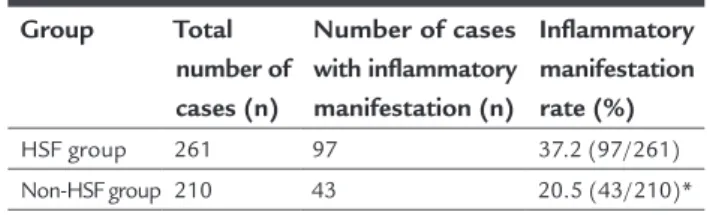
In this study, hysteroscopy was performed in 261 patients with HSF and 210 patients without HSF. The incidence rate of endometritis in these two groups of patients were 37.2% (97/261) and 20.5% (43/210), respectively. As shown in Table 1, the incidence rate of endometritis in the pa-tients with HSF is significantly higher than that of papa-tients without HSF.
FIGURE 5 Result of total RNA electrophoresis extracted from endometrial tissues.
18S
28S
Sample hole
FIGURE 4 Electrophoresis track: 1. IL-2/GAPDH of endometrium with normal manifestation. 2. IL-2/GAPDH of endometrium with inflammatory manifestation. 3. pUC Mix Marker.
1 2 3
111 bp
190 bp
331 bp 404 bp 501/489 bp 692 bp
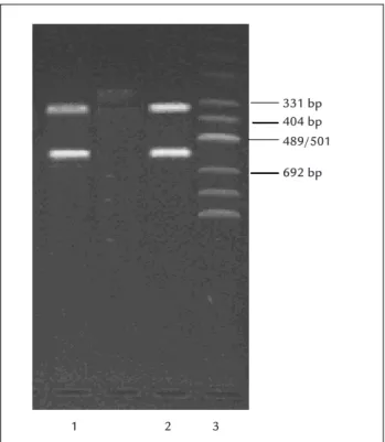
FIGURE 3 Electrophoresis track: 1. TNF-α/GAPDH of endometrium with normal manifestation. 2. TNF-α/GAPDH of endometrium with inflammatory manifestation. 3. pUC Mix Marker.
331 bp 404 bp 489/501
692 bp
2 3
TABLE 1 The ratio of endometritis in three groups of patients evaluated by hysteroscopy.
Group Total number of cases (n)
Number of cases with inflammatory manifestation (n)
Inflammatory manifestation rate (%)
HSF group 261 97 37.2 (97/261)
Non-HSF group 210 43 20.5 (43/210)* p<0.05 * compared with HSF group.
Sixty (60) patients from HSF group and 50 patients from non-HSF group were regrouped according to the inflammatory or normal status after endometrial biopsy. There were 49 patients in the inflammatory manifestation group and 61 patients in normal group. RT-PCR technol-ogy was adopted to detect the expression of inflammatory cytokines TNF-α and IL-2 mRNA in endometrial tissue.
Result of RT-PCR detection of TNF-α and IL-2 mRNA in endometrium
• (1) The level of TNF-α mRNA expression in endome-trial tissue with inflammatory manifestation was higher than that seen in normal endometrium (p<0.01). The-re was significant diffeThe-rences between them (Table 2).
TABLE 2 RT-PCR detection of the levels of TNF-α expression
in two groups of subjects investigated.
Group Number
of cases
Relative content of TNF-α (%)
Inflammatory manifestation group 49 76.75±11.95 Normal manifestation group 61 23.45±9.75 p<0.01
• (2) The level of IL-2 mRNA expression in endometrial tissues with inflammatory manifestation was higher than in normal endometrium (p<0.01). There was significant difference between them (Table 3).
TABLE 3 RT-PCR detection of the levels of IL-2 expression
in two groups of subjects investigated.
Group Number
of cases
Relative content of IL-2 (%)
Inflammatory manifestation group 49 80.56±13.35 Normal manifestation group 61 35.12±8.35 p<0.01
D
ISCUSSIONEmbryo quality and endometrial receptivity are two key factors that influence the clinical outcome of pregnancy.
cav-ity but better ways to diagnose CE are still needed. Wheth-er CE affects embryo implantation is still controvWheth-ersial; the high incidence rate of CE in patients with embryo implantation failure is an indisputable fact.10,11
Inflammatory cytokines are important mediators for the occurrence of CE and both TNF-α and IL-2 play an important role in the process of inflammatory response.12 TNF-α is mainly expressed by mononuclear macrophages, CD4+Th1 cells and natural killer (NK) cells etc. in the immune system. Besides the expression in immune cells, TNF-α can also be expressed in reproductive tissues such
as ovary, salpinx, uterus, placenta etc. And it also takes part in the process of gametogenesis, embryonic develop-ment, follicle growth and steroid hormone synthesis etc., through autocrine and paracrine action. There must be a right amount of TNF-α in pregnant women to maintain pregnancy, but TNF-α at high concentrations may lead to a series of inflammatory lesions, stimulate the production of multiple inflammatory factors such as IL-1, IL-6, NO etc. and result in the occurrence of inflammation and damage in tissues by activating inflammatory cells and upregulating adhesion molecules, NO and oxygen free radicals. After detecting the levels of IL-6 (interleukin-6), IL-1β and TNF-α in menstrual blood of the patients di-agnosed as CE by hysteroscopy and histology in follicular phase of the previous menstrual cycle, Tortorella et al.13 believed that proinflammatory cytokines IL-6 and TNF-α can be used as a biomarker for CE. TNF-α also has a direct toxic effect on endometrium and harms the decidua ves-sel, which brings about contraction of vascular smooth muscle, embolism of the embryonic blood-supply system and tissue necrosis.14 IL-2 is the glycoprotein produced and secreted by activated T cells with a molecular weight of about 15kD. At the same time, it is necessary for T cell proliferation, as well as a key mediator of the immune response. It plays a key role in the cellular immune re-sponse and possesses a mechanism of promoting the biological activity of T cells and B cells.15 IL-2 may also be expressed by endometrial glandular cells but excessive expression of IL-2 may affect embryo implantation.16 IL-2 level reflects the body’s cellular immune state to some degree and IL-2 has a function of anti-tumor, anti-micro-bial infection, also inducing graft rejection, autoimmu-nity and immune regulation. Therefore, detection of IL-2 levels is a sensitive index to assess the immune activation status of the body. TNF-α and IL-2 are all cytokines se-creted by Thl cells and play a key role in the inflamma-tory response. Piccinni et al.17 found that Th1 cytokines can downregulate the expression of LIF, and IL-2 is an indispensable cytokine in embryo implantation. HSF, in
turn, may influence the endometrial receptivity by decreas-ing the expression of LIF in the endometrium durdecreas-ing the implantation window period.16 After hysteroscopy, we found that the incidence rate of the inflammatory mani-festation of endometria in patients with HSF and the expression of TNF-α and IL-2 in endometrial issues with inflammatory manifestation increased significantly, in-dicating that inflammatory cytokines such as TNF-α and IL-2 in the endometrium of patients with HSF were in-volved in the inflammatory manifestation of endome-trium. In the process of embryo implantation, the matrix was in a state of immune tolerance with Th2 type immune response in a dominant position. The high expression of TNF-α and IL-2 in endometrium of patients with HSF may also indicate that the local immune balance of the endometrium biases towards the Th1 type immune re-sponse, which affects embryo implantation.
Due to limitation of experimental conditions, we only tested the typical TNF-α and IL-2 instead of detecting a large number of inflammatory cytokines. Detection of endometrial receptivity only involves inflammatory response, which may result in probable error for the conclusion. Further studies on the impact of HSF on endometrial receptivity are still needed.
C
ONCLUSIONCE is related to HSF, and endometrial receptivity may be influenced by HSF.
R
EFERENCES1. Mayer WR, Castelbanm AJ, Somkuti S, Sagoskin AW, Doyle M, Harris JE, et al. Hydrosalpinges adversely affect markers of endometrial receptivity. Hum Reprod. 1997; 12(7):1393-8.
2. Savaris RF, Giudice LC. The influence of hydrosalpinx on markers of endometrial receptivity. Semin Reprod Med. 2007; 25(6):476-82. 3. Ajonuma LC, Ng EH, Chan HC. New insights into the mechanisms underlying
hydrosalpinx fluid formation and its adverse effect on IVF outcome. Hum Reprod Update. 2002; 8(3):255-64.
4. Ng EH, Chan CC, Tang OS, Ho PC. Comparison of endometrial and subendometrial blood flows among patients with and without hydrosalpinx shown on scanning during in vitro fertilization treatment. Fertil Steril. 2006; 85(2):333-8.
5. Zolghadri J, Momtahan M, Aminian K, Ghaffarpasand F, Tavana Z. The value of hysteroscopy in diagnosis of chronic endometritis in patients with unexplained recurrent spontaneous abortion. Eur J Obstet Gynecol Reprod Biol. 2011; 155(2):217-20.
6. Nardo LG, Sabatini L, Rai R, Nardo F. Pinopode expression on during human implantation. Eur J Obstet Gyneccol Reprod Biol. 2002; 101(2):104-8. 7. Daftary GS, Troy PJ, Bagot CN, Young SL, Taylor HS. Direct regulation of
beta 3-integrin subunit gene expression by HOXA10 in endometrial cells. Mol Endocrinol. 2002; 16(3):571-9.
8. Makrakis E, Pantos K. The outcomes of hysteroscopy in women with implantation failures after in-vitro fertilization: findings and effect on subsequent pregnancy rates. Curr Opin Obstet Gynecol. 2010; 22(4):339-43. 9. Cicinelli E, Resta L, Nicoletti R, Tartagni M, Marinaccio M, Bulletti C, et al.
10. Yang R, Du X, Wang Y, Song X, Yang Y, Qiao J. The hysteroscopy and histological diagnosis and treatment value of chronic endometritis in recurrent implantation failure patients. Arch Gynecol Obstet. 2014; 289(6):1363-9. 11. Cicinelli E, Matteo M, Tinelli R, Lepera A, Alfonso R, Indraccolo U, et al.
Prevalence of chronic endometritis in repeated unexplained implantation failure and the IVF success rate after antibiotic therapy. Hum Reprod. 2015; 30(2):323-30.
12. Terranova PF, Hunter VJ, Roby KF, Hunt JS. Tumor necrosis factor-alpha in the female reproductive tract. Proc Soc Exp Biol Med. 1995; 209(4):325-42. 13. Tortorella C, Piazzolla G, Matteo M, Pinto V, Tinelli R, Sabbà C, et al. Interleukin-6,
interleukin-1β, and tumor necrosis factor α in menstrual effluents as biomarkers of chronic endometritis. Fertil Steril. 2014; 101(1):242-7.
14. Nawroth PP, Stern DM. Modulation of endothelial cell hemostatic properties by tumor necrosis factor. J Exp Med. 1986; 163(3):740-5.
15. Shin HW, Rose-Gottron CM, Cooper DM, Hill M, George SC. Impact of high-intensity exercise on nitric oxide exchange in healthy adults. Med Sci Sports Exerc. 2003; 35(6):995-1003.
16. Lu S, Qiao J, Liu C, Li R. The expression of LIF and IL-2 in the endometrium at implantation window before and after HSF. Chinese J Clin Obstetrics Gynecol. 2009; 10(6):423-5.