Evaluation of metallic brackets adhesion after the use
of bleaching gels with and without amorphous calcium
phosphate (ACP): In vitro study
Sissy Maria Mendes Machado1, Diego Bruno Pinho do Nascimento2, Robson Costa Silva2,
Sandro Cordeiro Loretto3, David Normando4
Objective:To evaluate in vitro the effects of tooth whitening using gel with Amorphous Calcium Phosphate (ACP) on the bond strength of metal brackets. Methods: Thirty-six bovine incisors were sectioned at the crown-root interface, and the crowns were then placed in PVC cylinders. The specimens were divided into 3 groups (n = 12) according to whitening treatment and type of gel used, as follows: G1 (control) = no whitening; G2 = whitening with gel not contain-ing ACP (Whiteness Perfect - FGM), G3 = whitencontain-ing with gel containcontain-ing ACP (Nite White ACP - Discus Dental). Groups G2 and G3 were subjected to 14 cycles of whitening followed by an interval of 15 days before the bonding of metal brackets. Shear bond strength testing was performed on a Kratos universal test machine at a speed of 0.5 mm/min. After the mechanical test, the specimens were assessed to determine the adhesive remnant index (ARI). The results were subjected to ANOVA, Tukey’s test and Kruskal-Wallis test (5%). Results: Significant differences were noted between the groups. Control group (G1 = 11.10 MPa) showed a statistically higher shear bond strength than the groups that un-derwent whitening (G2 = 5.40 Mpa, G3 = 3.73 MPa), which did not differ from each other. There were no significant differences between the groups in terms of ARI. Conclusion: Tooth whitening reduces the bond strength of metal brackets, whereas the presence of ACP in the whitening gel has no bearing on the results.
Keywords:Tooth whitening. Dental bonding. Shear bond strength. Orthodontics. Tooth enamel.
How to cite this article: Machado SMM, Nascimento DBP, Silva RC, Lo-retto SC, Normando D. Evaluation of metallic brackets adhesion after the use of bleaching gels with and without amorphous calcium phosphate (ACP): In vitro
study. Dental Press J Orthod. 2013 May-June;18(3):101-6.
Submitted: October 25, 2010 - Revised and accepted: October 20, 2011
Contact address: Sissy Maria Mendes Machado Specialite Saúde Oral – Rua Diogo Móia, 295 CEP: 66.055-170 – Umarizal – Belém/Pará – Brazil E-mail: [email protected] 1 Professor, Specialization Course of Orthodontics, Brazilian Dental Association
- Pará (ABO-PA).
2 Student of the Specialization Course of Orthodontics, ABO-PA.
3 Associate Professor of Dentistry. Professor of the MSc Program of the School of Dentistry, Federal University of Pará (UFPa).
4 Associate Professor, Division of Orthodontics, School of Dentistry, Federal University of Pará (UFPa), Professor of the Specialization Program of Orthodontics, ABO-PA.
» The authors report no commercial, proprietary or financial interest in the products or companies described in this article.
Objetivo:avaliar, in vitro, a influência do clareamento dentário com gel contendo fosfato de cálcio amorfo (ACP) na resistência da união adesiva de braquetes metálicos. Métodos: trinta e seis dentes incisivos bovinos foram seccionados no limite coronorradicular e tiveram suas coroas incluídas em cilindros de PVC. Os corpos de prova foram divididos em três grupos (n = 12), de acordo com a realização do tratamento clareador e tipo de gel utilizado, sendo: G1 (controle) – sem clareamento; G2 – clareamento com gel sem ACP (Whiteness Perfect, FGM); G3 – clareamento com gel contendo ACP (Nite White ACP, Discus Dental). Os grupos G2 e G3 foram submetidos a 14 ciclos de clareamento, seguidos de intervalo de espera de 15 dias para a fixação adesiva dos braquetes metálicos. O ensaio mecânico de cisalhamento foi realizado em máquina universal Kratos, com velocidade de 0,5mm/min. Após o teste mecânico, os corpos de prova fo-ram avaliados quanto ao índice de remanescente adesivo (ARI). Os resultados fofo-ram submetidos à ANOVA, ao teste de Tukey e ao de Kruskall-Wallis (α = 5%). Resultados: diferenças significativas foram observadas entre os grupos testados.
O grupo controle G1 (11,1MPa) mostrou uma resistência ao cisalhamento estatisticamente superior aos grupos submetidos ao clareamento (G2 = 5,40MPa; G3 = 3,73MPa), os quais não diferiram entre si. Não se observou diferença significativa para o ARI entre os grupos estudados. Conclusão: o clareamento dentário reduz a resistência da união adesiva de braquetes metálicos, enquanto a presença de ACP no gel clareador não influencia os resultados encontrados.
INTRODUCTION
Human beings’ motivation towards body esthet-ics and beauty has been increasingly extended to the
smile.29 Dental esthetics is now a primary factor in
seeking dental treatment, thus rendering teeth whit-ening a very sought procedure. Therefore, today this procedure is usually performed prior to different treatments in dentistry, such as tooth alignment with orthodontic appliances.
To be successful, orthodontic treatment with fixed appliances depends, among other factors, on proper bonding of brackets and a lasting retention of these attachments to the teeth. The need to rebond orthodontic attachments can severely hinder treat-ment progress, thereby increasing biological and
financial costs.22 These attachments are placed on
the tooth enamel and are subject to a wide range of intraoral forces, which is often entirely delivered to the bonding adhesive layer and the adhesive/enamel
interface.6 Thus, any treatment of the tooth surface
using chemicals – such as whitening agents – could
potentially affect bond strength.22
Nowadays, teeth whitening is a widespread cos-metic procedure in society, with a number of whit-ening products available in the market. Among the existing techniques, at-home whitening, which in-volves low concentrations, has evolved into a very popular technique given its effectiveness and
con-venience.7 However, even with the use of low
con-centrations, several studies report changes in tooth
structure in the whitened areas.18,31 There are
dif-ferences in the degree of adverse effects caused by
whitening vital teeth.30
Although it has been found that whitening teeth with carbamide peroxide at 10% does not interfere with bond strength to the enamel when performed
prior to the bonding of brackets,2 there have been
re-ports18 of interferences with the mechanical bonding
of orthodontic appliances to enamel previously sub-jected to the whitening procedure. Remnants from the whitening material possibly interfere with the composite by changing or preventing the formation of tags, thus impairing mechanical bond strength.
Some of the noteworthy changes occur in the mineral content of the whitened teeth, which can generate: Increased porosity and permeability of the enamel, which reduces its microhardness, undermines
bond strength after whitening, and an increase in
dentin hypersensitivity during and after treatment.5
In an attempt to avoid or minimize the undesirable effects that may occur in the whitened tooth struc-ture dental products have been launched on the mar-ket with some changes in their composition.
Recently, whitening gels and amorphous calcium phosphate (ACP), or just calcium, have been added to the composition. Manufacturers claim that these sub-stances can supply minerals to the whitened tooth struc-ture and thereby prevent the emergence of porosities and erosions. Additionally, they contribute to reduc-ing trans- and postoperative sensitivity by decreasreduc-ing enamel permeability, inhibiting neural activation and/or
obliterating exposed and open dentinal tubules.11
How-ever, few studies have been published on the behavior of these whitening gels as well as how their use can influ-ence the bond strength of orthodontic appliances. The present study therefore aimed to use these new products as a means to investigate the potential effects of employ-ing them prior to bracket bondemploy-ing.
OBJECTIVE
To evaluate, in vitro, the bond strength of
orth-odontic brackets after tooth whitening with and without ACP through mechanical shear bond tests. The adhesive remnant index (ARI) of orthodontic brackets after the use of different dental whitening agents was also examined.
MATERIAL AND METHODS
This study was approved by the Ethics Commit-tee for Animal Research (CEPAN) of the Pará State University (UEPA) under file nº 067-2009.
The study included 36 recently extracted bovine teeth, all permanent mandibular incisors, supplied by a slaughterhouse in the city of Belem, Pará State (PA). The selection criteria required that each tooth enamel be intact, with no cracks and no prior use of chemi-cal agents. Teeth with anatomichemi-cal irregularities in their labial surfaces were also excluded from the study. The specimens were stored in aqueous solution, and the water was changed every 5 days at room temperature.
at low speed, the pulp was removed with endodontic curettes (Ice - SP). Subsequently the crowns were at-tached to 25 x 20 mm PVC cylinders with self-curing acrylic resin (JET, São Paulo, SP), so that the most prominent portion and central labial surface of the teeth was exposed perpendicularly to the cylinder. This position was obtained with the aid of a square by determining a 90° angle between the labial surface of the crown and the cylinder base.
Prophylaxis was performed on the labial surface of the teeth with a rubber cup and pumice (fine-grained and without fluoride - JET-São Paulo/SP) for 10s and then each specimen was rinsed with water/air sprays for an equal time length.
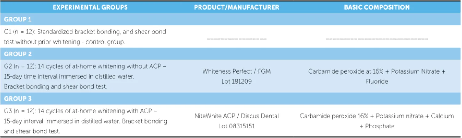
Groups
The specimens were randomly divided into three groups (n = 12) according to whether or not they had been whitened, and the type of whitening gel (Table 1). The gels were applied as recommended by the manufac-turers on the labial surface of the enamel in a layer about 0.5 mm thick, which corresponded to one cycle. Upon completion of each gel application cycle the specimens were washed with air/water jets for 30 seconds, and thereafter immersed in distilled water renewed weekly and stored at 34° C room temperature on average. Four-teen cycles were carried out spanning a 15-day time in-terval since the end of the whitening treatment. Only then were the brackets bonded to the teeth.
Standard Edgewise metal brackets with slots 0.022 x 0.028-in (Abzil, 3M/Unitek, São José do Rio Preto/SP, Brazil) were bonded to the maxillary central incisors. The size of the bracket base, as informed by
the manufacturer, was 14.35 mm.2 The base featured
metal mesh type mechanical retention. Transbond XT adhesive system (3M Unitek, Monrovia, CA, USA) was used for bonding the brackets. The brackets were bonded to the tooth surface with the slot parallel to the base of the cylinder, following the manufacturer’s protocol.
To perform the shear bond strength test of the spec-imens a Kratos TRCV59DUSB universal mechanical testing machine (Jundiaí, SP, Brazil) was utilized at a
speed of 0.5 mm/min (ISO 11405:2003)14 with a chisel
tip. Shear bond strength results were obtained in kgf, converted to N, and divided by the bracket base area
(14.35 mm2), yielding the results in MPa.
After conducting the test, the labial surface of each specimen was evaluated by stereomicroscopy (Opton, Germany) with 8x magnification to measure Adhesive Remnant Index (ARI), as recommended by Årtun and
Bergland,1 where 0 = no composite remnant adhered to
the enamel, 1 = less than half of composite adhered to the enamel, 2 = more than half of composite adhered to enamel, 3 = all composite adhered to the enamel.
Statistical analysis
All data were analyzed for normality by the Shapiro-Wilk test was used for normality analysis of all data. Data were considered normal after excluding an outlier (Group 2). If this specimen had been included the data would have been considered abnormal.
Shear bond strength test results were subjected to analysis of variance (ANOVA) at 5% significance level, and subsequently Tukey’s test to compare the control group with the other treatments, and also between the experimental groups (G2 and G3). Kruskal-Wallis test at 5% was used in evaluating ARI scores.
EXPERIMENTAL GROUPS PRODUCT/MANUFACTURER BASIC COMPOSITION
GROUP 1
G1 (n = 12): Standardized bracket bonding, and shear bond
test without prior whitening - control group. _________________ _____________________________
GROUP 2
G2 (n = 12): 14 cycles of at-home whitening without ACP – 15-day time interval immersed in distilled water.
Bracket bonding and shear bond test.
Whiteness Perfect / FGM Lot 181209
Carbamide peroxide at 16% + Potassium Nitrate + Fluoride
GROUP 3
G3 (n = 12): 14 cycles of at-home whitening with ACP – 15-day interval immersed in distilled water. Bracket bonding and shear bond test.
NiteWhite ACP / Discus Dental Lot 08315151
Carbamide peroxide 16% + Potassium nitrate + Calcium + Phosphate
Nevertheless, it is noteworthy that in vitro tests
exhibit numerous differences compared with in vivo
tests. The key difference lies in the fact that the forces which occur during mastication are compressive in nature and pose a greater risk of damage to enamel than forces applied to the bracket/adhesive during
shear bond tests.13 These peculiarities of laboratory
tests should be emphasized as they improve variable control. Conversely, clinical studies do not allow these variables to be controlled, which warrants fur-ther studies on this topic.
In actuality there is no such thing as a pure shear
force in vivo, given that the different components
combine to influence the bond. Moreover, in vitro
re-sults undergo significant effects, as shown by Swift Jr.,
Perdigão;29 Bishara and Sulieman.4 Despite the above,
this investigation followed the methodology adopted by the ISO/TS11405 standard.
As regards the type of substrate used, bovine teeth have long been considered a good alternative since not only are they more readily available but their enamel structure is similar to that of human teeth. Assessments made by comparative studies involving different substrates show that the bond strength of bo-vine enamel is quite acceptable, although their bond
strength may be lower compared to human teeth.9,21
Another noteworthy aspect concerns the storage medium adopted in the present study (distilled wa-ter). In bond strength tests of whitened substrates the storage medium used for the specimens plays a role as relevant as it is controversial. Artificial saliva, which RESULTS
ANOVA analysis of variance results with p values are depicted in Table 2.
Differences in bond strength between groups are displayed in Figure 1, which shows a significant dif-ference between Group 1 – control (not whitened), and the groups after whitening (Group 2 – conven-tional whitening product, and Group 3 – whitening product containing ACP). No significant differences were found between the whitened groups.
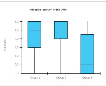
The value of the Kruskal-Wallis statistical test for ARI was p = 0.0509, indicating a statistically insignifi-cant difference. Figure 2 shows ARI in both groups.
DISCUSSION
A number of studies have evaluated the bond strength of orthodontic brackets bonded to
differ-ent surfaces.3,20,27 In this context, preference has
been given to central incisor brackets as they fea-ture a flatter base surface, which adapts more eas-ily to the surfaces being tested. These surfaces are normally flat since specimens are seldom fabricated with crown contours, which explains the brackets used in this experiment.
Table 2 - P values of the statistical test between experimental groups.
Figure 1 - Shear bond strength values, means and standard deviations of groups 1, 2 and 3.
Figure 2 - Adhesive remnant index (ARI), median and quartile in groups 1, 2 and 3.
Groups p value
Groups 1 and 2 < 0.05 Groups 1 and 3 < 0.01 Groups 2 and 3 n.s.
0 0,0
5
0,5 10
1,0 15
1,5 20
2,0
Shear Bonding test Adhesive remnant index (ARI)
Var Y MPa
Group 1
Group 1 = control Group 2 = whitening Group 3 = whitening with ACP
ARI sc
or
es
Group 1
Group 2 Group 3 Group 2 Group 3
25
that a longer time interval should be allowed to elapse prior to bonding any orthodontic appliances.
Furthermore, removal of the remaining resin from the tooth surface does not pose a challenge since re-moval of adhesive remnants from tooth surfaces after removal of the fixed orthodontic appliance is a rou-tine procedure. Thus, the choice of bonding mate-rials depends on a careful assessment of their clini-cal properties. This finding concerning the adhesive remnant index (ARI) is of great interest to the ortho-dontist, who can thus choose materials that respond clinically by presenting a greater amount of adhesive remnants on the tooth surface after removal of the brackets. This should ensure greater safety, prevent-ing enamel fractures and preservprevent-ing tooth integrity.
Although the use of hydrogen peroxide at 35% significantly reduces the amount of resin on the tooth
surface after debonding,31 this study did not reveal
any differences in terms of ARI in both whitened and non-whitened teeth, with and without ACP.
Proper orthodontic treatment requires scientific knowledge and special technical skills. It is also para-mount that orthodontists be instructed about the wide array of materials of different types and manufacturers currently available for clinical use. There are various products of different origins, both domestic and im-ported, manufactured specifically for direct bonding of orthodontic attachments to enamel. It is therefore ex-tremely important that these results be applied in clinical practice in order to optimize professional orthodontic treatment, thus avoiding frequent bond failures due to poor bond strength between brackets and tooth enamel.
CONCLUSION
Significant differences were found in the bond strength of metal brackets between whitened and non-whitened teeth. There was a considerable re-duction in bond strength in the groups that were subjected to tooth surface whitening. Both whit-ened groups (2 and 3) failed to achieve a clinically efficient bond strength, especially group 3 (whit-ened with ACP), underscoring the need to restore the tooth surface and remove all chemical whiten-ing agents prior to bondwhiten-ing the brackets. As regards ARI, there was no statistically significant difference between the 3 groups tested.
has been employed in different studies,8,10,16,17,28 is
pri-marily aimed at making laboratory tests resemble as closely as possible the clinical conditions actually ex-perienced in routine practice.
As to the mechanical test, it is a known fact that orthodontic brackets require a bond strength capable of withstanding masticatory forces and activation of the
mechanics utilized.21 The minimum acceptable bond
strength for routine orthodontic procedures ranges
from 6 to 8 Mpa.21 The results showed unacceptable
mean bond strength values for whitened teeth.
Thus, artificial saliva has often been blamed for the lack of differences in bond strength after
whiten-ing, according to published studies.15,19 This finding
could be attributed to the remineralizing effect of
sa-liva cited by Souza,28 who argued that enamel samples
treated with 10% carbamide peroxide and stored in artificial saliva display smaller spaces in between the
hydroxyapatite crystals. However, other studies29,30
claim that this potential remineralizing effect of saliva has not yet been properly explained.
Therefore, in order to avert a possible potentiat-ing (remineralizpotentiat-ing), or deleterious effect by artificial saliva, which could interfere with the analysis of the results, a storage medium. i.e., distilled water, was chosen as it exerts little or no effect on bond strength.
Residues derived from the degradation of per-oxide or oxygen leads to a lower amount of shorter resinous tags compared to teeth not subjected to
whitening,7,18 resulting in lower bond strength as
hydrogen peroxide negatively affects the curing of
adhesive systems.12,14,16,23,30
Therefore, teeth whitening can change the min-eral structure of tooth enamel and, hence, affect the
bond between this substrate and adhesive systems.24,25
It is thus necessary to wait for a post-whitening peri-od long enough for the enamel’s mineral structure to be restored, and for the residual oxygen of the whit-ening agent to be completely removed. Although the literature shows significant variation in the length of the post-whitening period recommended before
bonding brackets (24h to 4 weeks), Cavalli8 showed
1. Artun J, Bergland S. Clinical trials with crystal growth conditioning as an alternative to acid-etch enamel pretreatment. Am J Orthod. 1984;85(4):333-40.
2. Bishara SE, Ostby AW. Bonding and debonding from metal to ceramic: research and its clinical application. Semin Orthod. 2010;16(1):24-36. 3. Bishara SE, VonWald L, Laffoon JF, Warren JJ. Effect of a self-etch primer/
adhesive on the shear bond strength of orthodontic brackets. Am J Orthod Dentofacial Orthop. 2001;119(6):621-4.
4. Bishara SE, Sulieman AH, Olson M. Effect of enamel bleaching on the bonding strength of orthodontic brackets. Am J Orthod Dentofacial Orthop. 1993;104(5):444-7.
5. Bitter NC. Bleaching agents. J Am Dent Assoc. 1999;130:26.
6. Buonocore MG. Simple method of increasing the adhesion of acrylic filling materials to enamel surface. J Dent Res. 1955;34(6):849-53.
7. Cal Neto JOAP, Miguel JAM. Uma análise dos testes in vitro de força de adesão em Ortodontia. Rev Dental Press Ortod Ortop Facial. 2004;9(4):44-51. 8. Cavalli V, Reis AF, Giannini M, Ambrosano GM. The effect of elapsed time
following bleaching on enamel bond strength of resin composite. Oper Dent. 2001;26(6):597-602.
9. Cavalli V, Arrais CA, Giannini M, Ambrosano GM. High concentrated carbamide peroxide bleaching agent effect on enamel surface. J Oral Rehabil. 2004;31(2):155-9.
10. Cavalli V, Carvalho RM, Giannini M. Influence of carbamide peroxide-based bleaching agents on the bond strength of resin-enamel/dentin interfaces. Braz Oral Res. 2005;19(1):23-9.
11. Dishman MV, Covey DA, Baughan LW. The effects of peroxide bleaching on composite to enamel bond strength. Dent Mater. 1994;10(1):33-6. 12. Dunn WJ. Shear bond strength of an amorphous
calcium-phosphate-containing orthodontic resin cement. Am J Orthod Dentofacial Orthop. 2007;131(2):243-7.
13. Eliades T, Brantley WA. Orthodontics materials, scientific and clinical aspects. 1st ed. Stuttgart: Thieme; 2001.
14. International Organization for Standardization. Dental materials. [Access 2011 may 17]. Available from: http://www.iso.org.
15. Josey AL, Meyers IA, Romaniuk K, Symons AL. The effect of vital bleaching technique on enamel surface morphology and the bonding of composite resin to enamel. J Oral Rehabil. 1996;23(4):244-50.
16. Loretto SC, Braz R, Lyra AMVC, Lopes LM. Influence of photopolymerization light source on enamel shear bond strength after bleaching. Braz Dent J. 2004;15(2):133-7. Epub 2005 Mar 11.
REFERENCES
17. Kalili T, Caputo AA, Mito R, Sperbeck G, Matyas J. In vitro toothbrush abrasion and bond strength of bleached enamel. Pract Periodontics Aesthet Dent. 1991;3(5):22-4.
18. Miles PG, Pontier JP, Bahiraei D, Close J. The effect of carbamide peroxide bleach on the tensile bond strength of ceramic brackets: an in vitro study. Am J Orthod Dentofacial Orthop. 1994;106(4):371-5.
19. Murchison DF, Charlton DG, Moore BK. Carbamide peroxide bleaching: effects on enamel surface hardness and bonding. Oper Dent. 1992;17(5):181-5. 20. Newman GV. Epoxy adhesives for orthodontic attachments: progress report.
Am J Orthod. 1965;51(12):901-12.
21. Perdigão J, Francci C, Swift EJ, Ambrose WW, Lopes M. Ultra-morphological study of the interaction of dental adhesives with carbamide peroxide-bleached enamel. Am J Dent. 1998;11(6):291-301.
22. Oesterle LJ, Shellhart WC, Belanger GK. The use of bovine enamel in bonding studies. Am J Orthod Dentofacial Orthop. 1998;114(5):514-9.
23. Øgaard B, Fjeld M. The enamel surface and bonding in Orthodontics. Semin Orthod. 2010;16:37-48.
24. Oshiro M, Yamaguchi K, Takamizawa T, Inage H, Watanabe T, Irokawa A, et al. Effect of CPP-ACP paste on tooth mineralization: an FE-SEM study. J Oral Sci. 2007;49(2):115-20.
25. Perdigão J, Francci C, Swift EJ Jr, Ambrose WW, Lopes M. Ultra-morphological study of the interaction of dental adhesives with carbamide peroxide-bleached enamel. Am J Dent. 1998;11(6):291-301.
26. Reynolds IR. A review of direct orthodontic bonding. Br J Orthod. 1975;2(3):171-8.
27. Spyrides GM, Perdigão J, Pagani C, Araújo MA, Spyrides SM. Effect of whitening agents on dentin bonding. J Esthet Dent. 2000;12(5):264-70. 28. Souza MAL. Clareamento caseiro de dentes: ação do peróxido de carbamida
sobre dentes e mucosa bucal [tese]. Porto Alegre (RS): Pontifícia Universidade Católica; 1993.
29. Swift EJ Jr, Perdigão J. Effects of bleaching on teeth and restorations. Compend Contin Educ Dent. 1998;19(8):815-20; quiz 822.
30. Türkün M, Kaya AD. Effect of 10% sodium ascorbate on the shear bond strength of composite resin to bleached bovine enamel. J Oral Rehabil. 2004;31(12):1184-91.