Prevalence of Café-au-Lait Spots in children with solid tumors
Anna Claudia Evangelista dos Santos
1,2,*, Benjamin Heck
3, Beatriz De Camargo
3,4and
Fernando Regla Vargas
1,2,51
Departamento de Genética e Biologia Molecular, Universidade Federal do Estado do Rio de Janeiro
(UNIRIO), Rio de Janeiro, RJ, Brazil.
2
Departamento de Genética, Universidade Federal do Rio de Janeiro (UFRJ), Rio de Janeiro, SP, Brazil.
3
Departamento de Pediatria, AC Camargo Cancer Center, São Paulo, SP, Brazil.
4
Departamento de Oncologia Pediátrica, Instituto Nacional de Cancer, Rio de Janeiro, RJ, Brazil.
5Laboratório de Epidemiologia de Malformações Congênitas, Fundação Oswaldo Cruz,
Rio de Janeiro, RJ, Brazil.
Abstract
Cafe-au-lait maculae (CALM) are frequently observed in humans, and usually are present as a solitary spot. Multiple CALMs are present in a smaller fraction of the population and are usually associated with other congenital anomalies as part of many syndromes. Most of these syndromes carry an increased risk of cancer development. Previous stud-ies have indicated that minor congenital anomalstud-ies may be more prevalent in children with cancer. We investigated the prevalence of CALMs in two samples of Brazilian patients with childhood solid tumors, totaling 307 individuals. Additionally, 176 school children without diagnosis of cancer, or of a cancer predisposing syndrome, were investi-gated for the presence of CALMs. The prevalence of solitary CALM was similar in both study groups (18% and 19%) and also in the group of children without cancer. Multiple CALMs were more frequently observed in one of the study groups (Z = 2.1). However, when both groups were analyzed together, the significance disappeared (Z = 1.5). The additional morphological abnormalities in children with multiple CALMs were analyzed and compared to the findings observed in the literature. The nosologic entities associated with CALMs are reviewed.
Keywords: café-au-lait maculae; pediatric solid tumors; birth defects; nosology.
Received: January 22, 2015; Accepted: June 08, 2015.
Introduction
Café-au-lait maculae (CALM) are named after their typical coffee-and-milk hue, and a color only slightly dar-ker than the surrounding skin. There are two main types of CALMs. The most common type has fairly regular and clearly demarcated margins (“coast of California”). They may range in size from a few millimeters to several centi-meters and may be present as solitary or as multiple spots. There is a second, less frequent type of CALM that has a much more irregular margin (“coast of Maine”),and is usu-ally larger and solitary (Aase, 1990). At birth, a single CALM is observed in 5% of Caucasians and up to 15% of Americans of African descent. Three or more spots are ob-served in 2% of the population (Aase, 1990). CALMs may occur as isolated findings, or may be associated with minor
and/or major congenital anomalies, as part of many syndromes. CALMs are typically observed in neurofibro-matosis 1 (NF1), an autosomal dominant condition charac-terized by the presence of multiple neurofibromas, as well as other non-tumoral manifestations, like multiple CALMs, which are present in almost all adult patients with this dis-ease. CALMs are usual findings in many other monogenic diseases, as well as in chromosomal abnormalities that are associated with mosaicism. This is the case with chromo-somal rings, which are usually unstable during cell divi-sion, resulting in chromosomal mosaics. Interestingly, many of these genetically determined conditions that pres-ent with CALMs do also carry increased predisposition to cancer development. For this reason, we set out to investi-gate the prevalence of CALMs in children with and without cancer. Two groups of children with a pediatric solid tu-mor, resident in two states in southeastern Brazil (Rio de Ja-neiro and São Paulo), were independently examined for the presence of cutaneous pigmentary changes. A third group of children without cancer was also examined.
DOI: http://dx.doi.org/10.1590/1678-4685-GMB-2015-0024
Send correspondence to Fernando R. Vargas, Birth Defects Epide-miology Laboratory, Pav. Leonidas Deane, Fundação Oswaldo Cruz, Av. Brasil, 4365, 21040-360 Rio de Janeiro, RJ, Brazil. E-mail: [email protected]
*current affiliation: Programa de Genética, Instituto Nacional do Câncer, Rio de Janeiro, RJ, Brazil.
Methods
Two study groups of children (ages 0-18 years) with current or previous diagnosis of a pediatric solid tumor were examined by a medical geneticist trained in dysmor-phology. Study groups 1 and 2 are composed of children with current or previous diagnosis of a pediatric solid tu-mor: study group 1 (226 individuals) is from Rio de Janeiro (RJ); study group 2 (81 individuals) is from Sao Paulo (SP). The two cities are located in contiguous states in southeast-ern Brazil. Additionally, a third group (Study group 3) from Rio de Janeiro (RJ), and is composed of 176 school children without diagnosis of cancer or of a cancer predisposing syndrome. Age distribution among the three study groups did not show statistical differences. Both groups of children with cancer are part of a larger study aimed at investigating the prevalence of major and minor congenital anomalies in children with cancer. The same protocol for description of minor anomalies and morphologic variants based on the studies of Merkset al.(2003, 2006) was applied in all three study groups. The presence of two or more café-au-lait maculae was defined as “multiple CALMs”. Analysis of the frequency of CALMs between the different study groups was accomplished through estimation of Z scores. We considered Z > 1.96 (two standard deviations) as the threshold for significance. Children with hematologic ma-lignancies were not included in the study. Data on the size (diameter) of each individual café-au-lait spot, as well as ethnic background of the affected individuals were not col-lected in the present study. The present study was approved by the local Institutional Review Board of all involved in-stitutions (83/08). Subjects were included in the study after discussion and signature of the informed consent by their parents and/or legal guardian.
Results and Discussion
In the present study, two study groups of patients with pediatric solid tumors were independently investigated for the presence of CALM by two trained dysmorphologists. The same protocol was applied in a third group of children without diagnosis of cancer or of a cancer predisposing syndrome.
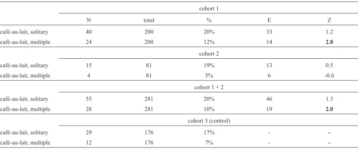
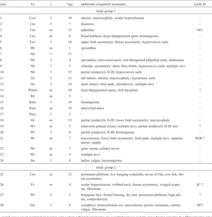
Our study identified 28 children with multiple CALMs, 25 of which were associated with other congenital anomalies. The frequency of solitary CALMs was similar in the study groups 1 and 2 (children with solid tumors), and did not differ from that observed among school chil-dren without cancer (Z = 1.2 and Z = 0.5, respectively). The frequency of multiple CALMs, however, was higher in group 1 (12%) than in group 2 (5%), and also than that of the group of children without cancer (Z = 2.8). However, when both study groups 1 and 2 were analyzed together, the difference with the group without cancer became of border-line significance (Z = 2.0). These data are shown in Table 1. The additional findings observed in each patient are shown in Table 2. Table 3 shows the prevalence of each congenital anomaly within the patient group. A known syndromic di-agnosis could be suspected in four patients. Additional pa-tients presented multiple findings compatible with the clinical presumption of a dysmorphic, private syndrome (Table 2). Table 4 shows the main nosologic entities associ-ated with multiple CALMs.
Burwellet al.(1982) observed a single café-au-lait spot in 20% and multiple spots in 6% of 732 school chil-dren. Merkset al.(2006) observed 13% of single spots and 3.3% of multiple spots in healthy children. According to Tekinet al.(2001), single spots may be observed in up to 27% in children under 10 years of age.
Table 1- Observed numbers of solitary and multiple CALMs in study groups 1, 2 and 3, along with expected numbers and Z scores for study groups 1, 2
and 1 + 2.
cohort 1
N total % E Z
café-au-lait, solitary 40 200 20% 33 1.2
café-au-lait, multiple 24 200 12% 14 2.8
cohort 2
café-au-lait, solitary 15 81 19% 13 0.5
café-au-lait, multiple 4 81 5% 6 -0.6
cohort 1 + 2
café-au-lait, solitary 55 281 20% 46 1.3
café-au-lait, multiple 28 281 10% 19 2.0
cohort 3 (control)
café-au-lait, solitary 29 176 17% -
-café-au-lait, multiple 12 176 7% -
Multiple café-au-lait spots were observed in cancer predisposing syndromes like neurofibromatosis 1, neuro-fibromatosis 2, tuberous sclerosis, McCune-Albright syn-drome, Fanconi anemia, among others (Evanset al., 1992; Giampietro et al., 1993; Roach et al., 1998; Kim et al., 1999; Ferneret al., 2007). More recently, café-au-lait spots were observed in 60% of carriers of biallelic mutations in mismatch repair (MMR) genes (Wimmer et al., 2014), characteristic of constitutional mismatch repair deficiency
syndrome (CMMRDS). These authors developed a score for investigation of this condition that includes the presence of CALMs and suggested that a score of three or more points warrants investigation of CMMRDS (Wimmer et al., 2014).
Merkset al.(2005) studied 1,073 children with can-cer and observed major abnormalities in 26.8% of the pa-tientsvs.15.5% in controls (p = 0.001), and minor anoma-lies in 65.1% of the patients vs. 56.2% in controls
Table 2- Additional birth defects, congenital anomalies, and morphologic variants detected by ectoscopy in children with pediatric solid tumor and
mul-tiple CALMs. Cases are stratified by tumor and by cohort.
case Tu s Age additional congenital anomalies syndr dx
study group 1
1 Ccrc f 10 obesity, macrocephaly, ocular hypertelorism
-2 Cns f 7 diastema
-3 Cns m 13 ephelides NF1
4 Cns m 6 broad halluces; hypo/depigmented spots; hemangioma
-5 Ewi f 18 upper limb asymmetry; thorax asymmetry; hyperconvex nails
-6 Hb m 1 epicanthus
-7 Nb f 17 -
-8 Nb f 4 epicanthus; anteverted nares; anti-Mongoloid palpebral slant; strabismus
-9 Nb f 3 clinodac; asymmetry; short, thin, brittle, hyperconvex nails; multiple nevi
-10 Nb f 15 partial syndactyly II-III; hyperconvex nails
-11 Os f 13 tall stature; obesity; macrocephaly; hypoplastic nails
-12 Os f 14 short stature; fetal pads; clinodactyly; multiple nevi
-13 Pnsmt m 10 hypo/depigmented spots; cleft lip/palate ?
14 Rb m 5 -
-15 Rms f 14 hemangioma
-16 Rms m 10 anteverted nares
-17 Rms f 2 -
-18 SS m 15 partial syndactyly II-III; lower limb asymmetry; macrocephaly
-19 Wt m 3 transverse palmar crease; multiple nevi; partial syndactyly II-III feet ?
20 Wt f 6 partial syndactyly II-III; hemangioma
-21 Wt m 7 macrostomia, lower limb asymmetry; fetal pads; multiple nevi,
supernu-merary nipple
SGB ?
22 Wt m 7 genu varum; solitary nevus
-23 Wt m 9 multiple nevi
-24 Sts f 8 hallux valgus; haemangioma
-study group 2
25 Cns m 12 prominent philtrum; low hanging columella; nevus of Ota; cow lick;
tho-rax asymmetry
-26 Fs m 9 ocular hypertelorism, webbed neck, thorax asymmetry, winged
scapu-lae, fibromata
JC ?
27 Wt f 5 triangular face, frontal bossing, dry hair, prominent philtrum, high
pal-ate, camptodactyly
?
28 Gct f 9 synophrys, heterochromia iris, microstomia, pectus carinatum, cubitus
valgus, fibromata
NF1
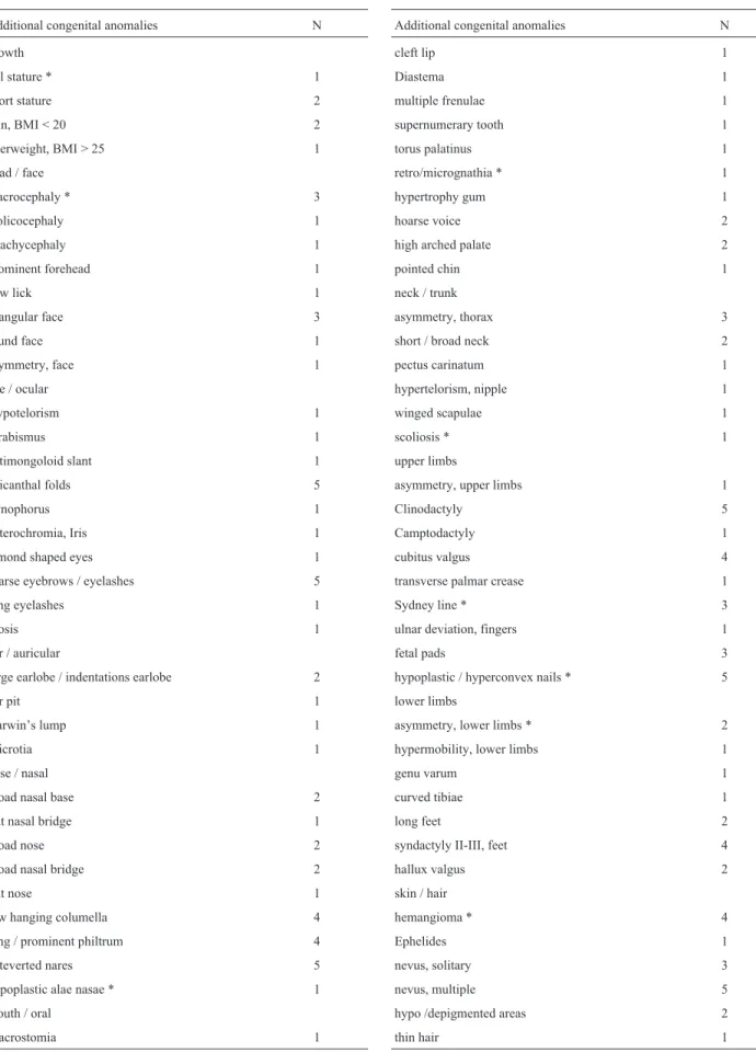
Table 3- Birth defects, congenital anomalies, and morphologic variants observed in children with pediatric solid tumor and multiple CALMs. Variants
marked with an asterisk (*) have been previously associated with pediatric cancer (Merkset al., 2008).
Additional congenital anomalies N
growth
tall stature * 1
short stature 2
thin, BMI < 20 2
overweight, BMI > 25 1
head / face
macrocephaly * 3
Dolicocephaly 1
Brachycephaly 1
prominent forehead 1
cow lick 1
triangular face 3
round face 1
asymmetry, face 1
eye / ocular
Hypotelorism 1
Strabismus 1
antimongoloid slant 1
epicanthal folds 5
Synophorus 1
heterochromia, Iris 1
almond shaped eyes 1
sparse eyebrows / eyelashes 5
long eyelashes 1
Ptosis 1
ear / auricular
large earlobe / indentations earlobe 2
ear pit 1
Darwin’s lump 1
Microtia 1
nose / nasal
broad nasal base 2
flat nasal bridge 1
broad nose 2
broad nasal bridge 2
flat nose 1
low hanging columella 4
long / prominent philtrum 4
anteverted nares 5
hypoplastic alae nasae * 1
mouth / oral
Macrostomia 1
Additional congenital anomalies N
cleft lip 1
Diastema 1
multiple frenulae 1
supernumerary tooth 1
torus palatinus 1
retro/micrognathia * 1
hypertrophy gum 1
hoarse voice 2
high arched palate 2
pointed chin 1
neck / trunk
asymmetry, thorax 3
short / broad neck 2
pectus carinatum 1
hypertelorism, nipple 1
winged scapulae 1
scoliosis * 1
upper limbs
asymmetry, upper limbs 1
Clinodactyly 5
Camptodactyly 1
cubitus valgus 4
transverse palmar crease 1
Sydney line * 3
ulnar deviation, fingers 1
fetal pads 3
hypoplastic / hyperconvex nails * 5
lower limbs
asymmetry, lower limbs * 2
hypermobility, lower limbs 1
genu varum 1
curved tibiae 1
long feet 2
syndactyly II-III, feet 4
hallux valgus 2
skin / hair
hemangioma * 4
Ephelides 1
nevus, solitary 3
nevus, multiple 5
hypo /depigmented areas 2
(p = 0.001). Three or more minor anomalies were detected in 15.2% of the patients and in 8.3% of the controls (p = 0.001). A known cancer predisposing syndrome could be detected or suspected in 7.2% of the patients. The same authors (Merks et al., 2008) observed 17 morphological abnormalities that occurred more frequently in pediatric cancer patients than in controls: blepharophimosis, asym-metric lower limbs, Sydney crease, broad foot, isolated short metacarpals, short distal phalanx of the thumb, port-wine stain, hyperconvex nails, retrognathia, hypoplastic alae nasae, prominent ears, broad hand, scoliosis, hyper-telorism, tall stature, macrocephaly, and microcephaly.
In-terestingly, CALMs are not included in this group. Two of these 17 congenital anomalies, blepharophimosis and asymmetric lower limbs, showed patterns of non-random association with other minor anomalies. The so-called “ble-pharophimosis pattern” consisted of the preferential associ-ation of blepharophimosis, increased anterior-posterior an-gulation of the spine, patchy hypopigmentation of the skin, and multiple CALMs. The asymmetric lower limb pattern consists of asymmetric lower limbs, tall stature, midface hypoplasia, ptosis, and pectus carinatum or excavatum.
It is worthy of note that, from 17 congenital anoma-lies described by Merkset al.(2008), eight were observed
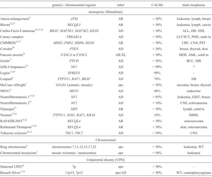
Table 4- Main syndromes associated with CALMs.
gene(s) / chromosomal regions inher CALMs main neoplasias
monogenic (Mendelian)
Ataxia-telangectasia9
ATM AR > 50% leukemia, lymph, breast
Bloom22,25 RECQL3 AR > 50% leukemia, lymph, carcin
Cardio-Facio-Cutaneous14,15,18
BRAF,MAP2K1,MAP2K2,KRAS AD > 30% ALL, HB, NHL
Carney complex PRKAR1A AD < 50% LCCSCT, PMS, endo tu
CMMRDS16,27 MHS2,PMS2,MSH6,MLH1 AR > 50% CRC, CNS, PST
Cowden20
PTEN AD < 50% breast, thyroid, skin
Fanconi anemia26 FANCAtoFANCL AR/XL < 50% MDS, AML, solid tu
Gorlin17
PTCH AD < 50% BCC, MB
Jaffe-Campanacci23
NF1 AD > 99% ?
Legius13,24 SPRED1 AD 99%
-Leopard5
PTPN11,RAF1,BRAF AD 70% NB
McCune-Albright7
GNAS1(somatic, mosaic) spo > 50% sarcoma, breast, thyroid
MEN14 MEN1 AD 40% endocrine
Neurofibromatosis 113,24
NF1 AD > 95% leukemia, GIST, breast
Neurofibromatosis 221 NF2 AD < 10% CNS, schwannoma
Nijmegen25
NBN AR < 50% lymph, solid tu
Noonan15,18
PTPN11,SOS1,RAF1,KRAS AD 10% JMML
RAPADILINO19,22 RECQL4 AR < 50% osteosarcoma
Rothmund-Thompson11,22
RECQL4 AR < 50% skin, osteosarcoma
Tuberous sclerosis10,12 TSC1,TSC2 AD < 50% CNS
Chromosomal
Ring chromosome8 chromosomes 7,11,12,15,17,22 spo > 50% leukemia, WT
Chromosomal mosaicism2 mosaic trisomies / monosomies spo < 50% leukemia
Uniparental disomy (UPD)
Maternal UPD78 7p spo < 50%
-Russell-Silver1,3,6 11p15, 7p13 spo/AD < 50% WT, craniopharyngioma
Legend: AD = autosomal dominant; ALL = acute lymphoid leukemia; AML = acute myeloid leukemia; AR = autosomal recessive; BCC = basal cell car-cinoma; carcin = carcar-cinoma; CMMRDS = congenital mismatch repair deficiency syndrome; CNS = central nervous system; CRC = colorectal cancer; GIST = gastrointestinal stromal tumor; HB = hepatoblastoma; inher = inheritance; JMML = juvenile myelomonocytic leukemia; MB = medulloblastoma; MDS = myelodysplastic syndrome; MEN1 = multiple endocrine neoplasia 1; NB = neuroblastoma; NHL = non-Hodgkin lymphoma; spo = sporadic; WT Wilms tumor; XL = X-linked. References: 1 - Bruckheimer and Abrahamov, 1993; 2 - Carellaet al., 2010; 3 - Chitayatet al., 1988; 4 - Darlinget al., 1997; 5 - Digilioet al., 2006; 6 - Drazninet al., 1980; 7 - Dumitrescu and Collins, 2008; 8-Fujinoet al., 2010; 9 - Greenbergeret al., 2013; 10 - Józwiaket
in at least one patient from our sample: tall stature, macro-cephaly, ocular hypertelorism, hypoplastic alae nasae, re-trognathia, scoliosis, hyperconvex nails, asymmetric lower limbs, and hemangioma. These anomalies are marked with an asterisk in Table 3. We did not observe any patient with the complete pattern described by Merks et al. (2008). However, two patients presented the association of multi-ple CALMS and hypopigmented / depigmented spots, sug-gestive of the “blepharophimosis pattern” (Table 3). It is important to take into consideration that we have included only patients with pediatric solid tumors in the study, while the sample studied by Merkset al.(2008) was composed of all pediatric malignancies. In fact, almost half (438 out of 1,073) of the children studied by Merkset al.(2008) were carriers of hematological malignancies (non-Hodgkin lym-phoma, Hodgkin lymlym-phoma, acute lymphoblastic leuke-mia, or acute myeloid leukemia).
In summary, our study showed that the frequency of solitary CALMs is not significantly increased in children with pediatric solid tumors. Multiple CALMs were more frequently observed in one of our studied samples (study group 1, Z = 2.8). However, the combined analysis of the studied samples (study groups 1 and 2) showed only bor-derline significance (Z = 2.0). Hence, the identification of multiple CALMs in a child with cancer warrants further morphologic evaluation, given the great number of cancer predisposing syndromes associated with multiple CALMs.
Acknowledgments
FRV is the recipient of grants from Conselho Nacio-nal de Desenvolvimento Científico e Tecnológico (CNPq; grant 486599/2012-4) and Fundação de Amparo à Pesquisa do Estado do Rio de Janeiro (FAPERJ, grant E26/110.535/2012).
References
Aase JM (1990) Diagnostic Dysmorphology. Plenum Medical Book Company, London and New York, 299 p.
Bruckheimer E and Abrahamov A (1993) Russell-Silver syn-drome and Wilms tumor. J Pediatr 122:165-166.
Burwell RG, James NJ and Johnston DI (1982) Café-au-lait spots in school children. Arch Dis Child 57:631-632.
Carella M, Spreafico F, Palumbo O, Storlazzi CT, Tabano S, Miozzo M, Miglionico L, Calvano S, Sindici G, Gamba B,et al.(2010) Constitutional ring chromosome 11 mosaicism in a Wilms tumor patient: Cytogenetic, molecular and clinico-pathological studies. Am J Med Genet (A) 152:17561763. Chitayat D, Friedman JM, Anderson L and Dimmick JE (1988)
Hepatocellular carcinoma in a child with familial Russell-Silver syndrome.Am J Med Genet (A) 31:909-914. Darling TN, Skarulis MC, Steinberg SM, Marx SJ, Spiegel AM
and Turner M (1997) Multiple facial angiofibromas and collagenomas in patients withmultiple endocrine neoplasia type 1. Arch Dermatol 133:853-857.
Digilio MC, Sarkozy A, de Zorzi A, Pacileo G, Limongelli G, Mingarelli R, Calabro R, Marino B and Dallapiccola B
(2006) LEOPARD syndrome: Clinical diagnosis in the first year of life. Am J Med Genet (A) 140:740-746.
Draznin MB, Stelling MW and Johanson AJ (1980) Silver-Russell syndrome and craniopharyngioma. J Pediatr 96:887-889. Dumitrescu CF and Collins MT (2008) McCune-Albright
syn-drome. Orphanet J Rare Dis 3:12.
Evans DG, Huson SM, Donnai D, Neary W, Blair V, Newton V, Strachan T and Harris R (1992) A genetic study of type 2 neurofibromatosis in the United Kingdom. II. Guidelines for genetic counselling. J Med Genet 29:847-852.
Ferner RE, Huson SM, Thomas N, Moss C, Willshaw H, Evans DG, Upadhyaya M, Towers R, Gleeson M, Steiger C,et al.
(2007) Guidelines for the diagnosis and management of in-dividuals with neurofibromatosis 1. J Med Genet 44:81-88. Fujino H, Fujita N, Hamamoto K, Oobu S, Kita M, Tanaka A, Matsubara H, Watanabe K, Heike T and Adachi S (2010) Ring/marker chromosome derived from chromosome 7 in childhood acute megakaryoblastic leukemia with mono-somy 7. Int J Hematol 92:386-390.
Giampietro PF, Adler-Brecher B, Verlander PC, Pavlakis SG, Da-vis JG and Auerbach AD (1993) The need for more accurate and timely diagnosis in Fanconianemia: A report from the International Fanconi Anemia Registry. Pediatrics 91:1116-1120.
Greenberger S, Berkun Y, Ben-Zeev B, Levi YB, Barziliai A and Nissenkorn A (2013) Dermatologic manifestations of ata-xia-telangiectasia syndrome. J Am Acad Dermatol 68:932-936.
Józwiak S, Schwartz RA, Janniger CK, Michalowicz R and Chmielik J (1998) Skin lesions in children with tuberous sclerosis complex: Their prevalence, natural course, and di-agnostic significance. Int J Dermatol 37:911-917.
Kim IS, Kim ER, Nam HJ, Chin MO, Moon YH, Oh MR, Yeo UC, Song SM, Kim JS, Uhm MR,et al.(1999) Activating mutation of GS alpha in McCune-Albright syndrome causes skin pigmentation by tyrosinase gene activation on affected melanocytes. Horm Res 52:235-240.
Larizza L, Roversi G and Volpi L (2010) Rothmund-Thompson syndrome. Orphanet J Rare Dis 5:2.
Leung AKC and Robson WLM (2007) Tuberous sclerosis com-plex: A review. J Pediatr Health Care 21:108-114.
Maertens O, De Schepper S, Vandesompele J, Brems H, Heyns I, Janssens S, Speleman F, Legius E and Messiaen L (2007) Molecular dissection of isolated disease features of mosaic neurofibromatosis type 1. Am J Hum Genet 81:243-251. Makita Y, Narumi Y, Yoshida M, Niihori T, Kure S, Fujieda K,
Matsubara Y and Aoki Y (2007) Leukemia in Cardio-facio-cutaneous (CFC) syndrome: A patient with a germline muta-tion in BRAF proto-oncogene. J Pediatr Hematol Oncol 29:287-90.
Merks JHM, van Karnebeek CDM, Caron HN and Hennekam RCM (2003) Phenotypic abnormalities: Terminology and classification. Am J Med Genet (A) 123:211-230.
Merks JHM, Huib N, Caron HN and Hennekam RCM (2005) High incidence of malformation syndromes in a series of 1,073 children with cancer. Am J Med Genet (A) 134:132-143.
Merks JM, Özgen H, Koster J, Zwinderman AH, Caron HN and Hennekam RCM (2008) Prevalence and patterns of morpho-logical abnormalities in patients with childhood cancer. JAMA 299:61-69.
Ohtake A, Aoki Y, Saito Y, Niihori T, Shibuya A, Kure S and Matsubara Y (2011) Non-Hodgkin lymphoma in a patient with cardiofaciocutaneous syndrome. J Pediatr Hematol Oncol 33:e342-6.
Poley JW1,Wagner A, Hoogmans MM, Menko FH, Tops C, Kros JM, Reddingius RE, Meijers-Heijboer H, Kuipers EJ, Dinjens WN,et al.(2007) Biallelic germline mutations of mismatch-repair genes: A possible cause for multiple pedi-atric malignancies. Cancer 109:2349-2356.
Ponti G, Tomasi A, Pastorino L, Ruini C, Guarneri C, V Des-mond, Mandel VD, Seidenari S and Pellacani G (2012) Di-agnostic and pathogenetic role of café-au-lait macules in nevoid basal cell carcinoma syndrome. Hered Cancer Clin Pract 10:15.
Rauen KA, Tidyman WE, Estep AL, Sampath S, Peltier HM, Bale SJ and Lacassie Y (2010) Molecular and functional analysis of a novel MEK2 mutation in cardio-facio-cutaneous syn-drome: Transmission through four generations. Am J Med Genet (A) 152:807-814.
Roach ES, Gomez MR and Northrup H (1998) Tuberous sclerosis complex consensus conference: Revised clinical diagnostic criteria. J Child Neurol 13:624-628.
Sandoval C, Dunbar J, Ozkaynak M and Jayabose S (2012) Osteosarcoma following growth hormone therapy in recur-rent acute lymphoblastic leukemia and Rapadilino syn-drome. Pediatr Hematol Oncol 29:270-271.
Scheper MA, Nikitakis NG, Sarlani E, Sauk JJ and Meiller TF (2006) Cowden syndrome: Report of a case with immuno-histochemical analysis and review of the literature. Oral Med Pathol Radiol Endod 101:625-631.
Seminog OO and Goldacre MJ (2013) Risk of benign tumours of nervous system, and of malignant neoplasms, in people with neurofibromatosis: Population-based record-linkage study. Br J Cancer 108:193-198.
Siitonen HA, Sotkasiira J, Biervliet M, Benmansour A, Capri Y, Cormier-Daire V, Crandall B, Hannula-Jouppi K, Henne-kam R, Herzog D,et al.(2009) The mutation spectrum in
RECQL4diseases. Eur J Hum Genet 17:151-8.
Stewart DR, Brems H, Gomes AG, Ruppert SL, Callens T, Wil-liams J, Claes K, Bober MB, Hachen R, Kaban LB,et al.
(2014) Jaffe-Campanacci syndrome, revisited: Detailed cli-nical and molecular analyses determine whether patients have neurofibromatosis type 1, coincidental manifestations, or a distinct disorder. Genet Med 16:448-459.
Tekin M, Bodurtha JN and Riccardi VM (2001) Café au lait spots: The pediatrician’s perspective. Pediatr Rev 22:82-90. Wang X, Levin AM, Smolinski SE, Vigneau FD, Levin NK and
Tainsky MA (2012) Breast cancer and other neoplasms in women with neurofibromatosis type 1: A retrospective re-view of cases in the Detroit metropolitan area. Am J Med Genet (A)158:3061-3064.
Weemaes CM, Hustinx TW, Scheres JM, van Munster PJ, Bak-keren JA and Taalman RD (1981) A new chromosomal in-stability disorder: The Nijmegen breakage syndrome. Acta Paediatr Scand 70:557-564.
Wimmer K, Kratz CP, Vasen HF, Caron O, Colas C, Entz-Werle N, Gerdes AM, Goldberg Y, Ilencikova D, Muleris M,et al.
(2014) Diagnostic criteria for constitutional mismatch repair deficiency syndrome: Suggestions of the European consor-tium ‘care for CMMRD’ (C4CMMRD). J Med Genet 51:355-365.
Associate Editor: Maria Isabel Achatz