https://doi.org/10.1590/0004-282X20180130
VIEW AND REVIEW
Neurocysticercosis as a probable risk factor
for hippocampal sclerosis
Neurocisticercose como provável fator de risco para esclerose hipocampal
Gagandeep Singh
1,2, Josemir W. Sander
2,3Human brain infestation with the larval stage of the
tape-worm
Taenia solium
, known as neurocysticercosis (NCC), is
endemic in many low-income countries but is also
recog-nized in high-income countries (e.g., the United States)
1. It is
estimated that between two and eight million people
world-wide have NCC
2. Population studies in
T. solium-
endemic
regions of South-Central America, India and parts of Africa
have identified NCC as the putative risk factor in roughly one
third of epilepsy cases
3,4,5,6.
Seizures are the most common clinical presentation
and are estimated to occur in up to 80% of people with
symptomatic NCC
7. Follow-up studies have suggested that
the prognosis for seizure control is good overall, though
seizures might recur on antiepileptic drug withdrawal
8,9.
Rarely, however, NCC might be associated with
difficult-to-treat epilepsy
10,11,12,13. The substrates underlying chronic,
often drug-resistant, epilepsy are a matter of considerable
speculation as well as ongoing investigation. Perilesional
gliosis, seen in unconventional magnetic resonance (MR)
sequences (e.g., magnetization transfer imaging) but
occult in routine MR sequences, has been associated with
poorly-controlled epilepsy
13,14. This, however, needs to be
1Dayanand Medical College, Department of Neurology, Ludhiana, India,
2NIHR University College London Hospitals Biomedical Research Centre, UCL Queen Square Institute of Neurology, London WC1N 3BG, United Kingdom; 3Stichting Epilepsie Instellingen Nederland (SEIN), Achterweg 5, Heemstede, Netherlands.
Correspondence: Josemir W. Sander; Box 29, 33 Queen Square, London WC1N 3BG, UK; E-mail: l.sander@ucl.ac.uk
Conflict of interest: GS has received research grants from the Indian Council of Medical Research. JWS has received research funding from Eisai, GSK and UCB, personal fees from Eisai and UCB outside the submitted work.
Received 04 June 2018; Received in final form 06 August 2018; Accepted 09 August 2018.
ABSTRACT
Neurocysticercosis is one of the most common risk factors for epilepsy but its association with drug-resistant epilepsy remains uncertain.
Conjectures of an association with drug-resistant epilepsy have been fueled by reports of an association between calcific neurocysticercosis
lesions (CNL) and hippocampal sclerosis (HS) from specialized epilepsy centers in
Taenia solium
-endemic regions. The debate arising from
these reports is whether the association is causal. Evidence for the association is not high quality but sufficiently persuasive to merit further
investigation with longitudinal imaging studies in population-based samples from geographically-diverse regions. The other controversial
point is the choice of a surgical approach for drug-resistant epilepsy associated with CNL-HS. Three approaches have been described:
standard anteromesial temporal lobectomy, lesionectomy involving a CNL alone and lesionectomy with anteromesial temporal lobectomy
(for dual pathology); reports of the latter two approaches are limited. Presurgical evaluation should consider possibilities of delineating the
epileptogenic zone/s in accordance with all three approaches.
Keywords: Drug resistant epilepsy; neurocysticercosis; epilepsy.
RESUMO
A neurocisticercose é um dos mais comuns fatores de risco para a epilepsia, mas sua associação com a epilepsia resistente a medicamentos
(DRE) permanece incerta. Conjecturas de uma associação com a DRE têm sido alimentadas por relatos de uma associação entre lesões
de neurocisticercose calcária (CNL) e esclerose hipocampal (HS) de centros especializados em epilepsia em regiões endêmicas de
Taenia
solium
. O debate que surge desses relatórios é se a associação é causal. Se bem as evidências para a associação não são de alta qualidade,
são suficientemente persuasivas para merecer mais investigação com estudos longitudinais de imagens em amostras de base populacional
de regiões geograficamente diversas. O outro ponto controverso é a escolha da abordagem cirúrgica para a DRE associada à CNL-HS.
Três abordagens têm sido descritas: lobectomia temporal ântero-mesial padrão, lesionectomia envolvendo apenas CNL e lesionectomia
com lobectomia temporal ântero-mesial (para patologia dupla); os relatórios das duas últimas abordagens são limitados. A avaliação
pré-cirúrgica deve considerar as possibilidades de delinear a (s) zona (s) epileptogênica (s) de acordo com as três abordagens.
corroborated in larger-scale studies. If confirmed, the find
-ing might be relevant to cases of apparent cryptogenic
epilepsy with normal imaging in people with evidence of
previous exposure
to T. solium
. An association between
cal-cified NCC lesion(s) (CNL) and mesial temporal lobe epi
-lepsy with hippocampal sclerosis (HS) has also been
sug-gested, although some believe it to be purely coincidental
15.
Conversely, the finding of an association between NCC and
HS might have a biological basis and implications for the
burden of surgically-remediable epilepsy, in endemic and
nonendemic regions
16,17. Two issues merit consideration: (1)
is the association between NCC and HS tenable and, if so, is
it causal?; and (2) could the association influence the choice
of surgical approaches to anti-epileptic drug-resistant
epi-lepsy associated with CNL-HS? Here, we examine published
data concerning the possible association between CNLs
and HS to clarify these issues.
METHODS
Search strategy
We searched Pubmed, LILACS, CABI Abstracts and the
databases of two epilepsy journals (Epilepsia and Epileptic
Disorders) in September 2016 for published reports
(includ-ing abstracts) of cohort, case-control and cross-sectional
studies and small series and one-off case reports of an asso
-ciation between NCC and HS. We used the search terms,
“neurocysticercosis” OR “cysticercosis” AND “temporal lobe
epilepsy” OR “mesial temporal sclerosis” OR “hippocampal
sclerosis”. Reference lists of the retrieved articles were hand
searched for further references. We reviewed abstracts
pub-lished in conference proceedings pubpub-lished in the two
epi-lepsy journals but were unable to obtain additional
informa-tion required from most authors.
Extracted information
We extracted information on: the age of epilepsy onset;
presence of, and age at, antecedent events; gender; and
distribution of spikes (bilateral vs. unilateral;
tempo-ral vs. extratempotempo-ral). In those who had surgery we also
extracted information on histological characteristics of
the excised hippocampi; and postsurgical seizure
out-come. Surgical approaches were classified as (i)
ral lobectomy (including standard anteromesial
tempo-ral lobectomy and selective amygdalohippocampectomy)
alone; (ii) temporal lobectomy plus resection of a CNL (of
which temporal lobectomy and resection of a temporal
lobe CNL was a special subgroup); and (iii) resection of
CNL alone. The number of subjects with Engel’s Class I
outcome following surgery in each of the three groups of
surgical approaches was noted
18.
Analysis and statistics
Three analyses were undertaken: (i) comparison of the
reported frequency of CNLs in samples of people with HS
and other epilepsies or neurological disorders; (ii) differ
-ences in various demographic and electroclinical parameters
including postsurgical seizure outcome between two groups
(people with HS with CNLs and those with HS alone); and
(iii) concordance between the laterality of HS and CNLs on
imaging studies.
RESULTS
Analysis of retrieved studies
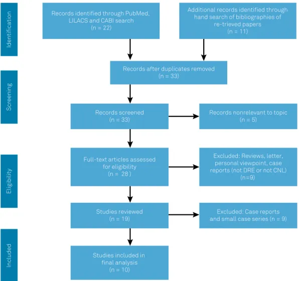
The literature search produced 33 abstracts (Figure 1).
Review of abstracts yielded five articles that were deemed
not relevant to the topic, another five published reviews
and two personal viewpoints/letters. Seven reports alluded
to isolated cases or small case series
10,12,19,20,21,22,23. Another
two case reports, one describing a degenerating
cysticer-cus (hence not CNL) in the amygdala and another with a
frontal CNL but not fulfilling criteria for drug-resistant
epilepsy, were reviewed but not included
24,25. Data for the
formal analysis were extracted mainly from the remaining
studies although isolated reports were examined as well.
Many of the reports were from the same center and
over-lapped in time. It is possible that these reports (Table 1)
partly covered the same set of subjects
11,15,16,26,27,28,29,30. Hence,
the most relevant or recent publications with the most
pro-tracted recruitment period from each center were included
for analysis)
11,27,28,29.
Association between HS and calcification
In five hospital-based studies that reported the fraction
of HS samples with CNLs, the proportion varied from 27% to
52% (median: 37%)
15,16,28,30,31. The proportion of CNLs in cor
-responding samples of epilepsies other than HS at the same
centers was 6-15% (median: 14%)
16,30,31.
Differences between HS with CNLs and HS alone:
Demographic and electro-clinical features
A small Indian observational study reported an older age
at onset of epilepsy in people with HS with CNLs in compari
-son with HS alone but this finding was not replicated in the
larger Brazilian study
11,27(Table 2). Likewise, two Brazilian
groups compared gender distribution in people who had HS
with CNLs, with HS alone
27,30. One group consistently reported
an excess of females in people who had HS with CNLs, but
this was not corroborated by an observational study from a
different center in the same Brazilian state
15,16,27,30.
Some have commented on the absence of an initial
precipitating illness in people who had HS with CNLs
12,16.
Others have found a lower frequency of an initial
those with HS alone
11,29, a finding not corroborated by still
others
31. An Indian study noted that while roughly a fifth of
individuals with HS with CNLs had childhood febrile sei
-zures, nearly another half reported afebrile seizures in the
first decade of life
29.
An earlier Brazilian study found no difference in the pro
-portion of those with unilateral versus bitemporal spikes but
a subsequent re-analysis from the same center with more
cases, as well as another Indian study, noted that the finding
of bitemporal spikes correlated with the presence of CNL in
individuals with HS
15,29,32.
Number and laterality of CNL and HS
Within CNL-HS groups in published reports, the propor
-tion of solitary calcifica-tion varied from 25% to 67%
11,28,29,30,31.
The case series from India had the highest proportion of soli
-tary calcifications
11,29. Overall, 21–83% of the calcifications
(regardless of whether they were solitary or multiple)
11,15,31and 85-100% of solitary calcifications
11,28,29,30were found
exclusively ipsilateral to the diseased hippocampus. Four
studies reported the lobar location of the CNLs and in these
reports the frequency of ipsilateral temporal lobe CNLs var
-ied between 9% and 67%.
11,15,28,31In some cases, varying from
3% to 33%, the CNL was located within or in close proximity
to the hippocampus (Figure 2 a-d).
11,12,15,28Choice of surgical approach and postsurgical
outcome
One small report of four cases, on whom a standard
temporal lobectomy was performed, reported seizure
free-dom in only one individual with CNL-HS.
11In comparison,
an Engel Class I outcome was reported in nearly three
quar-ters of a cohort of 126 people on whom standard temporal
lobectomy was performed
27(Table 3).
A lesionectomy alone
was performed on seven people and postoperative seizure
freedom documented in four.
11,23In one of these cases, who
continued to have seizures after lesionectomy, the seizures
were related to new parenchymal cysticerci emerging after
surgery.
24Only three individuals with an extratemporal CNL
and HS evaluated with invasive EEG were reported; all had
seizure onsets from the CNL as well as the hippocampus and
were seizure-free following standard temporal lobectomy
Records identified through PubMed,
LILACS and CABI search
(n = 22)
Additional records identified through
hand search of bibliographies of
re-trieved papers
(n = 11)
Records after duplicates removed
(n = 33)
Records screened
(n = 33)
Records nonrelevant to topic
(n = 5)
Full-text articles assessed
for eligibility
(n = 28 )
Excluded: Reviews, letter,
personal viewpoint, case
reports (not DRE or not CNL)
(n=9)
Studies reviewed
(n = 19)
Excluded: Case reports
and small case series (n = 9)
Studies included in
final analysis
(n = 10)
Identification
Screening
Eligibility
Included
Note: 1. Search terms: (“Neurocysticercosis”[Mesh] OR Cysticercosis[TW]) AND (“mesial temporal sclerosis”[TW] OR “hippocampal sclerosis”[TW]); 2. Refer to Table 1 for classification of the reports included in the review.
with lesionectomy (excision of the CNL).
11,16Lastly, a subset
comprising people in whom the CNL was located within or
in close proximity to the hippocampus, including the
para-hippocampal and fusiform gyri, the insula and the
tempo-ral pole (3–33% of all operated cases with HS and CNL) was
put together
11,12,15,20. All were seizure-free following temporal
resections, which included the CNL.
DISCUSSION
Is there an association between NCC and HS?
A growing number of reports supports the
plausibil-ity of an association between NCC and HS
10,11,15,16,19-29,31. The
small numbers of participants in most studies, variations in
methods used and outcomes assessments, however, limit
the application of quantitative meta-analytic approaches to
clarify the association. Additionally, the available reports are
either retrospective or cross-sectional studies, mostly
origi-nating from specialized centers in São Paulo, Brazil, thereby
introducing a potential referral bias, which could confound
the interpretation
16,30,31. Several studies have compared the
frequency of CNLs on CT scans between groups with HS and
other epilepsies
16,30,31. One compared the frequency of CNLs
in people with HS with those with headache
30. The compar
-ator groups (i.e., epilepsies other than those with HS and
headache) are not ideal as there are documented
associa-tions between CNLs and epilepsy and headache in
T. solium
cysticercosis-endemic areas
33,34. It might be pertinent to
compare the frequency of CNLs in mesial temporal lobe epi
-lepsy associated with HS with other temporal lobe epilepsies
and extratemporal lobe epilepsies but this does not seem to
Table 1.
Classification of published reports [excluding case reports and small (n = < 5) series] retrieved for this review.
Variable
Author, year
Study design
Study location
Comment
Studies of association
between HS and CNL
Velasco et al., 2006
16Hospital-based
cross-sectional study
Sao Paulo, Brazil,
Referral centre for
epilepsy surgery
Compared prevalence of CNLs
among patients with HS with
other etiologies of epilepsy
Oliveira et al., 2014
31Hospital-based
cross-sectional study
Sao Paulo, Epilepsy
clinic-based
Compared prevalence of CNLs
among patients with HS with
other etiologies of epilepsy
Taveira et al., 2015
30Case control study,
hospital-based
Sao Paulo, Referral
centre for epilepsy
surgery
Compared prevalence of CNLs
among s patients with HS with
other etiologies of epilepsy
Del Brutto et al., 2015
35Population-based,
exposed-unexposed
study
Ecuador
Determined prevalence ratio of
hippocampal atrophy among
patients with CNLs with those
with no evidence of NCC
Del Brutto et al., 2017
36Studies comparing
demographic features
of CNL-HS with HS
alone
*Bianchin et al., 2015
28Hospital-based series
Sao Paulo, Brazil
Bianchin et al., 2013
27Leite et al., 2000
15Rathore et al., 2012
29Hospital-based series
Trivandrum, India
Rathore et al., 2013
11Studies describing
surgical outcome in
CNL-HS/CNL alone
Bianchin et al., 2013
27Hospital-based series
Sao Paulo, Brazil
Leite et al., 2000
15Rathore et al., 2013
11Hospital-based series
Trivandrum, India
*The studies listed in this cell were published at different periods of time from the same center and comprised overlapping sets of cases. Hence, the latest reports with the largest number27,28 were used for analysis. Case reports and small series of cases are not represented in this table.
Table 2.
Comparison of demographic and electroclinical
features of CNL-HS and HS alone.
Author, year
HS alone
CNL-HS
Age of onset (First unprovoked seizure) (mean SD)
Rathore et al., 2012
2910 ± 6 years
16 ± 7 years
Bianchin et al., 2013
279 ± 9 years
10 ± 8 years
Gender distribution (Proportion of femal.es)
Bianchin et al., 2013
2754%
62%
Oliveira et al., 2015
3170%
67%
Antecedent febrile seizures (initial. precipitating illness)
{Proportion; (%)}
Chandra et al., 2010
12Nil
Rathore et al., 2012
2953%
22%
Oliveira et al., 2015
3123%
27%
Proportion of bitemporal. spikes(%)
Leite et al., 2000
1510%
11%
Figure 2.
Hippocampal sclerosis with a spatially-proximated CNL. Note (a) T2 oblique coronal image showing left HS, (b) T1
oblique coronal image demonstrating left hippocampal atrophy, (c) T2 oblique coronal image showing proximate CNL (arrow)
with surrounding hyperintensity presumed to be gliosis and (d) axial computed tomography image clearly demonstrating the left
temporal CNL.
Table 3.
Surgical procedures and outcomes following surgery for drug-resistant epilepsy in people with CNLs with or without HS.
Surgical procedure
Reference (Author, year)
Number of cases
operated on
Number of cases with
postoperative seizure
freedom
Standard anteromesial temporal
lobectomy (with/without CNL in the
surgical field)
Rathore et al., 2013
117
4 (57%)
Leite et al., 2000
1532
26 (81%)
Bianchin et al., 2013
27126
94 (75%)
Lesionectomy alone
Ooi et al., 2011
23
1
0
Rathore et al., 2013
116
#4
Anteromesial temporal lobectomy with
resection of extratemporal CNL
Rathore et al., 2013
112
2
Including one case with HS and five with no imaging evidence of HS (Ref. 11).
A
B
have been undertaken. The association should also be exam
-ined in a population-based sample, a major undertaking in
resource-limited settings. A single, small population-based
survey in Ecuador evaluated the association between CNLs
and hippocampal atrophy, suggesting that CNLs or their
precursors, i.e., active NCC or associated seizures, might be
causally associated with hippocampal injury
35. The majority
of participants, however, did not have epilepsy, a defining fea
-ture of HS. When stratified according to age, the association
became significant only in those aged above 68 years
36. This
age-specific association is in marked contrast to the usual
much younger age of onset of typical HS
37-39. Sclerosis of the
hippocampus on T2 or FLAIR MR images, crucial to the rec
-ognition of HS, was also not reported.
Demographic and electroclinical characteristics of
CNL-HS
It might be argued that, if indeed there were a causal
asso-ciation between CNLs and HS, the CNLs might modify some
of the demographic, electroclinical and prognostic
character-istics of HS. Among various demographic attributes, a gender
predilection has been reported from a single Brazilian cen
-ter with an excess of women among people with CNL-HS
15,26.
Gender-related differences in host inflammatory responses
to cysticerci have been suggested as the basis for this
obser-vation.
40The findings of a gender predilection have not been
replicated in a report from a different center in Brazil, nor
in India
29,31. There is also no documented gender predilection
for HS alone as no differences have been reported in large
HS series
37,38,39. Other demographic features, e.g., age of onset
of habitual seizures, in CNL-HS appear to be similar to HS
alone, usually in the first two decades of life. Some reports
also covered the frequency of an antecedent initial
precipi-tating illness in people with CNL-HS
11,12,16,29. Febrile seizures
have been reported in 82% of people with HS alone
37,39, while
antecedent, early life afebrile seizures are extremely rare.
There have been reports of a lower frequency, or even com
-plete absence, of antecedent febrile seizures in people with
CNL-HS
12,29,32. Conversely, afebrile seizures in the first decade
of life, reported with a high frequency (44%) in people with
CNL-HS from India may represent acute seizures related to
active parenchymal NCC
29. It is also possible that the lower
frequencies of febrile seizures may partly be accounted for by
a recall bias. More studies from different geographical loca
-tions are clearly desirable to clarify the gender predilection as
well as other possible distinctive features of CNL-HS, if any.
Location and laterality of CNLs in relation to HS
We surmise that causality is more likely if the CNL is
located ipsilateral to the side of HS and within the ipsilateral
hemisphere, in proximity to the hippocampus. The likelihood
of publication bias should be taken in to account in
inter-preting published data regarding the location of the CNLs.
Besides, the analysis of the laterality and location of CNLs in
relation to HS is complicated by the multiplicity of CNLs with
distribution across both hemispheres, making it difficult to
decide which of the lesions is the culprit; in some reports a
large proportion of the CNLs are multiple.
15,28In comparison,
a solitary CNL offers the opportunity of analyzing the lateral
-ity and lobar (or sublobar) location of the CNL in relation to
the HS. Overall, the majority (roughly three quarters) of the
reported solitary CNLs are located ipsilateral to the HS.
11,15,28,31In a number of isolated case reports and in some of the larger
series, the CNL may be located within, or in close proxim
-ity to, the hippocampus followed by the ipsilateral temporal
lobe.
11,12,15,20This seems to suggest that the proximity of the
CNL to the hippocampus and related structures might be a
factor in the development of HS. It is plausible that an inflam
-matory response to the CNL engenders an insult to the proxi
-mate hippocampus, thereby provoking a cascade of events
eventually leading to the development of HS.
Is the association causal?
Even as an association between NCC and HS is plausible,
we need to consider whether the association is causal.
41To
infer causality, more studies in different geographic regions,
endemic and nonendemic, are required to gauge the strength
and consistency of the association. Support for a biological
gradient in the association is provided by the predilection of
solitary CNLs distributed ipsilaterally and within the same
hemisphere, in proximate locations to the diseased
hippo-campus. These findings may be implicit with the argument
that acutely degenerating cysticercus or associated seizures
might constitute the initial precipitating illness for HS. It
is also desirable to conduct experiments in animal models
with, for example, cysticercus extracts injected into different
locations in the cerebral hemispheres, to determine the effect
on hippocampal structure and function.
42Choice of surgical approach
An assessment of seizure outcome following surgery
is precluded by the limited number of reports, varied use
of presurgical evaluation strategies and time periods and
methods of outcome assessment. It appears, however, that
surgical outcome following standard temporal lobe
pro-cedures in terms of seizure freedom in CNL-HS is similar
to HS alone.
15,16,27Surgical failure with standard
anterome-sial temporal lobectomy following noninvasive
presurgi-cal workup has also been anecdotally documented,
imply-ing that no one-fits-all approach can be recommended.
11Other approaches, including lesionectomy (removal of
CNL alone) and combined approaches (i.e., lesionectomy
in addition to the temporal lobectomy;
cf
, dual pathology)
have been employed in the surgical management of
drug-resistant epilepsy associated with CNL-HS.
11It is
origins from the hippocampus and CNL, is suspected dur
-ing noninvasive presurgical workup, then recourse to
inva-sive monitoring appears appropriate and justified despite
limitations of cost and availability in regions where NCC
is endemic. In situations where the CNL is located close
to the hippocampal structures, it is reasonable to excise
the lesion along with other temporal lobe structures. Since
different clinical scenarios exist, a case-by-case approach
should be followed in evaluating and managing people
with CNL-HS. Indeed, the categorization of CNLs in asso
-ciation with HS described in a recent review compliments
the approach presented here.
43Future directions
If CNLs and HS were causally associated, the underlying
basis might be an interaction between the cysticercus
gran-uloma and the hippocampus structure and function or an
effect mediated through kindling by repeated seizures. The
fact that CNLs can be found on imaging studies in as many
as 10–18% of asymptomatic individuals in
T. solium
-endemic
communities
44,45might be utilized in dissecting out a direct
effect of cysticercus granulomas versus an effect mediated
through seizures, and it might be worthwhile to study the
prevalence of hippocampal abnormalities in people with
asymptomatic CNLs.
The association between NCC and HS merits further
investigation as it has far-reaching implications for
under-standing not only the burden and etiology of epilepsies in
resource-poor countries but also, overall, the phenomenon
of epileptogenesis.
CONCLUSIONS
The available evidence is at best hypothesis-generating,
providing preliminary support for an association between
CNLs and HS. The association should be investigated fur
-ther in population-based samples to avoid selection bias.
Prospective, longitudinal follow-up imaging studies of the
hippocampal structure and volume in people presenting
with a first seizure associated with NCC are desirable. The
longitudinal design would help in establishing
temporal-ity between cause (NCC infestation) and effect (HS). Future
studies should also be located in geographically disparate
T
.
solium
-endemic regions.
ACKNOWLEDGMENTS
This work was done at the NIHR University College
London Hospitals Biomedical Research Centre, which receives
a proportion of funding from the UK Department of Health’s
Comprehensive Biomedical Research Centers funding scheme.
JWS receives support from the Dr. Marvin Weil Epilepsy
Research Fund and the UK Epilepsy Society. We are grateful to
Dr. Gail S. Bell for thoroughly reviewing the manuscript.
References
1. Del Brutto OH. Neurocysticercosis: a review. Sci World J. 2012;2012:159821. https://doi.org/10.1100/2012/159821
2. World Health Organization. Taenaisis/cysticercosis: fact sheet. Geneve: World Health Organization; 2017 [cited 2017 May 3]. Available from: http://www.who.int/mediacentre/factsheets/ fs376/en/
3. Montano SM, Villaran MV, Ylquimiche L, Figueroa JJ, Rodriguez S, Bautista CT et al. Neurocysticercosis: association between seizures, serology, and brain CT in rural Peru. Neurology. 2005 Jul;65(2):229-33. https://doi.org/10.1212/01.wnl.0000168828.83461.09
4. Medina MT, Aguilar-Estrada RL, Alvarez A, Durón RM, Martínez L, Dubón S et al. Reduction in rate of epilepsy from neurocysticercosis by community interventions: the Salamá, Honduras study. Epilepsia. 2011 Jun;52(6):1177-85. https://doi.org/10.1111/j.1528-1167.2010.02945.x
5. Singh G, Bawa J, Chinna D, Chaudhary A, Saggar K, Modi M et al. Association between epilepsy and cysticercosis and toxocariasis: a population-based case-control study in a slum in India. Epilepsia. 2012 Dec;53(12):2203-8. https://doi.org/10.1111/epi.12005
6. Rajshekhar V, Raghava MV, Prabhakaran V, Oommen A, Muliyil J. Active epilepsy as an index of burden of neurocysticercosis in Vellore district, India. Neurology. 2006 Dec;67(12):2135-9. https://doi.org/10.1212/01.wnl.0000249113.11824.64
7. Carabin H, Ndimubanzi PC, Budke CM, Nguyen H, Qian Y, Cowan LD et al. Clinical manifestations associated with neurocysticercosis: a systematic review. PLoS Negl Trop Dis. 2011 May;5(5):e1152. https://doi.org/10.1371/journal.pntd.0001152
8. Carpio A, Hauser WA. Prognosis for seizure recurrence in patients with newly diagnosed
neurocysticercosis. Neurology. 2002 Dec;59(11):1730-4. https://doi.org/10.1212/01.WNL.0000036320.69823.EA
9. Del Brutto OH, Santibañez R, Noboa CA, Aguirre R, Díaz E, Alarcón TA. Epilepsy due to neurocysticercosis: analysis of 203 patients. Neurology. 1992 Feb;42(2):389-92. https://doi.org/10.1212/WNL.42.2.389
10. Singla M, Singh P, Kaushal S, Bansal R, Singh G. Hippocampal sclerosis in association with
neurocysticercosis. Epileptic Disord. 2007 Sep;9(3):292-9. https://doi.org/10.1684/epd.2007.0122
11. Rathore C, Thomas B, Kesavadas C, Abraham M, Radhakrishnan K. Calcified neurocysticercosis lesions and antiepileptic drug-resistant epilepsy: a surgically remediable syndrome? Epilepsia. 2013 Oct;54(10):1815-22. https://doi.org/10.1111/epi.12349
12. Chandra PS, Bal C, Garg A, Gaikwad S, Prasad K, Sharma BS et al. Surgery for medically intractable epilepsy due to postinfectious etiologies. Epilepsia. 2010 Jun;51(6):1097-100. https://doi.org/10.1111/j.1528-1167.2010.02538.x
13. Pradhan S, Kathuria MK, Gupta RK. Perilesional gliosis and seizure outcome: a study based on magnetization transfer magnetic resonance imaging in patients with neurocysticercosis. Ann Neurol. 2000 Aug;48(2):181-7.
14. de Souza A, Nalini A, Kovoor JM, Yeshraj G, Siddalingaiah HS, Thennarasu K. Perilesional gliosis around solitary cerebral parenchymal cysticerci and long-term seizure outcome: a prospective study using serial magnetization transfer imaging. Epilepsia. 2011 Oct;52(10):1918-27. https://doi.org/10.1111/j.1528-1167.2011.03189.x
15. Leite JP, Terra-Bustamante VC, Fernandes RM, Santos AC, Chimelli L, Sakamoto AC et al. Calcified neurocysticercotic lesions and postsurgery seizure control in temporal lobe epilepsy. Neurology. 2000 Nov;55(10):1485-91. https://doi.org/10.1212/WNL.55.10.1485
16. Velasco TR, Zanello PA, Dalmagro CL, Araújo D Jr, Santos AC, Bianchin MM et al. Calcified cysticercotic lesions and intractable epilepsy: a cross sectional study of 512 patients. J Neurol Neurosurg Psychiatry. 2006 Apr;77(4):485-8. https://doi.org/10.1136/jnnp.2005.078675
17. Singh G, Burneo JG, Sander JW. From seizures to epilepsy and its substrates: neurocysticercosis. Epilepsia. 2013 May;54(5):783-92. https://doi.org/10.1111/epi.12159
18. Durnford AJ, Rodgers W, Kirkham FJ, Mullee MA, Whitney A, Prevett M et al. Very good inter-rater reliability of Engel and ILAE epilepsy surgery outcome classifications in a series of 76 patients. Seizure. 2011 Dec;20(10):809-12. https://doi.org/10.1016/j.seizure.2011.08.004
19. Kobayashi E, Guerreiro CA, Cendes F. Late onset temporal lobe epilepsy with MRI evidence of mesial temporal sclerosis following acute neurocysticercosis: case report. Arq Neuropsiquiatr. 2001 Jun;59 2-A:255-8. https://doi.org/10.1590/S0004-282X2001000200021
20. Chung CK, Lee SK, Chi JG. Temporal lobe epilepsy caused by intrahippocampal calcified cysticercus: a case report. J Korean Med Sci. 1998 Aug;13(4):445-8. https://doi.org/10.3346/jkms.1998.13.4.445
21. Silva AV, Martins HH, Marques CM, Yacubian EM, Sakamoto AC, Carrete Junior H et al. Neurocysticercosis and microscopic hippocampal dysplasia in a patient with refractory mesial temporal lobe epilepsy. Arq Neuropsiquiatr. 2006 Jun;64(2A):309-13. https://doi.org/10.1590/S0004-282X2006000200026
22. Son EI, Yi SD, Lee SW, Lee HC, Yim MB, Kim IH. Surgery for seizure-related structural lesions of the brain with intraoperative acute recording(ECoG) and functional mapping. J Korean Med Sci. 1994 Oct;9(5):409-13. https://doi.org/10.3346/jkms.1994.9.5.409
23. Ooi WW, Wijemanne S, Thomas CB, Quezado M, Brown CR, Nash TE. Short report: A calcified Taenia solium granuloma associated with recurrent perilesional edema causing refractory seizures: histopathological features. Am J Trop Med Hyg. 2011 Sep;85(3):460-3. https://doi.org/10.4269/ajtmh.2011.11-0221
24. Lee DJ, Owen CM, Khanifar E, Kim RC, Binder DK. Isolated amygdala neurocysticercosis in a patient presenting with déjà vu and olfactory auras. Case report. J Neurosurg Pediatr. 2009 Jun;3(6):538-41. https://doi.org/10.3171/2009.2.PEDS08140
25. Hasan MS, Basri HB, Hin LP, Stanslas J. Surgical remotion of a cysticercotic granuloma responsible for refractory seizures: A case report. Surg Neurol Int. 2011;2(1):177-9. https://doi.org/10.4103/2152-7806.90698
26. da Gama CN, Kobayashi E, Li LM, Cendes F. Hippocampal atrophy and neurocysticercosis calcifications. Seizure. 2005 Mar;14(2):85-8. https://doi.org/10.1016/j.seizure.2004.10.005
27. Bianchin MM, Velasco TR, Coimbra ER, Gargaro AC, Escorsi-Rosset SR, Wichert-Ana L et al. Cognitive and surgical outcome in mesial temporal lobe epilepsy associated with hippocampal sclerosis plus neurocysticercosis: a cohort study. PLoS One. 2013 Apr;8(4):e60949. https://doi.org/10.1371/journal.pone.0060949
28. Bianchin MM, Velasco TR, Wichert-Ana L, Araújo Junior D, Alexandre Junior V, Scornavacca F et al. Neuroimaging observations linking neurocysticercosis and mesial temporal lobe epilepsy with hippocampal sclerosis. Epilepsy Res. 2015 Oct;116:34-9. https://doi.org/10.1016/j.eplepsyres.2015.07.001
29. Rathore C, Thomas B, Kesavadas C, Radhakrishnan K. Calcified neurocysticercosis lesions and hippocampal sclerosis: potential dual pathology? Epilepsia. 2012 Apr;53(4):e60-2. https://doi.org/10.1111/j.1528-1167.2011.03386.x
30. Taveira MO, Morita ME, Yasuda CL, Coan AC, Secolin R, Costs ALC et al. Neurocysticercotic calcifications and hippocampal sclerosis: A case-control study. PLoS One. 2015;10: https://doi.org/10.1371/journal.pone.0131180
31. Oliveira MC, Martin MG, Tsunemi MH, Vieira G, Castro LH. Small calcified lesions suggestive of neurocysticercosis are associated with mesial temporal sclerosis. Arq Neuropsiquiatr. 2014 Jul;72(7):510-6. https://doi.org/10.1590/0004-282X20140080
32. Bianchin MM, Velasco TR, Araujo Junior D, Alexandre V Jr, Wichert-Ana L, Vera C et al. Clinical and electrophysiological differences between mesial temporal lobe epilepsy and mesial temporal lobe epilepsy plus neurocysticercosis [Abstract]. Epilepsia. 2006;4 suppl 4:244-5.
33. Cruz ME, Cruz I, Preux PM, Schantz P, Dumas M. Headache and cysticercosis in Ecuador, South America. Headache. 1995 Feb;35(2):93-7. https://doi.org/10.1111/j.1526-4610.1995.hed3502093.x
34. Garg RK, Kar AM, Singh MK. Prednisolone-responsive headache in patients with solitary cysticercus granuloma and seizures. Headache. 2004 Apr;44(4):365-9. https://doi.org/10.1111/j.1526-4610.2004.04096.x
35. Del Brutto OH, Salgado P, Lama J, Del Brutto VJ, Campos X, Zambrano M et al. Calcified neurocysticercosis associates with hippocampal atrophy: a population-based study. Am J Trop Med Hyg. 2015 Jan;92(1):64-8. https://doi.org/10.4269/ajtmh.14-0453
36. Del Brutto OH, Issa NP, Salgado P, Del Brutto VJ, Zambrano M, Lama J et al. The association between neurocysticercosis and hippocampal atropy is related to age. Am J Trop Med Hyg. 2017 Jan;96(1):243-8. https://doi.org/10.4269/ajtmh.16-0689
37. Cendes F, Andermann F, Dubeau F, Gloor P, Evans A, Jones-Gotman M, et al. Early childhood prolonged febrile convulsions, atrophy and sclerosis of mesial structures, and temporal lobe epilepsy: an MRI volumetric study. Neurology. 1993 Jun;43(6):1083-7. https://doi.org/10.1212/WNL.43.6.1083
38. Cendes F. Febrile seizures and mesial temporal sclerosis. Curr Opin Neurol. 2004 Apr;17(2):161-4. https://doi.org/10.1097/00019052-200404000-00013
39. French JA, Williamson PD, Thadani VM, Darcey TM, Mattson RH, Spencer SS, et al. Characteristics of medial temporal lobe epilepsy: I. Results of history and physical examination. Ann Neurol. 1993 Dec;34(6):774-80. https://doi.org/10.1002/ana.410340604
40. Rangel R, Torres B, Del Bruto O, Sotelo J. Cysticercotic encephalitis: a severe form in young females. Am J Trop Med Hyg. 1987 Mar;36(2):387-92. https://doi.org/10.4269/ajtmh.1987.36.387
41. Höfler M. The Bradford Hill considerations on causality: a counterfactual perspective. Emerg Themes Epidemiol. 2005 Nov;2(1):11. https://doi.org/10.1186/1742-7622-2-11
42. Stringer JL, Marks LM, White AC Jr, Robinson P. Epileptogenic activity of granulomas associated with murine cysticercosis. Exp Neurol. 2003 Oct;183(2):532-6. https://doi.org/10.1016/S0014-4886(03)00179-1
43. Escalaya AL, Burneo JG. Epilepsy surgery and neurocysticercosis: assessing the role of the cysticercotic lesion in medically-refractory epilepsy. Epilepsy Behav. 2017 Nov;76:178-81. https://doi.org/10.1016/j.yebeh.2017.01.029
44. Cruz ME, Schantz PM, Cruz I, Espinosa P, Preux PM, Cruz A et al. Epilepsy and neurocysticercosis in an Andean community. Int J Epidemiol. 1999 Aug;28(4):799-803. https://doi.org/10.1093/ije/28.4.799