Revista Científica Eletrônica de Medicina Veterinária é uma publicação semestral da Faculdade de Medicina veterinária e Zootecnia de Garça – FAMED/FAEF e Editora FAEF, mantidas pela Associação Cultural e Educacional de Garça ACEG. CEP: 17400-000 – Garça/SP – Tel.: (0**14) 3407-8000 www.revista.inf.br – www.editorafaef.com.br – www.faef.edu.br.
TOXOPLASMOSIS: MORPHOLOGIC AND MORPHOMETRIC EVALUATION OF MYELIN SHEATH OF SPINAL CORD FROM
NONSYMPTOMATIC SEROPOSITIVE DOGS
CARVALHO, Alessandra Cristina Francischini de
Departamento de Morfologia e Fisiologia Animal, Faculdade de Ciências Agrárias e Veterinárias, Universidade Estadual Paulista “Júlio de Mesquita Filho” - UNESP,
Jaboticabal, SP, Brasil.
PACHECO, Maria Rita
Departamento de Morfologia e Fisiologia Animal, Faculdade de Ciências Agrárias e Veterinárias, Universidade Estadual Paulista “Júlio de Mesquita Filho” - UNESP,
Jaboticabal, SP, Brasil.
BARALDI ARTONI, Silvana Martinez
Departamento de Morfologia e Fisiologia Animal, Faculdade de Ciências Agrárias e Veterinárias, Universidade Estadual Paulista “Júlio de Mesquita Filho” - UNESP,
Revista Científica Eletrônica de Medicina Veterinária é uma publicação semestral da Faculdade de Medicina veterinária e Zootecnia de Garça – FAMED/FAEF e Editora FAEF, mantidas pela Associação Cultural e Educacional de Garça ACEG. CEP: 17400-000 – Garça/SP – Tel.: (0**14) 3407-8000 www.revista.inf.br – www.editorafaef.com.br – www.faef.edu.br.
ABSTRACT
The aim of this work was to analyze the morphology and morphometry of myelin sheath of canine spinal cord. Twenty indefinite-breed adult dogs were used; ten hygid dogs, with negative serology for toxoplasmosis, were used as control group (group 1) and ten dogs nonsymptomatic but seropositive for toxoplasmosis were used as group 2. The morphologic analysis pointed no differences between the groups. In the morphometric study, axonal myelin sheath thickness was evaluated. The results were statistically analyzed by Student’s t test and no statistic difference was observed for canine myelin sheath thickness between the groups studied.
Key words: morphology, morphometry, spinal cord, dogs, toxoplasmosis.
INTRODUCTION
Inflammatory diseases of Central Nervous System (CNS) are important causes of neurologic disorders in dogs. Many of them are caused by infectious agents, such as Canine Distemper Virus (CDV), rabies, protozoa, fungi, as well as those of unknown origin (MORETTI et al., 2002).
Canine neurologic diseases caused by protozoa are uncommon (HASS et al., 1989; THOMAS, 1998). Encephalitis caused by these agents, in dogs, most often are caused by Toxoplasma gondii (T. gondii) and Neospora caninum (PAIXÃO; SANTOS, 2004).
NERVOUS TISSUE
The nervous tissue has, basically, two kinds of cells, neurons and glial cells or neuroglia (ROSS et al., 1995; CHRISMAN, 1997; GEORGE; CASTRO, 1998; MACHADO, 2002; JUNQUEIRA ; CARNEIRO, 2008).
Most of the neurons posses an axon, which is a long and slender projection originated in the cell body or in a main dendrite, in a region named hillock and it has almost no chromidial substance. (GEORGE; CASTRO, 1998; MACHADO, 2002; JUNQUEIRA; CARNEIRO, 2008). The axon presents variable length, depending on
Revista Científica Eletrônica de Medicina Veterinária é uma publicação semestral da Faculdade de Medicina veterinária e Zootecnia de Garça – FAMED/FAEF e Editora FAEF, mantidas pela Associação Cultural e Educacional de Garça ACEG. CEP: 17400-000 – Garça/SP – Tel.: (0**14) 3407-8000 www.revista.inf.br – www.editorafaef.com.br – www.faef.edu.br. the kind of neuron, and it might have, in human species, from some millimeters to more than one meter (MACHADO, 2002; JUNQUEIRA; CARNEIRO, 2008).
According to Zhang et al. (1995) there is a reduction in number and size of axons due to aging process.
CANINE TOXOPLASMOSIS
Since it was described in 1910 by Mello in Italy (VIDOTTO, 1992) and by Carini (1911) in Brazil, life cycle and different modes of transmission have been extensively discussed in veterinarian literature (HASS et al, 1989; LAPPIN, 2004). Dogs are considered very receptive animals for this zoonosis, probably, due to their carnivorous eating habit, what facilitates the ingestion of tissues contaminated by cysts and the contact with sporulated oocysts in contaminated soil. (GERMANO, 1985).
The infection has been noticed in cats and dogs in many countries, what demonstrates its cosmopolitan feature. Severe and fatal cases have been reported, although clinical manifestations are uncommon due to the efficiency of T. gondii as a intrecellular parasite (DUBEY; BEATTLE, 1988; HASS et al., 1989; LINDSAY, 1990; GUIMARÃES et al., 1992; PAIXÃO; SANTOS, 2004).
Ataxia, convulsions, tremors, cranial nerve disorders, progressive paresis and paralysis are the most common manifestations of nervous toxoplasmosis (AVERILL; LAHUNTA, 1971; NESBIT et al., 1981; SUTER et al., 1984; BRAUND et al., 1988; DAVIDSON, 2000; LAPPIN, 2004).
Uncolored, necrosed areas and areas with cerebellar atrophy were observed in CNS macroscopically (DUBEY; LAPPIN, 1998).
The most common histopathological alterations in the CNS are characterized by non suppurative meningoencephalomyelitis associated with vasculitis, necrosis, malacia and gliosis with possible involvement of peripheral nerves (DUBEY; BEATTLE, 1988; DUBEY; LAPPIN, 1998; GIRALDI et al., 2002).
It is known that important histopathological changes are observed in the CNS with clinical toxoplasmosis, so this study aimed to analyze the occurrence of early changes in spinal cord neuroglia structures of apparently healthy dogs, but with high serological reactivity to toxoplasmosis, in order to better investigate and document the possible histological changes.
Revista Científica Eletrônica de Medicina Veterinária é uma publicação semestral da Faculdade de Medicina veterinária e Zootecnia de Garça – FAMED/FAEF e Editora FAEF, mantidas pela Associação Cultural e Educacional de Garça ACEG. CEP: 17400-000 – Garça/SP – Tel.: (0**14) 3407-8000 www.revista.inf.br – www.editorafaef.com.br – www.faef.edu.br.
MATERIAL AND METHODS
Animals
For the proposed objectives, twenty indefinite-breed adult dogs weighing from 7 to 15 kg were used, being ten healthy, with negative serology for toxoplasmosis, used as control group (group 1) and ten nonsymptomatic dogs, with levels of reactivity for T. gondii above 8, detected through indirect immunoenzymatic assay (ELISA), used as reagent group (group 2).
Experimental procedure
The serology for T. gondii was performed in Department of Veterinarian Pathology of FCAVJ-UNESP, by indirect immunoenzymatic assay (ELISA), method developed and standardized by the Department itself, with the technique described by Domingues et al. (1998). The reactivity of serums was analyzed according to ELISA levels (from 0 to 9) and the animals used in this study were those that presented reactivity 8 or 9.
After the formation of groups 1 and 2, fragments of spinal cord corresponding to cervical, thoracic and lumbar area were collected and fixed in Bouin’s solution during 24 hours and processed, as usual, to be included in paraffin.
After microtomy, at interval of 100µm, histological sections, of 5µm thickness, were stained with hematoxylin-eosin (HE) and Masson's trichrome (MT) techniques. Slices of 15µm thickness were stained by silver impregnation because this technique is chosen to better identify microglia and oligodendrocytes. All three techniques were performed according to Tolosa (2003).
The glial fibrillary acidic protein (GFAP), subunit of the intermediary filaments of the cellular cytoskeleton, exists in the cytoplasm of astrocytes. Immunohistochemistry using primary antibody anti-GFAP is generally chosen to identify astrocytes in the CNS. Slices of 5µm thickness were stained by the immunohistochemistry PAP (peroxidase antiperoxidase) technique using the primary antibody anti-GFAP, according to Lemos and Alessi (1999). The glass slides were
Revista Científica Eletrônica de Medicina Veterinária é uma publicação semestral da Faculdade de Medicina veterinária e Zootecnia de Garça – FAMED/FAEF e Editora FAEF, mantidas pela Associação Cultural e Educacional de Garça ACEG. CEP: 17400-000 – Garça/SP – Tel.: (0**14) 3407-8000 www.revista.inf.br – www.editorafaef.com.br – www.faef.edu.br. examined by light microscopy to verify the morphology of structures and were photographed using a photo microscope model Olympus BX50.
The thickness and morphometry of myelin sheath were analyzed. The measurement, in µm, was done using the image analysis software, Image Pro-plus, joined to a binocular microscope, both by Carl Zeiss.
Statistic Analysis
The results were statistically analyzed by Student’s t test at 5% probability level (p<0.05), using Statistic Analysis Software SAS.
RESULTS
Morphologic results
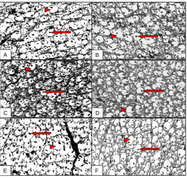
The transverse cut of the White matter from spinal cord revealed axons with different calibers, involved by myelin sheath, which thickness varied proportionally according to the axon diameter. This complex was evidenced with negative image, because the lipidic components were extracted during the histological routine and only the vestiges of protein web remained, giving to the myelin sheath a circular aspect (Fig. 01). No morphological differences were observed between the groups in the studied segments.
Revista Científica Eletrônica de Medicina Veterinária é uma publicação semestral da Faculdade de Medicina veterinária e Zootecnia de Garça – FAMED/FAEF e Editora FAEF, mantidas pela Associação Cultural e Educacional de Garça ACEG. CEP: 17400-000 – Garça/SP – Tel.: (0**14) 3407-8000 www.revista.inf.br – www.editorafaef.com.br – www.faef.edu.br. Figure 01 – Axons (arrows) photomicrographies, with different calibers, involved by myelin sheath (arrow head) of control group (group 1) and toxoplasmosis seropositive group (group 2), from cervical (CR), thoracic (TR) and lumbar (LR) regions:
A) CR (group 1, TM,40X). B) CR (group 2, TM,40X). C) TR (group 1, TM,40X).
D) TR (group 2, TM,40X). E) LR (group 1, TM,40X). F) LR (group 2, TM, 40X).
Morphometric Results
Table 1 – Average values (m) of myelin sheath thickness of axons of control animals (group 1) and seropositive animals (group 2), in cervical, thoracic and lumbar areas of spinal cord. A B C D E F A e B e C e D e F e E e
Revista Científica Eletrônica de Medicina Veterinária é uma publicação semestral da Faculdade de Medicina veterinária e Zootecnia de Garça – FAMED/FAEF e Editora FAEF, mantidas pela Associação Cultural e Educacional de Garça ACEG. CEP: 17400-000 – Garça/SP – Tel.: (0**14) 3407-8000 www.revista.inf.br – www.editorafaef.com.br – www.faef.edu.br. MYELIN SHEATH THICKNESS
CERVICAL THORACIC LUMBAR
GROUP 1 5,72±1,93 a 6,53±2,41 a 7,41±3,18 a
GROUP 2 5,71±1,95 a 6,76±2,24 a 8,04±2,78 a
Same letters in same column are not different by Student’s t test (p>0.05).
No significant difference (p>0.05) between the groups was observed. The highest average values were found in lumbar area and the lowest in cervical area. (table 01).
DISCUSSION
In this study about morphologic characteristics of myelin sheath and axon, in transverse cut, no differences were noticed in these structures in animals of group 2 when they were compared to the dogs in group 1.
Comparing the evidences of this research with the ones mentioned in literature by Di Fiori et al. (1982), Young and Heath (2001), Junqueira and Carneiro (2008), there are similarities among the descriptions of these morphologic characteristics. These authors also report that, in transverse cut of white matter of spinal cord, the axon presented different calibers, involved by myelin sheath, a lipoprotein complex, which thickness is proportional to the axon diameter.
As for the average value found in myelin sheath of axons from white matter, it was confirmed that, in the lumbar area, the biggest axons occurred in both experimental groups and, consequently, the highest average value for the myelin sheath, which is proportional to the axon diameter.
So, based on Chrisman (1997), Molenaar et al. (1997) and Carvalho et al. (2006), we presume that, probably, in this segment, it is necessary rapid nerve impulse conduction (saltatory conduction), starting the muscle twitch and creating moving force, which is indispensable for body displacement.
Revista Científica Eletrônica de Medicina Veterinária é uma publicação semestral da Faculdade de Medicina veterinária e Zootecnia de Garça – FAMED/FAEF e Editora FAEF, mantidas pela Associação Cultural e Educacional de Garça ACEG. CEP: 17400-000 – Garça/SP – Tel.: (0**14) 3407-8000 www.revista.inf.br – www.editorafaef.com.br – www.faef.edu.br. studied areas, between the groups, what indicates that the dogs of group 2 didn’t present changes in myelin sheath when compared to the dogs of group1.
CONCLUSION
The morphologic and morphometric results indicate that there weren’t any changes in myelin sheath and axon of animals belonging to group 2 when they were compared to the dogs belonging to group 1, in that occasion, what allow us to conclude that the characteristics of these structures agree with those described in classic literature.
REFERENCES
AVERILL, D.R.; LAHUNTA, A. Toxoplasmosis of the Canine Nervous System: Clinicopathological Findings in Four Cases. J. Am. Vet. Med. Assoc., Schaumburg, v. 159, p. 1134-1141, 1971.
BRAUND, K.G.; BLAGBURN, B.L.; TOIVIO-KINNUCAN, M.; AMLING, K.A.; PIDGEON, G.L. Toxoplasma polymyositis/polyneuropathy – a new clinical variant in two mature dogs. J. Am. Anim. Hosp. Assoc., Golden, v. 24, n. 1, p. 93-97, 1988.
CARINI, A. Infection spontanée du pigeon et du chien due au Toxoplasma cuniculi.
Bull. Soc. Pathol. Exot., Paris, v. 4, p. 518-519,1911.
CARVALHO, A. C. F.; PACHECO, M. R.; BARALDI-ARTONI, S. M.; MATEUS, O. Canine spinal cord neuron and axon myelin sheath morphometry. Anat. Histol.
Embryol. J. Vet . Med. Berlin, v.35, n.5, p. 284-286, 2006.
CHRISMAN, C.L. Introdução ao Sistema Nervoso. In: CHRISMAN, C.L. Neurologia
dos pequenos animais, São Paulo: Roca, 1997, 432p.
DAVIDSON, M. G. Toxoplasmosis. Vet. Clin. North Am. Small Anim. Pract., v. 30, n.5, p. 1051-1062, 2000.
Revista Científica Eletrônica de Medicina Veterinária é uma publicação semestral da Faculdade de Medicina veterinária e Zootecnia de Garça – FAMED/FAEF e Editora FAEF, mantidas pela Associação Cultural e Educacional de Garça ACEG. CEP: 17400-000 – Garça/SP – Tel.: (0**14) 3407-8000 www.revista.inf.br – www.editorafaef.com.br – www.faef.edu.br. DI FIORE, M.S.H.; MANCINI, R.E.; DE ROBERTIS, E.D.P. Tecido Nervoso. In: DI FIORE, M.S.H. Novo atlas de histologia, 5. ed. Rio de Janeiro: Guanabara Koogan, 1982. 335p.
DOMINGUES, L.M.; MACHADO, R.Z.; TINUCCI COSTA, M.; CARVALHO, C. S.; COSTA, A J.; MALHEIROS, E. B. Canine toxoplasmosis: a comparative evaluation of the detection of anti-toxoplasma gondii antibodies by the indirect immunoenzymatic assay (elisa) and the indirect immunofluorescence reaction (IFF). Rev. Bras. Parasitol.
Vet., v.7, n.2, p. 79-85, 1998.
DUBEY, J.P.; BEATTLE, C.P. Toxoplasmosis of animals and man. Boca Raton: CRC Press, 1988. 220p.
DUBEY, J. P.; LAPPIN, M. R. Toxoplasmosis and neosporosis. In: GREENE, C. E.
Infectious diseases of the dog and cat. 2 ed. Philadelphia: Saunders, 1998. cap. 80, p.
493-509.
GERMANO, P.M.L. Estudo sorológico da toxoplasmose canina, pela prova de imunofluorêscencia indireta, na cidade de Campinas em 1981, Rev. Fac. Med. Vet.
Zootec., São Paulo, v.22, p.53-58, 1985.
GEORGE, L.L.; CASTRO, R.R.L. Tecido nervoso. In: GEORGE, L.L.; CASTRO, R.R.L. Histologia Comparada, 2ª ed. São Paulo: Roca, 1998, 286p.
GIRALDI, J. H.; BRACARENSE, A. P. F. R.; VIDOTTO, O.; TUDURY, E. A.;
NAVARRO, I. T.; BATISTA, T. N. Serology and histopathology of Toxoplasma gondii
and Neospora caninum in dogs with neurologic disorders. Sem. Cienc. Agr., Londrina,
v.23, n.1, p. 9-14, 2002.
GUIMARÃES, A.M.; RIBEIRO, M.F.B.; LIMA, J.D.; CURY, M.C.; SPIEWAK, G. Freqüência de anticorpos anti-Toxoplasma gondii em cães de Belo Horizonte, Minas Gerais. Arq. Bras. Med. Vet. Zootec., Belo Horizonte, v. 44, n. 1, p. 67-68, 1992.
Revista Científica Eletrônica de Medicina Veterinária é uma publicação semestral da Faculdade de Medicina veterinária e Zootecnia de Garça – FAMED/FAEF e Editora FAEF, mantidas pela Associação Cultural e Educacional de Garça ACEG. CEP: 17400-000 – Garça/SP – Tel.: (0**14) 3407-8000 www.revista.inf.br – www.editorafaef.com.br – www.faef.edu.br. HASS, J. A.; SHELL, L.; SAUNDERS, G. Neurological manifestations of
toxoplasmosis: a literature review and case summary. J. Am. Anim. Hosp. Assoc., Lakewood, v. 25, p. 253-260, 1989.
JUNQUEIRA, J.; CARNEIRO, F. Tecido nervoso. In: JUNQUEIRA, J.; CARNEIRO, F. Histologia básica texto/atlas 11. ed. Rio de Janeiro: Guanabara Koogan, 2008. p. 153-181.
LAPPIN, M. R. Infecções protozoárias e mistas. In: ETTINGER, S. J.; FELDMAN, E. C. Tratado de medicina interna veterinária: doenças do cão e do gato, 5ed., Rio de Janeiro: Guanabara Koogan, 2004. p.433-435.
LEMOS, K. R., ALESSI, A. C. Astrócitos imunorreativos à proteína glial fibrilar ácida (GFAP) em sistema nervoso central de equinos normais e de equinos com
leucoencefalomalácia. Pesquisa Veterinária Brasileira, p.104-108, 1999.
LINDSAY, S. D. Serological prevalence of Neospora caninum and Toxoplasma gondii in dogs from Kansas. J. Helminthol. Soc., Washington, v.57, n.1, p.86-88, 1990.
MACHADO, C.R.S. Tecido nervoso. In: MACHADO, A. Neuroanatomia funcional. 2. ed. São Paulo: Atheneu, 2002, 363p.
MOLENAAR, G.J. O Sistema Nervoso. In: DYCE, K. M.; SACK, W. O.; WENSING, C. J. G. Tratado de anatomia veterinária. 2. ed. Rio de Janeiro: Guanabara Koogan, 1997, 663p.
MORETTI, L. D.; UENO, T. E.; RIBEIRO, M. G. Toxoplasmose em cães co-infectados com o vírus da cinomose. Sem. Cienc. Agr., Londrina, v.23, n.1, p. 85-91, 2002.
Revista Científica Eletrônica de Medicina Veterinária é uma publicação semestral da Faculdade de Medicina veterinária e Zootecnia de Garça – FAMED/FAEF e Editora FAEF, mantidas pela Associação Cultural e Educacional de Garça ACEG. CEP: 17400-000 – Garça/SP – Tel.: (0**14) 3407-8000 www.revista.inf.br – www.editorafaef.com.br – www.faef.edu.br. NESBIT, J.W.; LOURENS, D.C.; WILLIAMS, M.C. Spastic paresis in two littermate pups caused by Toxoplasma gondii. J. S. Afr. Vet. Assoc., Pretoria, v. 52, p. 243-246, 1981.
PAIXÃO, T. A.; SANTOS, R. L. Encefalite por Neospora caninum e Toxoplasma
gondii em cães. Clín. Vet., v.9, n.48, p. 44-52, 2004.
ROSS, M.H.; ROMRELL, L.J.; KAYE, G.I. Nervous tissue – In: ROSS, M.H.
Histology: a text and atlas 3.ed. Baltimore: Williams & Wilkins, 1995, 823p.
SUTER, M.M.; HAUSER, B.; PALMER, D.G.; OETTLI, P. Polymyositis – polyradiculitis due to toxoplasmosis in the dog: Serology and tissue biopsy as
diagnostic aids. Zentralbl. Veterinaermed., Hamburg, v. 31, n.10, p. 792-798, 1984.
THOMAS, W. B. Inflammatory diseases of the central nervous system in dogs. Clin.
Tech. Small An. Pract., v.13, n.3, p. 167-178, 1998.
TOLOSA, E.M.C. Manual de técnicas para histologia normal e patológica. São Paulo: Universidade de São Paulo, 2003.
VIDOTTO, O. Toxoplasmose: epidemiologia e importância da doença na saúde animal.
Sem. Ciênc. Agr., Londrina, v.13, n.1, p.69-75, 1992.
YOUNG, B.; HEATH, J.W. Sistema Nervoso Central. In: YOUNG, B. Wheater
histologia funcional, 4. ed. Rio de Janeiro: Guanabara Koogan, 2001, 415p.
ZHANG, C.; GOTO, N.; ZHOLE, M. Morphometry analyses and aging process of nerve fibers inthe human spinal psoterior funiculus. Okajimas Folia Anat. Jpn, v.72, n. 5, p.259-264, 1995.