1
Escola de Engenharia
Alícia Vanessa Fontes Rodrigues
Phenotypic profiling of several
strains of
Pseudomonas aeruginosa
:
identification of potential virulence
determinants
February 2014 P h e n o ty pic pr o filin g o f s e v e ra l s tr a in s o f P se u d o m o n a s a e ru gin o sa : id e n ti fic a tio n o f po te n tia l v ir u le n c e d e te rm in a n ts A líc ia V a n e ss a F o n te s R o d ri g u e s U M in h o | 2 0 1 4Alícia Vanessa Fontes Rodrigues
Phenotypic profiling of several
strains of
Pseudomonas aeruginosa
:
identification of potential virulence
determinants
Master Dissertation for Master’s degree in
Biomedical Engineering
Supervisor:
Doctor Maria Olívia Pereira
Co-supervisor:
Doctor Anália Lourenço
February 2014
iv
Nome: Alícia Vanessa Fontes Rodrigues
Endereço electrónico: aliciarodrigues_22@hotmail.com Número do Cartão de Cidadão: 13732739
Telefone: 918929819
Título da Dissertação: Phenotypic profiling of several strains of Pseudomonas aeruginosa: identification of potential virulence determinants
Orientador: Doutora Maria Olívia Pereira Co-Orientador: Doutora Anália Lourenço Ano de Conclusão: 2014
Designação do Mestrado: Mestrado Integrado em Engenharia Biomédica Área de Especialização: Engenharia Clínica
Escola: de Engenharia
Departamento: de Engenharia Biológica
DE ACORDO COM A LEGISLAÇÃO EM VIGOR, NÃO É PERMITIDA A REPRODUÇÃO DE QUALQUER PARTE DESTA DISSERTAÇÃO.
Universidade do Minho, __ /__ /__
v
This master dissertation was financed by project
PTDC/SAU-ESA/646091/2006/FCOMP-01-0124-FEDER-007480FCT, the Strategic Project PEst-OE/EQB/LA0023/2013, the Project “BioHealth - Biotechnology and Bioengineering approaches to improve health quality", Ref. NORTE-07-0124-FEDER-000027, co-funded by the Programa Operacional Regional do Norte (ON.2 – O Novo Norte), QREN, FEDER, the project “RECI/BBB-EBI/0179/2012 - Consolidating Research Expertise and Resources on Cellular and Molecular Biotechnology at CEB/IBB”, Ref. FCOMP-01-0124-FEDER-027462, FEDER.
vi
A
CKNOWLEDGEMENTSFirst and especially my thanks go to my supervisor, Dr. Maria Olívia Pereira for her support and guidance throughout my work and during the elaboration of this thesis. Also I like to thank all my laboratory colleagues of the Biofilm Group and all MOP group for the support and help provided in circumstances of great need and work. Among my peers, special thanks go to Ana Margarida for all the guidance, help and advices both inside and outside the laboratory.
Gostaria de agradecer aos meus colegas de curso, a muitos dos quais posso chamar verdadeiramente de amigos. Obrigada a todos por todos estes anos de verdadeiras aventuras, de partilha e de pura amizade. Somos e seremos sempre uma família.
Às minhas grandes amigas com quem posso sempre contar: Carla, Alice, Sara, Luísa e Sarinha. Por serem tão importantes, pelo laço de amizade tão forte que partilhamos e por estarem sempre comigo nos bons e menos bons momentos. São sem sombra de dúvida um grande pilar para mim.
Devo, no entanto, um enorme e especial obrigado à minha melhor amiga, Carla. Juntas enfrentámos tudo para sempre. És o meu anjo da guarda, a irmã que não escolhi. Obrigada por estares simplesmente presente e seres quem és para mim. E obrigada também à minha Tia Zeza por te pôr no meu caminho. Terão sempre um lugar especial no meu coração.
Um obrigado muito especial e sentido a toda a minha família. Muito especialmente para o meu grande Pai, homem da minha vida! Por todos os sacrifícios que fizeste pelas tuas meninas e por nunca desistires de nos dares o melhor apesar de todas a circunstâncias. És o maior!
Obrigado à minha princesa, bem mais precioso da minha vida, minha irmã Cristiana. És tudo para a mana, um grande orgulho, um grande apoio e minha maior razão de ser. Obrigada pelos desabafos e pelos teus conselhos, apesar de seres tão pequena és mesmo grande e fico feliz por fazer parte da tua grande pessoa.
Por fim, obrigada Mãe! Apesar de já não te encontrares presente, é em ti que procuro o apoio nos momentos de maior necessidade. O teu sorriso irá sempre iluminar a minha vida e o meu caminho. Serás sempre a razão e o motivo por detrás de todas as minhas escolhas e decisões.
vii
A
BSTRACTNumerous studies have described that bacteria display higher resistance to antimicrobials when growing in biofilm mode of growth, however the phenotypic and virulence factors changes associated with this increased resistance are still poorly understood.
The goal of this study was to enlarge the knowledge about the augmented biofilm-related resistance of Pseudomonas aeruginosa characterizing phenotypically P.
aeruginosa biofilms formed by different strains in order to find whether these bacterial
biofilms display a common profile. This bacterium is a common opportunistic pathogen that is often related with persistent infections that cause high rates of patient morbidity and mortality due to its intrinsic resistance to several antimicrobials and great adaptive resistance to environmental stimuli.
The experimental analysis consisted of a phenotypical examination comprising biofilm formation ability, susceptibility to antibiotics (ciprofloxacin and colistin), expression of several virulence factors (motility, toxin production, and slime production), and phenotypic switching. To best replicate real-world circumstances this work comprised the study of several P. aeruginosa strains: four reference strains (PAO1, ATCC 10145, CECT 111, and PA14) and three clinical isolates (PAI1, PAI2, and PAI3).
Biofilms are often related with augmented resistance to antimicrobial agents and are usually associated with chronic infections, which are difficult to eradicate. Therefore, in this study, antibiotic susceptibility was tested and related with P. aeruginosa strains biofilm-forming ability. Results showed that an improved biofilm-forming ability does not seem to be related with biofilm resistance since the seven P. aeruginosa strains showed identical resistance and susceptibility profiles against ciprofloxacin and colistin, respectively, with the exception of PAI3 which exhibited higher resistance to ciprofloxacin.
P. aeruginosa motility has been pointed out as a relevant factor for adhesion and
biofilm development and also increased virulence. Thus, the swimming, swarming and twitching abilities of P. aeruginosa were evaluated. P. aeruginosa strains presented specific ability to move along surfaces. However, it was not found a clear relationship between higher motility and biofilm-forming ability and increased resistance.
Similarly, slime production also contributes to biofilm development and is often implied in increased biofilm-resistance. Considering the results obtained, it was not found a
viii
evident relationship between slime production and both biofilm-forming ability and increased resistance.
The ability to produce and release extracellular toxins by P. aeruginosa is frequently related with higher levels of bacterial pathogenicity which can lead to severe consequences and even death. Among the several extracellular toxins, hemolysin and pyocyanin production were assessed in this study. Each P. aeruginosa strain displayed a different expression of these virulence factors. It was not possible to conclude if any P.
aeruginosa strain was more virulent than other based only on these results.
Phenotypic switching is an usual adaptive mechanism used by P. aeruginosa to increase their diversity and overcome exposure to stress conditions and environmental changes. Because colony morphology variation is the most visible characteristic of phenotypic switching, both planktonic and biofilm colonies were observed in order to establish where there is a relationship between bacterial mode of growth and colony morphology and also biofilm resistance. It was observed that phenotypic switching occurred when the P. aeruginosa strains changed their way of growth to biofilm-form. The rising of small-colony morphotypes was recorded for some P. aeruginosa strains (PAO1, ATCC 10145, PA14, PAI1, and PAI3) when growing in biofilm-form, a morphotype usually associated with biofilm resistance. However it was not found a clear relationship with the rising of these small-colony variants and biofilm resistance and further studies are needed to better understand and establish the relationship of biofilm resistance and these morphotype variants.
This work contributed to a further understanding of P. aeruginosa biofilm characteristics, including resistance features, virulence factors expression and phenotypic variation. It was concluded that there is not a general P. aeruginosa biofilm profile. Although resistance and susceptibility profiles against ciprofloxacin and colistin, respectively, of all strains were very similar, all virulence factors evaluated showed to be variable characteristic among all strains tested. In addiction it was not verified direct correlations between all virulence factors studied. Because there was recorded a variability in the virulence factors studied among all strains, no strain was considered more virulent than other given strain.
Furthermore, results also show that there is an urgent necessity to reexamine the current clinical guidelines. These guidelines should regard phenotypic switching, intrinsic
ix
responses of biofilms and virulence factors presented by P. aeruginosa biofilms, characteristics with therapeutical impact.
x
S
UMÁRIOAo longo dos últimos anos, vários estudos têm descrito que as bactérias demonstram uma maior resistência quando crescem em biofilmes. No entanto, as mudanças fenotípicas e os factores de virulência que apresentam e que se encontram associados com o aumento desta resistência são ainda pobremente compreendidos.
O objectivo do presente estudo foi alargar o conhecimento acerca do aumento da resistência associada aos biofilmes de Pseudomonas aeruginosa e caracterizando fenotipicamente biofilmes formados por diferentes estirpes com o intuito de compreender se estes apresentavam um perfil de biofilme idêntico. P. aeruginosa é um agente patogénico oportunista muito comum que frequentemente se encontra associado a infecções persistentes que causam elevadas taxas de morbidade e mortalidade nos pacientes infectados devido à sua resistência intrínseca e à sua elevada resistência adaptativa a estímulos ambientais.
A análise experimental consistiu na caracterização fenotípica de biofilmes, nomeadamente, a capacidade de formação de biofilme, susceptibilidade a antibióticos (ciprofloxacina e colistina), a expressão de vários factores de virulência e variação fenotípica. Para melhor replicar as condições reais este trabalho incluiu o estudo de várias estirpes de P. aeruginosa: quatro estirpes de referência (PAO1, ATCC 10145, CECT 111 e PA14) e três isolados clínicos (PAI1, PAI2 e PAI3).
Os biofilmes encontram-se frequentemente associados com resistência aumentada a antimicrobianos e estão tipicamente relacionadas a infecções crónicas, muito difíceis de erradicar. Assim, neste trabalho, a susceptibilidade a dois antibióticos de uso clínico (ciprofloxacina e colistina) foi testada e relacionada com a habilidade de formação de biofilme das estirpes de P. aeruginosa de maneira a melhor compreender a resistência associada a biofilmes. No entanto, os resultados observados demonstraram que uma melhor habilidade de formação de biofilme não confere uma maior resistência a antibióticos, pois todas as estirpes de P. aeruginosa estudadas apresentaram perfis de susceptibilidade idênticos relativamente a ambos os antibióticos testados, com a excepção da estirpe PAI3 que revelou maior resistência à ciprofloxacina do que as restantes estirpes.
A motilidade da P. aeruginosa tem sido apontada como um factor relevante para adesão, desenvolvimento de biofilme e virulência aumentada desta bactéria. Três tipos de motilidade comummente exibidos pela P. aeruginosa foram avaliados neste trabalho.
xi
Todas as estirpes de P. aeruginosa estudadas apresentaram habilidades específicas de se mover ao longo de superfícies. No entanto, não foi estabelecida uma relação clara entre uma maior motilidade e habilidade de formação de biofilme e resistência aumentada. A produção de matriz (“slime”) está também envolvida no desenvolvimento do biofilme e muitas vezes é implicada na resistência aumentada a antimicrobianos. Considerando os resultados obtidos, não foi encontrada evidência que relacione a produção de matriz com formação de biomassa de biofilme e um aumento da resistência.
A capacidade de produzir e libertar toxinas extracelulares pela P. aeruginosa é frequentemente relacionada com níveis mais elevados de patogenicidade que podem conduzir a graves consequências e ainda à morte do hospedeiro. Entre estas toxinas extracelulares foram estudadas as produções de hemolisina e piocianina. Cada estirpe de
P. aeruginosa estudada demonstrou uma diferente expressão destes factores de
virulência.
A variação fenotípica é um mecanismo adaptativo muito comum utilizado pela P.
aeruginosa para aumentar a sua diversidade e ultrapassar a exposição a condições de stress e mudanças ambientais. Como a variação da morfologia de colónias é a
característica da variação fenotípica mais visível, foram observadas colónias formadas por células planctónicas e de biofilme com o objectivo de estabelecer se há uma correlação entre o modo de crescimento bacteriano e variação da morfologia de colónias e, até, resistência associada a biofilmes. Foi observado que, de facto, a variação fenotípica ocorria quando as estirpes de P. aeruginosa cresciam em biofilme. Relativamente à resistência associada a biofilmes, foi registado o aparecimento de pequenas colónias (small-colony variants) em algumas estirpes de P. aeruginosa (PAO1, ATCC 10145, CECT 111, PA14, PAI1 e PAI3), um morfotipo usualmente relacionado com a resistência associada a biofilmes. No entanto, não foi encontrada a relação entre o aparecimento destes morfotipos com a resistência associada a biofilmes e são necessários estudos adicionais para melhor entender e estabelecer a relação entre a resistência associada a biofilmes e estes morfotipos.
Este trabalho contribuiu para uma melhor compreensão das características dos biofilmes formados pela P. aeruginosa, incluindo resistência, expressão de factores de virulência e variação fenotípica. Foi concluído que não existe um perfil de biofilme da P.
aeruginosa universal. Apesar dos perfis de resistência e susceptibilidade contra a
xii
estirpes, todos os factores de virulência avaliados demonstraram ser características variáveis entre todas as estirpes estudadas. Para além disso, não foi verificada uma correlação directa entre todos os factores de virulência estudados. Devido à variabilidade encontrada na expressão dos diferentes factores de virulência estudados, nenhuma estirpe foi considerada mais virulenta do que outra.
Em adição, os resultados demonstram que há uma necessidade urgente de rever as directrizes clínicas actuais. Estas directrizes devem ter em conta a variação fenotípica, a resposta intrínseca dos biofilmes e os factores de virulência expressos pelos biofilmes de P. aeruginosa, características que têm maior impacto a nível terapêutico.
xiii
TABLE OF CONTENTS
Acknowledgements iv
Abstract vii
Sumário x
Abbreviations and Acronyms xv
List of Figures xvi
List of Tables xvii
General Outline xviii
CHAPTER 1.THE INFLUENCE OF PSEUDOMONAS AERUGINOSA RESISTANCE AND VIRULENCE
IN CLINICAL ENVIRONMENTS 1
1.1 Bacterial Biofilms 3
1.2 Pathogenesis of Pseudomonas aeruginosa 5
1.3 Virulence Factors 6
1.3.1 Biofilm Resistance 6
1.3.2 Motility 8
1.3.3 Slime Production 9
1.3.4 Production of Extracellular Toxins 10
1.3.5 Phenotypic Switching 12
CHAPTER 2.MATERIALS AND METHODS 14
2.1 Bacterial Strains 15
2.2 Biofilm Formation 15
2.3 Quantification of Biofilm Formation 15
2.4 Quantification of the number of viable biofilm-cells 16
2.5 Susceptibility Testing 16
2.6 Motility Tests 17
2.7 Slime Production Assays 17
2.8 Extracellular Toxins Production 17
2.8.1 Hemolysis Assays 17
2.8.2 Pyocyanin Quantification 18
2.9 Phenotypic characterization of biofilm-cells and planktonic cells 18
xiv
CHAPTER 3.RESULTS AND DISCUSSION 19
3.1 Quantification of Biofilm Formation 20
3.2 Virulence characterization of biofilm-cells 22
3.2.1 Susceptibility testing of biofilm-cells 22
3.2.2 Motility Tests 24
3.2.3 Slime and Extracellular Toxins Production 29
3.2.4 Phenotypic switching 35
CHAPTER 4.CONCLUSION 41
REFERENCES 46
APPENDIX 53
APPENDIX A–COLONY CLASSIFICATION SYSTEM 53
APPENDIXB–PLANKTONIC COLONY MORPHOTYPES 56
xv
A
BBREVIATIONS ANDA
CRONYMSAbbreviation/Acronym: term
BHI: Brain Heart Infusion CA: Columbia Agar
CFU: Colony-forming Unit CIP: Ciprofloxacin
COL: Colistin CV: Crystal Violet
EUCAST: European Committee on Antimicrobial Susceptibility Testing HCl: Hydrochloric acid
MBC: Minimum Bactericidal Concentration MHB: Mueller-Hinton Broth
MIC: Minimum Inhibitory Concentration OD: Optical Density
PLC: Phospholipase C
PLcHR: Hemolytic Phospholipase C PLcN: Nonhemolytic Phospholipase C sCV: small-colony Variant
TSA: Trypic Soy Agar TSB: Trypic Soy Broth
xvi
L
IST OFF
IGURESFigure 1.1 Stages of biofilm development………...4 Figure 1.2 Mechanisms comprised in P. aeruginosa biofilm-associated resistance…………...7 Figure 1.3 Schematic showing the resistance mechanism of persister/phenotypic variant……...8 Figure 1.4 Model for the role of flagella and type IV pili in biofilm formation………...9 Figure 3.1 Biofilm mass (OD570) of P. aeruginosa strains used in this work……….20
Figure 3.2 Number of viable biofilm-cells of P. aeruginosa strains used in this work………..21 Figure 3.3 Diameter of the swimming migration zone formed by P. aeruginosa strains……...25 Figure 3.4 Diameter of the swarming migration zone formed by P. aeruginosa strains………26 Figure 3.5 Diameter of the twitching migration zone formed by P. aeruginosa strains……….28 Figure 3.6 Slime Production Assay……….30 Figure 3.7 Hemolysis Assay………33 Figure 3.8 Amount of pyocyanin produced (OD520) by P. aeruginosa planktonic cells……….34
Figure 3.9 Amount of pyocyanin produced (OD520) by P. aeruginosa biofilm cells…………..34
Figure 3.10 Number of colony morphotypes registered for P.aeruginosa planktonic cells……36 Figure 3.11 Number of colony morphotypes recorded for P.aeruginosa biofilm-derived cells.37 Figure 3.12 Small-colony variants (sCV) registered for P. aeruginosa biofilm-derived cells…39 Figure B.1Colony morphotypes registered for P. aeruginosa planktonic cells……….56 Figure C.1Colony morphotypes registered for P. aeruginosa biofilm-derived cells………….57
xvii
L
IST OFT
ABLESTable 1.1 Some human infections associated with biofilms and the microorganisms causing
them………2
Table 3.1 Minimum inhibitory concentration (MIC) and minimum bactericidal concentration (MBC)
values of ciprofloxacin (CIP) and colistin (COL) against P. aeruginosa strains used in this study……....23
Table 3.2 Classification of slime production of P. aeruginosa strains utilized in this
study……….29
Table 3.3 Classification of hemolysin production of P. aeruginosa strains utilized in this
study……….32
Table A.1 Morphological criteria of colony classification system……….53 Table A.2 Morphological concepts……….54
xviii
G
ENERALO
UTLINEUntil recent developments, acute infections were treated with antibiotics but these
infections were not considered to involve biofilms[1] . Most of the infections that affect
compromised hosts often comprise bacteria that are communal in the environment or are commensal with the human body such as Staphylococcus epidermidis and Pseudomonas
aeruginosa[1].
Biofilm-associated infections display some similar clinical characteristics. These
infections are characterized by persistent inflammation and tissue damage[2]. Bacterial
biofilms consist of a community of cells enclosed in an exopolysaccharide matrix and
attached to solid inert surfaces, such as medical devices, or in dead or living tissues[3].
These biofilms can be composed by single or many species.
Infections associated with biofilms are often slow to manifest apparent symptoms[1] and
are frequently related to hospital-acquired infections. When growing in biofilm forms
bacterial cells can exhibit a 10-1000 times higher resistance to antimicrobial agents[3]
and therefore biofilm-related infections are more difficult to eradicate. Given these evidences, it is clear that biofilms have a massive impact on medicine since they are associated with high patient morbidity and mortality and biofilm-based infections can increase costs linked with diagnostic and therapeutic practices. Therefore it is essential to rethink the current clinical guidelines and recognize the influence of biofilms in the treatment of infections.
P. aeruginosa is one of the major microorganisms involved in urinary, bloodstream,
pulmonary, soft tissue, and surgical site infections[4] in compromised individuals as
those in intensive care units. These infections are also associated with increase morbidity and mortality and therapeutic possibilities are nowadays becoming limited
due to the increased emergence of resistant strains[5].
This bacterium pathogenicity relies on its ability to easily adapt to diverse environmental conditions, expression of several virulence factors, and intrinsic resistance to several antimicrobial agents. Moreover P. aeruginosa pathogenicity can be increased when this bacterium grows under stress conditions and/or in biofilm form. Many processes for P. aeruginosa pathogenesis, antibiotic resistance and virulence, take place in the cell envelope and depend on components residing in the periplasmic space. Therefore, it is pivotal to analyze the phenotype of P. aeruginosa collected from different situations in order to identify some virulence and resistance factors of this
xix
bacterium as well as characterize its biofilm-forming ability and recognize patterns of susceptibility to selected antibiotics.
One of the main goals of this work is to deeply characterize virulence and antibiotic resistance profiles of P. aeruginosa cells recovered from biofilm in order to determine common features. In order to accomplish this goal biofilm-forming ability, susceptibility, slime production, production of extracellular toxins (hemolysin and pyocyanin), motility, and phenotypic switching of P. aeruginosa will be assessed. Ultimately, the impact of the P. aeruginosa characteristics studied on the therapy of infections caused by this bacteria will be discussed.
This thesis is organized into four chapters. The first chapter reviews some significant aspects of P. aeruginosa biofilm formation and virulence factors and resumes the state of the art understanding the antimicrobial resistance in biofilm cells. Chapter 2 summarizes the microorganisms, materials and methodology used in the experimental work. In Chapter 3 the results obtained in this study as well as the discussion of these results are presented. The final chapter involves the overall conclusions of the work
1
C
HAPTER1.
T
HEI
NFLUENCE OFP
SEUDOMONAS AERUGINOSA RESISTANCEAND VIRULENCE IN CLINICAL ENVIRONMENTS
This chapter resumes present knowledge on P. aeruginosa biofilm-associated virulence factors. Exceptional importance is given to biofilm resistance to antimicrobial agents, phenotypic variation, toxin production, and proteomic analysis of P. aeruginosa.
2
Bacteria growing in biofilm form are often the cause of chronic infections characterized by constant inflammation and severe tissue damage. Despite antibiotic treatment, biofilm-associated infections are difficult to eradicate and quite persistent causing high morbidity and mortality in compromised individuals. A vast number of bacterial species are known to cause these persistent infections (Table 1.1).
Table 1.1 Some human infections associated with biofilms and the microorganisms causing them.
(adapted from Costerton et al.[1])
Infection or disease Bacterial species
Dental Caries Acidogenic Gram-positive cocci (e.g. Streptococcus) Periodontitis Gram-negative anaerobic oral bacteria
Otitis media Nontypable strains of Haemophilus influenzae Musculoskeletal infections Gram-positive cocci (e.g. staphylococci) Necrotizing fasciitis Group A streptococci
Biliary tract infection Enteric bacteria (e.g. Escherichia coli)
Osteomyelitis Various bacterial and fungal species – often mixed Bacterial prostatitis E. coli and other Gram-negative bacteria
Native valve endocarditis Viridans group streptococci
Cystic fibrosis pneumonia P. aeruginosa and Burkholderia cepacia
Meloidosis Pseudomonas pseudomallei
Nosocomial infections
ICU pneumonia Gram-negative rods
Sutures Staphylococcus epidermidis and S. aureus
Exit sites S. epidermidis and S. aureus
Arteriovenous shunts S. epidermidis and S. aureus
Schleral buckles Gram-positive cocci
Contact lenses P. aeruginosa and Gram-positive cocci
Urinary catheter cystitis E. coli and other Gram-negative rods
Peritoneal dialysis peritonitis A variety of bacteria and fungi Intrauterine device Actinomyces israelii and many others
3 Hickman catheters S. epidermidis and Candida albicans
Central venous catheters S. epidermidis and others
Mechanical heart valves S. aureus and S. epidermidis
Vascular grafts Gram-positive cocci
Biliary stent blockage A variety of enteric bacteria and fungi Orthopedic devices S. aureus and S. epidermidis
Penile prostheses S. aureus and S. epidermidis
Among these bacteria P. aeruginosa emerge as one of the microorganisms causing
infection (Table 1.1). According to Hancock et al.[6], this bacterium is among one of the
major opportunistic pathogens that origins hospital-acquired infections, causing up to 9-10% of nosocomial infections. An intrinsic resistance and a high adaptive flexibility are the key mechanisms that contribute to P. aeruginosa subsistence. Because several strains of P. aeruginosa are becoming resistant to the antimicrobials traditionally used in the treatment of the infections caused by it, it is very important to understand the mechanisms that allow P. aeruginosa to cause these infections in order to establish new clinical guidelines for the treatment and eradication of P. aeruginosa related infections. For the reasons presented above, P. aeruginosa was selected for the development of this work among the several microorganisms that are known to cause biofilm-related persistent infections.
1.1BACTERIAL BIOFILMS
Biofilms can be defined as organized communities of planktonic cells embedded in an extracellular exopolysaccharide matrix which is attached to an inert or living surface,
such as medical devices[7, 8]. Biofilm formation is a complex process with a highly
regulated development[9]. P. aeruginosa biofilm formation can be divided in five
different stages (Figure 1.1): (i) reversible adhesion, (ii) irreversible attachment, (iii)
4
Biofilm formation and development is initiated once bacterial cells attach irreversibly to the surface. As bacterial cells start to replicate and produce extracellular components biofilm starts to increase its complexity hence formatting microcolonies (Figure 1.1 (iii)). Then biofilm differentiates and matures (Figure 1.1 (iv)), a stage characterized by
its complex biofilm architecture[10]. The growth potential of the mature biofilm is
restricted by numerous factors such as nutrient availability, nutrient perfusion within the biofilm, removal of waste products, and environmental physical and chemical
conditions, such as pH, oxygen perfusion and osmolarity[11].
As soon as the biofilm reaches a dynamic equilibrium and critical mass, some cells may detach from the biofilm and may colonize other surfaces (Figure 1.1 (v)). This stage occurs due to marked activity and cellular differentiation in the center of the mature biofilm causing bacterial cells to disperse from within the biofilm, a process called
“seeding dispersal”[12]. This process involves enzymes which degrade the
exopolysaccharide matrix and death of cell subpopulations within the biofilm[13, 14]. It
has been hypothesized that cell death disturbs biofilm architecture and living bacteria profit from nutrients released from the dead ones, aiding the change of surviving bacteria to the motile dispersal phenotype.
All stages of biofilm development are controlled by bacterial population density gene
expression regulated by quorum sense or cell-to-cell signaling molecules[15]. In P.
aeruginosa two quorum sensing systems were identified: lasR-lasI and rhlR-rhlI.
According to Davies et al.[16], both quorum sensing systems are related to biofilm
differentiation, although they are not connected to the initial phases of biofilm
Figure 1.1Stages of biofilm development. Stage (i) bacterial cells attach reversibly to the surface. Stage (ii) irreversal attachement. Stage (iii) microcolony formation. Stage (iv) maturation and diferentiation of the biofilm. Stage (v) dispersion of single motile cells from the cells. (adapted from Sauer et al[10])
5
formation. Furthermore, Sauer et al.[12], suggested that physiologic changes as well as
protein regulation alterations are also implied in biofilm development.
1.2PATHOGENESIS OF PSEUDOMONAS AERUGINOSA
P. aeruginosa is a Gram-negative bacterium and, also, an opportunistic pathogen of
plants, animals and humans[17, 18]. This bacterium is one of the leading cause of
life-threatening infections such as dermatitis, chronic wounds, urinary tract infections,
severe burn infections, ocular infections and respiratory tract infections[17,19]. Also it is
often related with the high morbidity and mortality infections in cystic fibrosis patients and immune compromised individuals.
As an opportunistic pathogen, to initiate an infection, P. aeruginosa usually needs a
substantial drop in the first line of the organism defenses[8]. This drop can have several
backgrounds, such as alterations in the mechanisms of the immunologic system, a break in the mucosal or cutaneous barriers, or a disruption in the normal mucosal flora
through the use of large spectrum antibiotics[20].
The pathogenicity of this bacterium is due to its ability to adapt phenotypically and to
its high degree of genomic flexibility[18]. Moreover, this microorganism is able to
augment its pathogenicity and virulence when exposed to stress conditions or when alternates its way of growth to biofilm. In the case of cystic fibrosis, the infected lungs provide a habitat where P. aeruginosa faces a diverse range of environmental barriers
which cause morphologic modifications and forces its installation in specific niches[21].
When exposed to these conditions in the lungs, this bacterium assumes a mucoid form, this is, converts to a form that produces alginate exopolysaccharides at a large scale
causing larger damages and a weak prognostic[22]. Therefore, similar to other bacteria
species, such as Pseudomonas tolaasi[23], P. aeruginosa adapts its phenotype
accordingly to regulatory environmental factors.
Besides phenotypic adaptation, P. aeruginosa is known to exhibit an intrinsic resistance to antimicrobials. This characteristic holds in clinical environment severe consequences for treatment procedures and infection control.
6
1.3VIRULENCE FACTORS
This bacterium exhibits several virulence factors that are crucial to its pathogenesis. Production of extracellular enzymes and exopolysaccharides, low outer-membrane permeability, and motility are among some of the virulence factors produced by P.
aeruginosa. Some of these aspects will have a special emphasis due to their importance
in the upcoming results of this work.
1.3.1 Biofilm Resistance
P. aeruginosa antimicrobial resistance is due to a combination of several factors: this
bacteria has an intrinsic resistance to antimicrobial agents due to a low outer-membrane permeability, it can become resistant trough mutations in its chromosomal genes responsible for the regulation of genes involved in resistance: it has a genetic ability to express a large repertoire of resistance mechanisms and, ultimately, it can acquire
resistance genes throughout plasmids, bacteriophages and transposons[24].
When growing in biofilm form, bacteria benefit from a distinctive type of resistance, which is different from the standard antimicrobial resistance mechanisms in P.
aeruginosa, referred above. Biofilms exhibit not only resistance to antimicrobial agents
but also to components of the host immune system. As a result, diseases that involving
biofilms are usually chronic and difficult to eradicate[15].
Bacterial biofilms can often exhibit a resistance up to 1000 times higher to antimicrobial
agents than planktonic bacteria[25], suggesting that some mechanisms associated with
antimicrobial resistance of biofilms can differ from those involved in antimicrobial
resistance in planktonic cells[15]. Therefore, infections associated with biofilms are
difficult to eradicate with antimicrobial treatment and susceptibility tests in vitro have shown a considerable resistance to killing.
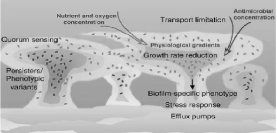
Biofilm-associated resistance to antimicrobials (Figure 1.2) may be the outcome from several mechanisms: (i) bacterial cells grow very slowly due to oxygen and nutrient diminution within the biofilm; (ii) the physiology of the biofilm can be changed by intercellular signals, which can lead to the expression and overproduction of efflux pumps and augment biofilm resistance to a plurality of drugs; (iii) the negative charge
7
of the biofilm matrix neutralizes and damages antimicrobial agents, preventing them to reach the bacterial cells within the biofilm, this mechanism is denominated low outer-membrane permeability; (iv) the antimicrobial concentration diminishes from the periphery to the center of the biofilm, impeding antimicrobial agents to kill bacterial cells localized within the biofilm; (v) genetic diversity contributes to the survival of some cells; (vi) induction of the general stress response and activation of quorum-sensing systems can add to biofilm resistance; and (vii) the emergence of phenotypic and persister variants[15, 26].
Brooun et al.[27] demonstrated that, though most of the cells present in a biofilm of P.
aeruginosa are effectively killed in low antibiotic concentrations, when raising the
concentration of the antibiotic a small fraction of the cell population does not suffer death. Thus, the authors conclude that only a small fraction of the biofilm cells are related to its high resistance to antibiotics. Therefore, that study shows that, unlike planktonic cells where this small fraction of the cell population would be eliminated by the immune system, biofilm cells are protected by the matrix and this subpopulation of resistant cells can be responsible for the regrowth of the biofilm after antibiotic
treatment. These cells are called persisters (Figure 1.3) and are not mutant[28].
In the same way, it is accepted that phenotypic variation of P. aeruginosa populations is intertwined with the resistance to high concentrations of antibiotics. Bacterial cells
Figure 1.2 Numerous mechanisms comprised in P. aeruginosa biofilm-associated resistance. The increase of bacterial density within the biofilm (indicated by darkening colors) regulates gradients of nutrient and oxygen concentration (indicated by narrowing arrow). Reduced antimicrobial penetration is caused by the biofilm matrix (indicated by narrowing arrow). Resistance mechanisms may include the activation of quorum-sensing systems, general stress response and overexpression of efflux pumps. The emergence of biofilm-specific phenotypes, persisters and phenotypic variants is also a well-known resistance factor. (adapted from Drenkard et al[15])
8
undergo phenotypic, physiologic and metabolic alterations soon after the adhesion to a
surface[15]. Drenkard et al.[29] suggested that the presence of phenotypic variants in
biofilms can be partially responsible for great levels of resistance to antimicrobials in P.
aeruginosa biofilms. In the same study, a protein that controls the conversion between
resistant and susceptible bacterial forms was identified. Therefore, the authors conclude that P. aeruginosa is able to experience transitory phenotypic alterations associated with its ability to form biofilms which allow it to increase its resistance to antibiotics both in
vivo and in vitro.
Although these two types of variants described represent a small fraction of the bacterial population that are able to ensure the persistence of P. aeruginosa biofilms throughout several cycles of antimicrobial treatment, persister cells only survive to killing by
antibiotics[30] while phenotypic variants grow normally in presence of high levels of
antibiotics[29]. Consequently, it is believed that different mechanisms are involved in the
susceptibility changes experimented by both these variants[15].
1.3.2 Motility
Motility can be considered one of the most interesting features exhibit by P. aeruginosa. This characteristic allows P. aeruginosa to move across different surfaces, permitting
bacteria to establish pathogenic and symbiotic associations with the host[31]. Advantages
of motility may include the aptitude to access ideal colonization niches within hosts, disperse in the environment, augment the nutrient uptake, and evade toxic substances
released[31]. Bacterial motility can be classified into different types: (i) swimming,
Figure 1.3 Schematic showing the resistance mechanism of persister/phenotypic variants. Antimicrobial treatment eradicates a great portion of biofilm susceptible population. A small fraction of resistant variants survives the antimicrobial treatment and is capable to re-start biofilme development once antimicrobial treatment is finished. (adapted from Drenkard et al[15])
9
which allows bacteria to move across aqueous surfaces and is a flagellum-mediated type of motility; (ii) swarming, refers to bacterial movement on semisolid medium and is, also, flagellum-mediated; and (iii) twitching, which is a surface motility mediated by
type IV pili and allows bacteria to move on solid environments[32-34].
Motility is known to be involved in biofilm development. In a former study, O’ Toole et
al.[9] concluded that factors associated with bacterial surface, such as flagella and type
IV pili, are essential to the P. aeruginosa biofilm development (Figure 1.4).
Prior to attachment P. aeruginosa cells swim across the surface in order to find the best colonization site. Therefore flagellum-mediated motility is important to the initial
attachment to the surface and to initiate the biofilm formation[7]. Swarming is a rapid
movement across the surface, allowing bacteria to migrate and disperse to other sites, also, it is thought to be involved in protecting the pathogens against the host
macrophages and is associated with secretion of toxins[35]. Type IV pili are essential
adhesins that promote initial attachment to a surface. In addition there are evidence that in P. aeruginosa biofilms they are associated with both initial attachment and
development of microcolonies[34]. In first stages of adhesion, microcolonies can also
move across the surfaces throughout swarming motility promoting interactions with the surface and with each other.
1.3.3 Slime Production
One of the virulence factors known to several bacteria species is slime production.
Some P. aeruginosa strains are also slime producers[37]. In other species such as
Figure 1.4 Model for the role of flagella and type IV pili in biofilm formation.
Flagella-mediated motility is significant for the development of bacterial monolayer in the surface. Type IV pili are associated with initial attachment to the surface and microcolony formation. Flagella also seem to be connected to rapid movement of microcolonies which is imperative to protect the bacterial cells and bacteria migration. (adapted from O’ Toole et al[9])
10
staphylococci, slime production promotes its adherence to surfaces, facilitating biofilm
development and survival[38].
As for P. aeruginosa, slime production seems to have a similar role as the ones described for different bacterial species, being essential in biofilm formation and resistance[39].
The term slime can be explained as extracellular material or the biofilm matrix. Most biofilms are only comprised in less than 10% of dry mass while the matrix accounts for
the remaining 90%[40]. The matrix is constituted by a conglomeration of different types
of biopolymers in which biofilm cells are embedded and these are responsible for
biofilm consistency and adhesion to surfaces[40]. Biofilm matrix also comprises a
mixture of DNA, fatty acids, and proteins[41]. Of all biopolymers, exopolysaccharides
have a noted importance since they represent a major fraction of the matrix[40].
P. aeruginosa produces at least three distinct exopolysaccharides that contribute to
biofilm formation and architecture: alginate, Pel, and Psl[40, 42]. Alginate is one of the extendedly studied exopolysaccharides and it is involved in microcolonies formation in
mucoid biofilms and responsible for mechanical stability of mature biofilms[40, 42].
Non-mucoid strains produce Pel and Psl instead of alginate and both are involved in biofilm establishment.
1.3.4 Production of Extracellular Toxins
One of the virulence factors of P. aeruginosa is its ability to produce and release several extracellular toxins. These toxins include pigments, phytotoxic factor, phospholipases
(hemolysins), proteolytic enzymes, enterotoxin, hydrocyanic acid, and exotoxin[36].
These extracellular toxins are often related to bacteria pathogenesis that can lead to severe consequences and even death of the host. Among those toxins, hemolysin and pyocyanin production will be described with more detail, due to their importance in the overall results of this work.
11
1.3.4.1 Hemolysin Production
Hemolysin is one of the earlier extracellular toxin released by P. aeruginosa. Hemolytic activity in many bacteria is considered an important virulence factor and often contributes to bacteria pathogenicity.
P. aeruginosa is known to produce two distinct types of phospholipases C (PLC’s). One
of them is hemolytic (PLcHR) and the other nonhemolytic (PLcN)[43]. Consequently it
is implied that PLcN has no pathogenic significance, while the production of PLcHR is a noted virulence factor.
Hemolytic PLC has severe effects on the host. Several studies showed that clinical isolates from the lungs are able to produce phospholipase C and cystic fibrosis patients
have circulating antibodies against PLC[44]. Also, PLcHR is capable of inducing
leukotriene and thromboxane release from host cells, which can be an explanation for
the inflammatory responses in P. aeruginosa infections[44].
In addition, the virulence associated with PLcHR seems to be connected to the suppression of neutrophil respiratory burst activity, suggesting that PLcHR is a significant toxin which facilitates pseudomonas survival in tissues despite the
abundance of neutrophils[43].
1.3.4.2 Pyocyanin Production
Pyocyanin is a blue-green phenazine-derived pigment produced by several P.
aeruginosa strains[45]. Production of this pigment is seen as one of the most significant
virulence factors exhibited by this bacterium[46].
Pyocyanin exhibits a pro-oxidant propriety. Cellular respiratory inhibition is certainly
the most significant toxic mechanism of pyocyanin against host cells[45]. Its ability to
alter the redox cycle and increase oxidative stress appears to be the fundamental effects
on host cells[45]. The increase of the oxidative stress will generate several consequences
which depend on toxin concentration and time of exposure[46].
Lau et al.[47] reported that the production of pyocyanin by P. aeruginosa is critical in mice lung infections causing tissue damage and necrosis while the progression of the
12
creates a redox potential gradient, called electroline, which increases iron availability, essential to biofilm development.
1.3.5 Phenotypic Switching
Bacteria are constantly confronted by environmental changes and adapting to these changes is vital to their survival. When these environmental alterations arise, a set of complex regulator mechanisms is activated allowing bacteria to survive. Often adaptation mechanisms include behavioral, physiological and genetic variations, as
phenotypic variation[49].
Phenotypic switching or phase variation is considered to be a reversible switch between two phenotypic states. Usually it arises in a small fraction of biofilm population and is
much more common than spontaneous mutations[49, 50]. Phenotypic switching is often
used to increase population diversity and escape the host immunologic response[50].
Therefore, phase variation is translated in different expression of one or more genes resulting in two subpopulations: one subpopulation missing or having diminished level
of phase variable gene(s) and the other expressing the gene completely[51]. A remarkable
feature of phenotypic switching is the interchange between ‘On’ and ‘Off’ phenotypes. Meaning that bacteria can exhibit one of phenotypic states but keep the ability to switch
to the other state[51] whenever different environment stimuli occur[49]. However, the
event that results in this interchange is arbitrary because it is impossible to guess which
bacterial cells will undergo the switch [51]. However several reports evidence that
environmental signals and intercellular regulatory networks are involved on phenotypic
switching mechanisms[49, 51, 52].
It is known that even in a homogeneous environment, isogenic bacterial population can display several phenotypes. This phenomena is called inherent phenotype heterogeneity and does not comprise changes in bacterial genes, it is a result of chemical reactions at
DNA level, such as alterations in the rates of protein synthesis and degradation[49, 53].
One of the most visible characteristic of phenotypic switching is colony morphology
variation[49]. Relationship between colony morphology and bacterial characteristics are
yet to be understood. However colony morphology is very significant in the sense that its macroscopic observation can lead to which bacterial features were probably
13
information about the correlation between colony morphology, virulence, and antimicrobial resistance, which is valuable to plan new therapeutic approaches.
Different kinds of colony morphotypes have been identified, such as small colony variants (sCV). These sCV are believed to be responsible for the increased virulence
and resistance in P. aeruginosa strains[54]. Small colony variants (sCV) have been
involved in persistent and recurrent human infections[49], for example sCV have been
isolated from the respiratory tract of patients infected with cystic fibrosis[54] and medical
devices. For P. aeruginosa a sCV is defined as a colony with a diameter of 3 mm or less.
In bacteria that grow in biofilm, different colony morphotypes have also been identified and phenotypic heterogeneity within biofilms is considered one of the main reasons of
biofilm resistance[49]. However, this is still a recent study field and is yet to be
completely understood. Biofilm-colony diversity is affected by the stage of biofilm development and the proportion of each colony morphotypes is influenced by external factors, showing evidence that these morphotypes are highly connected to mechanisms in biofilm establishment and biofilm survival, increase tolerance to antimicrobials and other stress factors[49, 55, 56].
14
C
HAPTER2.
M
ATERIALS ANDM
ETHODS15
2.1BACTERIAL STRAINS
In the present study a total of seven strains of P. aeruginosa were used. Among these strains, four were reference strains (PAO1, ATCC 10145, CECT 111, and PA14) and three clinical isolates (PAI1, PAI2, and PAI3). All strains were routinely cultured on Trypic Soy Broth (TSB, 30 g/L, Liofilchem) or Trypic Soy Agar (TSA; agar, 15 g/L, Liofilchem; TSB, 30 g/L, Liofilchem) and incubated at 37°C. Bacteria were preserved in criovials at -80 ± 2°C. Before each experiment, bacteria were grown on TSA plates for 24h at 37°C.
2.2BIOFILM FORMATION
The ability of P. aeruginosa to form biofilm was performed as previously described[7,
57]. P. aeruginosa strains were grown overnight on TSB at 37°C under agitation (120
rpm). Cell suspension was diluted in order to obtain the final concentration of 1 × 10
CFU/mL. Lastly, the 96-well microtitre plates were inoculated with 200 µL of the adjusted cellular suspension per well. After inoculation, plates were incubated aerobically at 37°C for 24 h under agitation (120 rpm).
After biofilm formation, the content of the plates was discarded and they were washed twice with sterile water in order to remove weakly attached cells and cell products which were in suspension. To remove biofilm-cells from the biofilm matrix, plates were subjected to an ultrasonic bath for 6 minutes.
2.3QUANTIFICATION OF BIOFILM FORMATION
P. aeruginosa strains biofilm formation was quantified by crystal violet (CV)[57].
Biofilms in 96-well microtitre plates were washed twice with sterile distilled water to remove unattached or weakly attached bacteria. Afterwards 200 µL per well of methanol were added and plates were allowed to stand for 15 minutes in order to fix the biofilm-cells to the walls of the well. Then, methanol was discarded and plates were left to dry for 5 minutes at room temperature. Biofilms were stained with 200 µL of pure CV () and plates were incubated at room temperature for approximately 5 minutes. Then, plates were rinsed thoroughly and repeatedly with tap water. Finally, the amount
16
of biofilm formed was quantified by solubilization of the CV in 200 µL of acetic acid (33% v/v). The optical density (OD) was measured at 570 nm using a microtiter plate reader (Tecan (Sunrise-Basic Tecan), Austria).
2.4QUANTIFICATION OF THE NUMBER OF VIABLE BIOFILM-CELLS
The number of viable cells obtained from biofilms was inferred through colony-forming unit (CFU). Cell suspensions obtained from biofilm removal were serial diluted, plated on TSA, and incubated overnight at 37 °C.
2.5SUSCEPTIBILITY TESTING
Susceptibility of P. aeruginosa biofilm-cells was evaluated determining the minimum inhibitory concentration (MIC) and the minimum bactericidal concentration (MBC). The antibiotics ciprofloxacin and colistin were used and purchased from Fluka and Sigma, respectively. Ciprofloxacin and colistin were employed in the current study because they are both used in the treatment of P. aeruginosa infections.
The MIC values were determined according to standard European Committee on Antimicrobial Susceptibility Testing (EUCAST) through broth microdilution
method[58]. The optical density at 640 nm of bacterial suspension was measured in order
to adjust the cellular concentration to 10 × 10ହ CFU/mL. To maintain viable cell
number concentration plates were inoculated within 30 minutes of standardizing the bacterial suspension. Working antibiotic solutions (diluted antibiotic in broth) were dispensed into plates at 50 µL per well with double of the desired final concentration. To each well containing 50 µL of diluted antibiotic agent in broth a volume of 50 µL of inoculum suspension was added. One of the wells only contained 100 µL of Mueller-Hinton (MHB, 22 g/L, Fluka) broth (antibiotic-free medium) to work as a negative
control. Therefore, the final inoculum concentration was 5 × 10ହ
CFU/mL. Plates were incubated overnight at 37°C under agitation (120 rpm).
For determination of MBC values, 10 µL were removed from the wells of the microdilution trays after incubation and were plated in TSA plates and incubated at 37 °C for 24 h.
17
2.6MOTILITY TESTS
Optical density of the biofilm-cell suspension was measured at 640 nm so that the final
concentration would be 1 × 10଼ CFU/mL. Swimming, swarming and twitching motility
assays were performed in TSA plates with agar concentration of 0.3 %, 0.5% and 1.5% (w/v), respectively. Plates were inoculated with 1 µL of cellular suspension and then incubated at 37 °C for 24 h. After incubation the circular turbid zone formed by the bacterial cells migrating away from the point of inoculation was measured in millimeters.
2.7SLIME PRODUCTION ASSAYS
To determine whether P. aeruginosa cells collected from biofilms produced slime, Brain Heart Infusion (BHI, 37 g/L, Liofilchem) with Congo Red (0.8 g/L, Sigma) plates were used. Afterwards, optical density at 640 nm was measured in order to adjust the
bacterial suspension to the final concentration of 1 × 10଼
CFU/mL. Then, BHI plates were inoculated with 1 µL of inoculum and incubated overnight at 37 °C.
The strong slime producers usually result in really dark colonies (almost black) with a dry crystalline consistency whereas the negative result was showed by almost pink colonies. Sometimes these pink colonies revealed darkening centers and these colonies indicated an intermediate result.
2.8EXTRACELLULAR TOXINS PRODUCTION
2.8.1 Hemolysis Assays
Resembling the previous assay, hemolytic activity of cells derived from P. aeruginosa biofilms was assessed using Columbia Agar (CA; agar, 15 g/L, Liofilmchem; Columbia broth, 43 g/L, Liofilchem) with sheep blood plates. Optical density at 640 nm was then
measured to regulate the biofilm-cell suspension to the final concentration 1 × 10଼
CFU/mL. Lastly, CA with blood plates were inoculated with 1 µL of cellular suspension and incubated overnight at 37 °C.
18
2.8.2 Pyocyanin Quantification
Pyocyanin levels were determined using a previously described[59] quantitative chemical
assay. Bacteria of P. aeruginosa planktonic and biofilm cultures were pelleted at 13000 rpm for 10 minutes. Then, 750 µL of cell-free supernatants containing pyocyanin were collected and mixed with the same amount of chloroform. Samples were mildly vortexed and centrifuged at 13000 rpm for 1 minute. Afterwards the inorganic phase was removed and 750 µL of 0.2M HCl (Panreac) was added to the organic phase. Samples undergone a brief vortexing and were centrifuged at 13000 rpm for 1 minute. After centrifugation the pink layer was collected and its optical density was measured at 520 nm. Ultimately, the pyocyanin concentration was determined multiplying the OD520 values with17.072 and results were expressed in µg/mL.
2.9PHENOTYPIC CHARACTERIZATION OF BIOFILM-CELLS AND PLANKTONIC
CELLS
To assess biofilm population diversity, bacteria derived from biofilms were serial diluted and plated on TSA and grown at 37 °C for 48 h. Colonies were observed by directly placing the petri plates under a magnifying glass (Olympus SZ-CTV) and photographed with a CCD camera (AVC, D5CE; Sony, Tokio, Japan). Each colony morphology identified was classified using ten parameters: form, margin, surface, texture, sheath, opacity, elevation, size, color and diameter (Appendix A). A phenotypic variant was considered when it differed in at least one of the referred morphological parameters.
2.10STATISTICAL ANALYSIS
Statistical analysis was performed using GraphPad Prism, version 5.0. Statistical significance values of the groups’ means of biofilm quantification, number of viable biofilm-cells, swimming, swarming, twitching and biofilm and planktonic pyocyanin were evaluated using one-way ANOVA. Following comparisons were performed using Turkey’s test. Statistical analyses performed were considered significant when p < 0.05.
19
C
HAPTER3.
R
ESULTS ANDD
ISCUSSIONThis Chapter displays the experimental results, observations, and remarks about these results.
20
In this study seven P. aeruginosa strains were tested. Among these four were reference strains (PAO1, CECT 111, ATCC 10145, and PA14) and three were clinical isolates (PAI1, PAI2 and PAI3). Ability to form biofilm, expression of several virulence factors, and antibiotic susceptibility were evaluated for all of the strains.
3.1QUANTIFICATION OF BIOFILM FORMATION
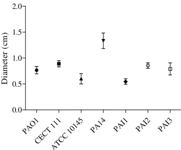
The biofilm formation ability of P. aeruginosa strains is shown in Figure 3.1.
PAO1 CECT 111 ATCC 1014 5 PA14 PAI1 PAI2 PAI3 0 1 2 3 4 B io fi lm b io m a ss ( O D 5 7 0 )
Figure 3.1 Biofilm mass (OD570) of P. aeruginosa strains used in this work. Bars represent the average of
four independent repeats ± SD.
It can be observed that PAO1 was the P. aeruginosa strain that formed the larger amount of biofilm biomass (p <0.05), in contrast with PA14 that formed the lowest quantity of biofilm biomass (p <0.05). Although is noted that PAO1 strain produced a larger biofilm biomass, differences between the quantity of biofilm biomass formed by this strain and CECT 111 and ATCC 10145 strains were not statistically significant (p >0.05). In fact, only the differences of biofilm biomass produced by PAO1 strain and PA14, PAI1, PAI2, and PAI3 strains were found to be statistically significant (p <0.05).
21
Concerning the amount of viable cells recovered from biofilms (Figure 3.2), it was noted that all P. aeruginosa strains formed biofilms with equal number of cells (p >0.05). PAO1 CECT 111 ATCC 1014 5 PA14 PAI1 PAI2 PAI3 1.0×1000 1.0×1001 1.0×1002 1.0×1003 1.0×1004 1.0×1005 1.0×1006 1.0×1007 1.0×1008 1.0×1009 C FU /m L
Figure 3.2 Number of viable biofilm-cells of P. aeruginosa strains used in this work. Bars represent the
average of three independent repeats ± SD.
Taking into account data related to biofilm biomass quantification and number of viable cell, it is hypothesized that the amount of viable cells does not seem to influence the amount of biofilm biomass that each P. aeruginosa strain is able to form.
It is well known that biofilms are the favored mode of bacterial growth in nature and
infectious diseases[60]. Biofilms are three-dimensional structures in which bacteria are
imbedded in a polysaccharide matrix, protein, and DNA[60]. Thus, it is understandable
that the amount of viable cells is just one of the many components that may contribute to the biofilm formation ability of P. aeruginosa. In this study, evidence that the amount of viable cells does not seem to be a relevant factor for the formation of biofilm was shown. Although all P. aeruginosa strains had similar amounts of viable cells, their biofilm formation ability was quite different among some strains. Therefore, the ability to form biofilms by P. aeruginosa must comprise other mechanisms, such as the cells capability to form the matrix components.
It is important to add that the crystal violet assay offers information about the adhesion of P. aeruginosa strains to a non-biological material. Therefore it can provide information about their adhesion onto medical devices, such as catheters, in which this
22
concluded that PAO1 is the P. aeruginosa strain which represents higher risk to cause a nosocomial and chronic infection. This fact emphasizes that the ability to form biofilm is only one of the factors involved in the development of a P. aeruginosa infection however when dealing with these infections this is only one of the virulence factors to be considered among several that can be exhibit by P. aeruginosa.
3.2VIRULENCE CHARACTERIZATION OF BIOFILM-CELLS
Biofilm-cells were subjected to an extensive phenotypic study in order to compare the expression of virulence factors of P. aeruginosa. Virulence factor expression is determinant for the success of P. aeruginosa survival in human environment. The display of these virulence factors allows P. aeruginosa to adapt to several host environments and to colonize different kinds of niches, being extremely important to the pathogenesis of this bacterium. Among the virulence factors exhibited by P. aeruginosa some can be enhanced like low outer-membrane permeability, motility and production of exopolysaccharides and extracellular enzymes. Some of these factors will be thoroughly studied in this work.
3.2.1 Susceptibility testing of biofilm-cells
Currently, the range of traditionally used antibiotics against pseudomonal infections includes ticarcillin, aminoglycosides, ceftazidime, carbapenems (with the exception of
ertapenem), ureidopenicillins, aztreonam, cefepime, ciprofloxacin and levofloxacin[61].
In the present study, two antibiotics were chosen to perform the susceptibility tests: ciprofloxacin, an empirical therapy for P. aeruginosa infections, and colistin, a
last-resort therapeutic option for such infections due to its severe toxicity[61].
Ciprofloxacin belongs to the fluoroquilonone drug class[6]. Ciprofloxacin is known to be
particularly effective against gram-negative bacteria. Its action mechanism includes the
inhibition of DNA replication and possibly transcription [62].
Colistin belongs to the polymyxins class[6]. Polymixins are positive charged and cyclic
peptides antibiotics resultant from several species of Paenibacillus polymyxa. This antibiotic class action mechanism comprises the disruption of the cellular membrane resulting in outflow of intracellular components.
23
MICs and MBCs were determined concerning the two antibiotics of clinical use (Table 3.1). P. aeruginosa strains were classified into two classes according to the EUCAST clinical breakpoints: susceptible (S) if MIC was lower than the clinical breakpoint and resistant (R) if MIC is found to be higher than the clinical breakpoint.
Table 3.1 Minimum inhibitory concentration (MIC) and minimum bactericidal concentration (MBC)
values of ciprofloxacin (CIP) and colistin (COL) against P. aeruginosa strains. S stands for susceptible strain and R stands for resistant strain according to the EUCAST clinical breakpoints. EUCAST clinical breakpoints (mg/L): (i) ciprofloxacin: S ≤ 0.5 and R > 1; (ii) colistin: S ≤ 4 and R > 4.
Strain
MIC (mg/L)
MBC (mg/L)
COL CIP COL CIP
PAO1 2 S 4 R 16 8 CECT 111 2 S 4 R 16 8 ATCC 10145 2 S 4 R 8 8 PA14 2 S 4 R 16 8 PAI1 2 S 4 R >16 16 PAI2 2 S 4 R 8 >16 PAI3 2 S 8 R >16 >16
All P. aeruginosa strains were found to be susceptible to colistin and resistant to ciprofloxacin. MIC value was found to be highest for ciprofloxacin (4 and 8 mg/L) and lowest for colistin (2 mg/L). All strains showed the same level of susceptibility against colistin. However PAI3 strain was found to be the most resistant strain among those used in this study (8 mg/L) against ciprofloxacin. Therefore, and since colistin susceptibility was the same for all strains studied, this strain was considered the most resistant P. aeruginosa strain studied.
Concerning the MBC values, the lowest and highest values registered was equal for both antibiotics 8 mg/L and >16 mg/L, respectively. Against colistin, PAI1 and PAI3 strains were the P. aeruginosa strains that showed highest MBC values (Table 3.1). On the other hand, against ciprofloxacin, PAI2 and PAI3 were the strains that registered the highest MBC values (Table 3.1). Although some differences were noted, it can be said