UNIVERSIDADE DO ALGARVE
IDENTIFICATION AND MOLECULAR
CHARACTERIZATION OF BONE-RELATED MICRORNAS:
FUNCTIONAL IMPLICATIONS
Vânia Cristina Palma Roberto
DOUTORAMENTO EM CIÊNCIAS BIOMÉDICAS
Tese orientada por:
Professora Doutora Leonor Cancela
Doutor Daniel Tiago
Doutor Kaare Gautvik
IDENTIFICATION AND MOLECULAR CHARACTERIZATION OF
BONE-RELATED MICRORNAS: FUNCTIONAL IMPLICATIONS
Declaração de autoria do trabalho
Declaro ser a autora deste trabalho, que é original e inédito. Autores e trabalhos consultados estão devidamente citados no texto e constam da listagem de referências incluída. Esta publicação não poderá ser reproduzida de forma parcial ou integral, nem poderá ser copiada de alguma maneira, incluindo electronicamente, mecanicamente, fotocopiada, gravada ou digitalizada, sem autorização escrita do autor.
_________________________________
Copyright Vânia Cristina Palma Roberto. "A Universidade do Algarve tem o
direito, perpétuo e sem limites geográficos, de arquivar e publicitar este trabalho através de exemplares impressos reproduzidos em papel ou de forma digital, ou por qualquer outro meio conhecido ou que venha a ser inventado, de o divulgar através de repositórios científicos e de admitir a sua cópia e distribuição com objectivos educacionais ou de investigação, não comerciais, desde que seja dado crédito ao autor e editor."
Our genes define who we are. By improving our knowledge about DNA/RNA, we can impact on our lives.
Agradecimentos
À Professora Leonor Cancela agradeço a oportunidade de conhecer o mundo da ciência e de me acolher no seu laboratório. Agradeço a sua orientação e o seu apoio no desenvolvimento deste trabalho.
Ao Daniel, obrigada pelo apoio e orientação ao longo do meu doutoramento. Obrigada pelos comentários e sugestões científicas que muito enriqueceram este manuscrito. E… obrigada por “partires pedra” comigo no mundo dos microRNAs.
À Fundação para a Ciência e Tecnologia pelo financiamento da minha bolsa de doutoramento (SFRH/BD/38607/2007), à Fundação Calouste Gulbenkian, através do programa ‘‘Na Fronteira das Ciências da Vida’’ pelo co-financiamento deste trabalho, ao CCMAR e ao Departamento de Ciências Biomédicas e Medicina pelo acolhimento.
À Natércia e ao Vincent, agradeço a disponibilidade para esclarecerem as minhas dúvidas sempre que vos pedi ajuda.
Ao Gavaia, obrigada por me apresentares os mundos da biologia molecular e da histologia, há uns anos atrás!
À Dr. Ana Teresa Maia e à Maria João, obrigada por me introduzirem no mundo do ChiP-assay.
A todos os membros do EDGE, obrigada por partilharem esta experiência comigo, pelos bons momentos e pela paciência nos dias em que o stress era uma constante. Um especial agradecimento ao corredor maravilha (o corredor mais procurado do laboratório!!!) pela interajuda e pelos momentos de descontracção… Ao corredor verde/azul pelos eppis sempre autoclavados… Obrigada especialmente à Íris, pela ajuda com as clonagens e o estudo da sintenia. À Andreia pela sua, sempre presente, boa disposição. Ao espanhol,
as minhas desculpas por o ter “expulsado” da bancada maravilha, a tua vida teria sido muito diferente! Obrigada ao Mike… por ser o Mike! À Sara por puxar sempre por mim! À Brigite, por todos os artigos que me enviaste e pela paciência com tantos e-mails!
À dona Fernanda, o meu obrigada pelo carinho e por estar sempre disponível para me ajudar em tudo o que precisei.
Aos meus “raios de sol”… Cátia, Rodrigo, Anabela, Joana, João e Inácio… Obrigada pela companhia, pelas longas conversas que tivemos, pelo carinho e pela força que me deram, principalmente neste último ano, mas… também pelos momentos de paródia que me ajudaram a manter sanidade mental! Sem vocês isto teria sido muitoooo mais difícil. João e Anabela, a procrastinação foi importante!
Ao João, obrigada por me apoiares sempre e por me obrigares a falar de ciência de um modo pouco científico… Obrigada pela compreensão nos momentos em que não pude estar presente… Sem ti, este percurso teria sido sem dúvida mais doloroso. Obrigada por tudo…
À minha irmã, por existir… por estar presente sempre que preciso… pela pressão dos últimos dias…
Aos meus pais, obrigada pelo carinho, apoio e incentivo de sempre. Por acreditarem em mim e por toda a força que sempre me transmitiram. Obrigada por me ensinarem que na vida nunca nos devemos comparar aos mais fracos mas sim aos mais fortes… E que isso faz de nós sempre pessoas melhores e maiores.
À minha filhota… a quem devo todo o tempo que não brinquei com ela, todos os momentos em que não pude vê-la crescer, todos os momentos que não pude viver com ela. Obrigada por me fazeres rir só porque sim… Obrigada por seres o meu alento… a minha força… o meu sol. Obrigada apenas por existires e seres quem és… Esta tese é dedicada a ti!
Abstract ………15
Resumo ……… 17
List of abbreviations ……….21
List of Figures and Tables ……….. 23
CHAPTER 1 General Introduction ………. 27
1.1. MICRORNAS ……… 29 1.1.1. Overview ………... 29 1.1.2. History of miRNAs ……….. 30 1.1.3. miRNA Genes ………. 32 1.1.4. Identification of miRNAs ………..34 1.1.5. Biogenesis of miRNAs ……… 35 1.1.5.1. miRNA Transcription ………. 35 1.1.5.2. miRNA Processing ……… 36
1.1.6. miRNA Mechanism of action ………...39
1.1.7. miRNA Functions ……… 42
1.1.8. Investigating miRNA functions ……… 44
1.1.8.1. miRNA Profiling ……… 44
1.1.8.2. miRNA Targets: Identification and Validation ……… 45
1.2. SKELETAL DEVELOPMENT AND MAINTENANCE ………. 49
1.2.1. Overview ………. 49
1.2.2. Control of Chondrogenesis ………. 50
1.2.2.1. Post-transcriptional Control of Chondrogenesis ……… 53
1.2.3. Control of Osteogenesis ………. 54
1.2.3.1. Post-transcriptional Control of Osteogenesis ……… 58
1.2.4. Control of Bone Remodelling ……….. 59
1.2.4.1. Post-transcriptional Control of Bone Remodelling ………. 61
1.3. ZEBRAFISH AS A MODEL ……… 63
1.3.1. Zebrafish and miRNAs ……… 64
1.4. OBJECTIVES ………... 67
CHAPTER 2 EVIDENCE FOR CONSERVATION OF MIR-223 IN ZEBRAFISH (DANIO RERIO): IMPLICATIONS FOR FUNCTION ………... 69
2.1. ABSTRACT………... 71
2.2. INTRODUCTION……….. 72
2.3. MATERIALS AND METHODS……… 74
2.3.1. Biological Material ………... 74
2.3.2. Total RNA extraction ………... 74
2.3.3. Marathon library and molecular cloning of zebrafish miR-223 primary transcript……… 74
2.3.5. In silico analysis ……….. 75
2.3.6. Prediction of miR-223 target transcripts ………..76
2.4. RESULTS ………..77
2.4.1. Identification of zebrafish miR-223 primary transcript and gene structure ……….. 77
2.4.2. Genomic context of zebrafish and human mir-223 genes ……….. 79
2.4.3. Precursor of miR-223 contains highly conserved domains in vertebrates ……….. 81
2.4.4. Zebrafish miR-223 expression analysis ……….. 85
2.4.5. Identification of zebrafish putative miR-223 targets ……… 88
2.5. DISCUSSION ………... 92
2.5.1. miR-223 is conserved throughout evolution: insights from gene and precursor analysis……… 92
2.5.2. MiR-223 expression is correlated with hematopoiesis in zebrafish ………... 94
2.5.3. Evidences for conservation of miR-223 targets and regulatory functions in mammals and zebrafish………. 95
2.6. CONCLUSIONS ………...97
2.7. SUPPLEMENTARY MATERIAL ……… 98
CHAPTER 3 miR-29a is an enhancer of mineral deposition in bone-derived systems ………... 101
3.1. ABSTRACT ………...103
3.2. INTRODUCTION ………. 104
3.3. MATERIALS AND METHODS ………...106
3.3.1. Cell culture maintenance ……….106
3.3.2. Extracellular matrix (ECM) mineralization ……… 106
3.3.3. RNA extraction and quantitative real-time PCR (qPCR) analysis ……… 107
3.3.4. Establishment of stable fish cell clones overexpressing miR-29a ……….. 107
3.3.5. Transient overexpression of miR-29a during ECM mineralization ……….. 108
3.3.6. Vector constructions ……… 108
3.3.7. Dual-Luciferase Reporter Assays ………... 109
3.3.8. Western Blot analysis ………. 109
3.3.9. Statistical analysis ………...110
3.4. RESULTS 110 3.4.1. miR-29 family conservation in vertebrates ………. 110
3.4.2. Zebrafish and mouse have similar miR-29 expression patterns ……… 113
3.4.3. miR-29a is up-regulated during ECM mineralization of ABSa15 cells ……… 114
3.4.4. miR-29a promotes ECM mineralization in ABSa15 cells ……… 116
3.4.5. Seabream sparc is putatively regulated by miR-29a in ABSa15 cells ……… 119
3.4.6. -catenin protein levels are up-regulated in ABSa15 cells overexpressing miR-29 ………... 121
3.5. DISCUSSION ………...123
3.5.1. miR-29 is a generally conserved family of miRNAs ……… 123
3.5.2. miR-29a promotes ECM mineralization ……….. 124
3.5.3. As in mammals, miR-29a is also capable to regulate SPARC and Wnt signalling in fish bone-derived cells 126 3.6. CONCLUSIONS ………...129
3.7. SUPPLEMENTARY MATERIAL ……… 130
CHAPTER 4 miR-214: A new player in chondrogenesis? ……….. 133
4.1. ABSTRACT ………... 135
4.2. INTRODUCTION ………. 136
4.3. MATERIALS AND METHODS ………...138
4.3.1. Analysis of miRNAs expression ……….. 138
4.3.2. In Situ Hybridization (ISH) ……….. 139
4.3.3. Identification and analysis of miR-214 promoter sequences ……….. 140
4.3.4. Cloning of zebrafish miR-214 5´ end ……….. 140
4.3.5. Plasmid Constructs ………. 141
4.3.6. Cell culture ……….. 142
4.3.7. Dual-Luciferase Reporter Assays ………... 142
4.3.8. miR-214 overexpression during ATDC5 cell differentiation ……… 143
4.4. RESULTS ………..143
4.4.1. miR-214 is expressed in the skeleton of zebrafish ………... 143
4.4.2. Comparative sequence analysis of the Dnm3os putative promoter region ………. 147
4.4.3. Cloning and identification of a functional Dnm3os promoter ……….. 150
4.4.4. Transcriptional regulation of miR-214 in skeletal-related cell lines ………. 154
4.4.5. miR-214 is down-regulated during ATDC5 chondrogenic differentiation ……… 156
4.4.6. miR-214 mitigates chondrogenic differentiation of ATDC5 cells ………. 157
4.5. DISCUSSION ………... 160
4.5.1. miR-214 is implicated in skeleton formation of mammals and zebrafish ……… 160
4.5.2. Cartilage and bone related TFs seem to coordinate miR-214 transcription ……… 163
4.5.3. miR-214, the most recent putative regulator of chondrogenesis ………... 166
4.6. CONCLUSIONS ………...170
4.7. SUPPLEMENTARY MATERIAL ……… 172
CHAPTER 5 General Conclusions and Future Perspectives ………. 175
5.1. OVERVIEW ………... 177
5.2. MIR-223 IS ASSOCIATED TO MAMMALIAN HEMATOPOIESIS AND OSTEOCLASTOGENESIS AND HAS CONSERVED FUNCTIONS IN ZEBRAFISH ……… 177
5.3. MIR-29A INDUCES ECM MINERALIZATION IN BONE-DERIVED SYSTEMS THROUGH CONSERVED MECHANISMS IN VERTEBRATES ………. 180
5.4. MIR-214 AFFECTS GENES WITH CRUCIAL FUNCTIONS IN CHONDROCYTE DIFFERENTIATION AND MINERALIZATION ………. 181
5.5. CONCLUDING REMARKS ……… 182
CHAPTER 6 References ………... 185
CHAPTER 7 Annex I - Mir-20a regulates in vitro mineralization and BMP signalling pathway by targeting BMP-2 transcript in fish ………..217
Abstract
MicroRNAs (miRNAs) are a conserved class of small RNAs providing a post-transcriptional mechanism for fine-tuning of intricate physiological and pathological cellular processes, such as those affecting development. Skeletogenesis however, was so far poorly investigated and mainly focused on mammalian models, with a general lack of knowledge concerning other vertebrates. We aimed at the identification of bone-related miRNAs and their characterization from an evolutionary perspective, using fish (mostly zebrafish) as model, in comparison to mammalian systems.
First, we focused on miR-223, a miRNA that was associated with bone remodelling. We demonstrated that miR-223 genomic organization/context and primary/secondary structures are largely maintained between human and zebrafish. As in mammals, miR-223 expression in zebrafish was highly correlated with hematopoietic events and osteoclastogenesis. Finally, miR-223 targets identified in mammals were also predicted in zebrafish, supporting a functional conservation of this miRNA.
In a second set of experiments, we studied the biological role of miR-29a, a bone-related miRNA that was fairly investigated in mammals, but with no mineralogenic effects yet demonstrated. We took advantage of our fish bone-derived systems to explore miR-29a mineralogenic effects through gain-of-function experiments. We demonstrated a strong stimulation of this process through a mechanism probably involving the canonical Wnt signalling. Once more, through bioinformatics analysis, patterns of expression and target prediction/validation, we provided evidences for miR-29 conservation throughout evolution.
Finally, we explored miR-214 putative roles on skeleton formation in vertebrates. Although our initial hypothesis of miR-214 involvement in osteogenesis was recently demonstrated by Wang et al. (2013), we proceeded with our investigation and finally showed that miR-214 is also associated with chondrogenesis. Overexpression of miR-214 in ATDC5 cells mitigated differentiation and down-regulated Mgp and Osteocalcin, probably by targeting Atf4.
This work provides novel evidence that some miRNAs have conserved functions across vertebrates and, probably, conserved regulatory mechanisms of action.
Resumo
Nos últimos anos, assistiu-se a uma marcante expansão na área da biologia molecular, devendo-se isto principalmente à descoberta de pequenas moléculas de RNA não codante e ao seu modo peculiar de intervir na regulação genética. Dentro deste grupo de moléculas, os microRNAs (miRNAs) são, definitivamente, a classe melhor compreendida, o que se comprova pelo crescimento exponencial do número de trabalhos publicados desde a sua descoberta. Os miRNAs, na sua forma matura, são RNAs com aproximadamente 22 nucleótidos (nt), altamente conservados em vertebrados e que asseguram um controlo apertado de vários processos celulares através de uma regulação pós-transcricional. Esta regulação ocorre através da ligação específica do miRNA à 3’UTR do RNA mensageiro (mRNA). Neste mecanismo, destaca-se o envolvimento do complexo RISC (RNA-induced silencing complex; associado ao miRNA), a complementaridade da denominada região “seed” (extremidade 5’ do miRNA) ao mRNA, e o consequente bloqueio da tradução ou degradação do mRNA. Desta forma, cada miRNA pode regular centenas de genes transcritos, e de facto, hoje em dia pensa-se que a maioria dos genes humanos são controlados por miRNAs. Assim, os miRNAs são considerados não só importantes reguladores de múltiplos processos biológicos, incluindo desenvolvimento, diferenciação e apoptose celular, mas também responsáveis por vários processos patológicos, como o cancro, onde se observou que inúmeros miRNAs têm a sua expressão desregulada. Assim, a caracterização dos miRNAs (a vários níveis) é fundamental para a compreensão das suas funções, permitindo alargar também o conhecimento dos processos biológicos e patológicos onde estão envolvidos.
Apesar do conhecimento sobre miRNAs ter aumentado francamente nos últimos anos, o papel dos miRNAs na formação e homeostasia do osso ainda está pouco caracterizado, e a maioria dos estudos tem abordado principalmente esta forma de regulação em mamíferos, havendo assim uma lacuna de conhecimento na regulação destes processos noutros vertebrados. Neste sentido, este trabalho focou-se na identificação de miRNAs potencialmente envolvidos na regulação do osso e na sua caracterização numa
perspectiva evolutiva, usando o peixe (essencialmente o peixe-zebra) como modelo, e em comparação com mamíferos.
Numa primeira abordagem, focámos a nossa investigação no estudo do miR-223, um miRNA anteriormente associado à diferenciação celular da linhagem hematopoiética e ao processo de remodelação óssea. Neste estudo, demonstramos que a organização e contexto genómicos do miR-223 estão preservados em vertebrados, verificando-se uma conservação das estruturas primária e secundária do pre-miR-223 em 46 espécies. Este estudo mostra ainda que a expressão deste miRNA se correlaciona com determinadas fases do desenvolvimento do peixe-zebra onde a hematopoiese e a osteoclastogénese são eventos predominantes. Além disso, este estudo mostra que o miR-223 apresenta uma expressão elevada no principal órgão hematopoético de peixes e ratinhos adultos (rim anterior e medula óssea, respectivamente), sugerindo que a função hematopoiética também se encontra conservada. Por último, através de análise bioinformática demonstrámos que a regulação de genes alvo do miR-223 em mamíferos também deverá estar mantida em peixe-zebra.
Na secção seguinte estudámos o papel biológico do miR-29a, cujo efeito osteogénico em mamíferos se encontra bem caracterizado, mas sem nenhum fenótipo mineralogénico ainda associado. Neste estudo utilizámos uma linha celular derivada do osso de peixe previamente desenvolvida no nosso laboratório e com capacidade de mineralização in vitro. A fim de explorar os efeitos mineralogénicos do miR-29a foram realizadas experiências de ganho de função. O aumento dos níveis endógenos deste miRNA resultaram num incremento da mineralização da matriz extra-celular, o que provavelmente terá sido devido a uma aceleração da diferenciação celular pelo potenciamento da via de sinalização Wnt, tal como evidenciado pela acumulação de um dos seus principais componentes, a -catenina. Além disso, foi demonstrada a conservação da função deste miRNA através de estudos baseados em homologia de sequências, análise de sintenia, padrão de expressão tecidular e na manutenção da regulação do SPARC, um alvo previamente descrito em mamíferos. Reforçou-se assim a ideia de que o miR-29a é um regulador crucial na diferenciação de osteoblastos, induzindo um aumento da mineralização em sistemas in vitro.
Finalmente, explorámos a hipótese do miR-214 ser regulador da formação do esqueleto/osso, em vertebrados. Apesar da nossa primeira hipótese, que consistia no envolvimento do miR-214 na osteogénese, ter sido entretanto demonstrada através do trabalho realizado por Wang et al. (2013), continuámos com este estudo, tentando demonstrar um potencial envolvimento deste miRNA na condrogénese, um processo essencial na formação do esqueleto de vertebrados. Através do padrão de expressão espacial e temporal do miR-214 durante o desenvolvimento do peixe-zebra, verificou-se uma clara associação com estruturas cartilagíneas. Adicionalmente, demonstrámos que a região reguladora (promotor) do transcrito primário deste miRNA se encontra conservada em oito vertebrados, assim como os locais de ligação de factores de transcrição (associados à condrogénese e/ou osteogénese) identificados. De acordo com a análise funcional deste promotor, concluiu-se que esta região reguladora (quer de peixe-zebra quer de humano) é activada e regulada de forma semelhante em condrócitos e osteoblastos. Por último, verificou-se que a sobreexpressão do miR-214 nas células ATDC5, um modelo in vitro para a condrogénese, atenua a diferenciação condrocítica, possivelmente através da regulação do gene Atf4. O decréscimo simultâneo de dois marcadores ósseos, a Mgp e a osteocalcina, aquando da sobreexpressão deste miRNA sugere que a mineralização dos condrócitos poderá estar comprometida nesta condição. Assim, propomos que o miR-214 desempenha um papel fundamental na formação do esqueleto de vertebrados, não apenas pela regulação da osteogénese, mas também pelo controlo da condrogénese, promovendo assim a normal e equilibrada formação de estruturas ósseas e cartilagíneas.
No seu conjunto, estes estudos evidenciam uma conservação na função e mecanismos de regulação de muitos dos miRNAs identificados em vertebrados. Este conhecimento é bastante importante, por exemplo para a investigação de tratamento de patologias, uma vez que permite a utilização de modelos alternativos no rastreio de potenciais alvos terapêuticos, com particular destaque para as vias reguladas por miRNAs. Nesta perspectiva, em doenças como por exemplo a osteoporose, onde se verifica uma perda de massa óssea, terapias que estimulem a acção de miRNAs que promovam a osteoblastogénese ou que inibam a osteoclástogénese, são atractivas e com
grande potencial na estimulação da formação óssea ou na redução da reabsorção óssea excessiva, respectivamente.
Palavras-chave: miRNAs; conservação; peixe-zebra; regulação; expressão;
List of abbreviations
Ago2 - Argonaute 2
AP2alpha - transcription factor AP-2 alpha Atf4 - activating transcription factor 4 BMP - bone morphogenetic protein BS - binding site
Col10a1 - collagen type X alpha 1 Col2a1 - collagen type II alpha 1
DGCR8 - DiGeorge syndrome critical region in gene 8 dpf - days post fertilization
dsRNA - double-stranded RNA ECM – Extracellular matrix GFP - green fluorescent protein hpf - hours post fertilization ISH - in situ hybridization kb – kilobase pairs
Mgp - Matrix Gla Protein miRNA – microRNA mRNA - messenger RNA
MSCs - mesenchymal stem cells ncRNA - non-coding RNA
nt - nucleotide Oc – Osteocalcin Osx or Sp7 - Osterix Pol II - RNA Polymerase II pre-miRNA - miRNA precursor
pri-miRNA – primary microRNA transcripts qPCR - real time-PCR
RACE - rapid amplification of cDNA ends RISC – RNA induced silencing complex RNAi - RNA interference
SP1 - specific protein 1 transcription factor ssRNA - single-stranded antisense RNA TFBS - transcription factor binding site TFs - transcription factors
TNAP - alkaline phosphatase U6 – U6 small nuclear RNA UTR - untranslated region WT - wild type
List of Figures and Tables
Figure 1.1. Graphical representation of the number of publications in the
PubMed database per year
Figure 1.2. Genomic organization of miRNA genes
Figure 1.3. Representation of a canonical intergenic pri-miRNA Figure 1.4. Biogenesis of miRNAs
Figure 1.5. Representation of pri-miRNAs structure and Drosha cleavage
Figure 1.6. Putative mechanisms for miRNA-mediated post-transcriptional
regulation
Figure 1.7. Representation of miRNA:mRNA interaction in animals
Figure 1.8. Endochondral bone formation and bone remodeling in mammalian
systems
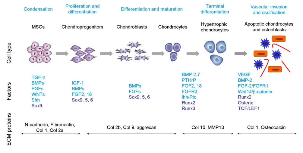
Figure 1.9. Sequence of multi-step events during chondrogenesis Figure 1.10. Sequence of multi-step events during osteogenesis
Figure 1.11. Sequence of multi-step events during osteoclast differentiation
Figure. 2.1. Structural organization of mir-223 gene
Figure. 2.2. Schematic representation of the genomic context of human and
zebrafish mir-223 genes using data from the Ensembl project
Figure 2.3. Conserved features of miR-223 hairpin among vertebrate species Figure 2.4. Pri-miR-223 secondary structure conservation among vertebrates Figure 2.6. Analysis of mature miR-223 expression levels in zebrafish
Figure. 2.5. Analysis of mature miR-223 expression levels during development
of zebrafish
Table 2.1. Mammalian target genes of miR-223 analysed in this study
Table 2.2. Resume of in silico analysis of zebrafish miR-223 putative target
genes
Supplementary Table 2.1. List of primers used in this study
Supplementary Figure 2.1. Pairwise percent identities of pre-miR-223
Figure 3.1. Gene synteny analysis in vertebrates relative to the host genes
Figure 3.2. Relative miR-29a expression in (a) zebrafish and (b) mouse adult
tissues
Figure 3.3. Relative miR-29a expression during ABSa15 cell differentiation Figure 3.4. Effect of miR-29a stable overexpression in ABSa15 cells
undergoing ECM mineralization
Figure 3.5. Effect of transient miR-29a overexpression in Absa15 cells
undergoing ECM mineralization
Figure 3.6. Prediction of miR-29a binding sites (BS) in the 3’UTR of gilthead
seabream SPARC and respective functional characterization in ABSa15 cells
Figure 3.7. Levels of -catenin protein production in wild type ABSa15 cells (WT) and stable clone overexpressing miR-29a (OE-miR-29)
Figure 3.8. Proposed mechanism for miR-29a action on osteogenic
differentiation by stimulation of canonical Wnt signalling
Supplementary Table 3.1. List of primers and oligoduplexes used in this study Supplementary Figure 3.1. Sequence alignment of mature miRNAs from
miR-29 family
Supplementary Figure 3.2. Relative miR-29c expression in mouse adult
tissues
Figure 4.1. Relative expression of mature miR-214 during developmental
stages of zebrafish
Figure 4.2. Detection of mature miR-214 in zebrafish larvae by miRNA specific
in situ hybridization
Figure 4.3. Relative expression of mature miR-214 in zebrafish (a) and mouse
(b) adult tissues
Figure 4.4. Schematic representation of Dnm3os promoter region and
identification and localization of conserved TFBS
Figure 4.5. Transcriptional regulation of zebrafish Dnm3os putative promoter Figure 4.6. Transcriptional regulation of human Dnm3os putative promoter Figure 4.7. Relative expression of miR-214 during ATDC5 cell differentiation Figure 4.8. Relative gene expression of different genes associated with
Figure 4.9. Effect of miR-214 overexpression in ATDC5 chondrocyte
differentiation
Figure 4.10. Proposed regulatory mechanism for miR-214 effect in
chondrogenic differentiation
Supplementary Table 4.1. List of primers and oligoduplexes used in this study Supplementary Figure 4.1. Relative expression of mature miR-199 during
developmental stages of zebrafish
Supplementary Figure 4.2. Effect miR-214 overexpression on Hedgehog
CHAPTER 1
General Introduction
CHAPTER 1 General Introduction
1.1. MicroRNAs
1.1.1. Overview
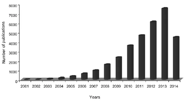
In the last years, only few research areas of biology have witnessed such a remarkable expansion as that observed for RNA molecular biology. Although this breakthrough has occurred through many fronts, one of the areas where major progresses were obtained concerned the discovery of the mode of action and impact of small non-coding RNAs (ncRNAs) on the regulation of genes and genomes. Since the first discovery of double-stranded RNA (dsRNA) ability to regulate gene expression by antisense base-pairing to target messenger RNA (mRNA), a process named RNA interference (RNAi) (Fire et al., 1998), an enormous increase in the number of identified small ncRNAs was observed, and these have been found in animals, plants, fungi and viruses. Despite the existence of various classes of small ncRNAs, these molecules are generally divided in three main categories based on their biogenesis, structure, associated effector proteins and biological functions (Bartel, 2009; Moazed, 2009): (i) short interfering RNAs (siRNAs), which are processed by Dicer from long dsRNAs into duplexes of 21-25 nucleotides (nt) in length and act through the RNAi pathway to regulate gene expression (Ambros et al., 2003; Reinhart and Bartel, 2002); (ii) piwi-interacting RNAs (piRNAs) which are longer RNAs of about 25-30 nt that interact with Piwi proteins (Aravin et al., 2007); and (iii) microRNAs (miRNAs) (Lagos-Quintana et al., 2001; Lau et al., 2001; Lee and Ambros, 2001; Lee et al., 1993), which will be described in detail in the next sections. MiRNAs are the best understood of these three classes, and the emergent perception of their importance led to a boost on publications in the past years with over 32500 scientific reports currently recorded and from them more than 30000 were published during the course of the work here presented (last checked on the 23 of June 2014 at PubMed database,
Figure 1.1. Graphical representation of the number of publications in the PubMed database per year. The keywords microRNA, microRNAs and miRNA were searched in the title
and/or abstract of articles available. Note that for 2014, the number of publications refers to publications until the 23 of June 2014.
A perspective on animal miRNA discovery, genomics, biogenesis, mechanisms and functions will be described in the following sections.
1.1.2. History of miRNAs
The central dogma of molecular biology which considered that RNA molecules acted as simple messengers between DNA, encoding cellular instructions, and proteins, the end-products which executed those instructions (Crick, 1970), started to be questioned when researchers realized that ncRNA molecules could interfere with gene expression. The biological phenomenon of antisense control mechanisms was first recognized in the late 70s and early 80s when scientists found that exogenous oligonucleotides with a complementary sequence to ribosomal RNA could prevent ribosome function in Echerichia coli (Eckhardt and Lührmann, 1979; Jayaraman et al., 1981). More then ten years later, Ambros and colleagues discovered that lin-4, a gene known to be essential for developmental timing of the nematode worm Caenorhabditis
elegans larvae, did not encode a protein, but rather produced a pair of small
RNAs, one containing 22 nt and another containing ~61 nt sequence with a predicted stem loop structure and proposed to be the precursor of the shorter (~22 nt) molecule (Lee et al., 1993). Both RNAs were found to have antisense
complementarity with multiple sites in the 3’-untranslated region (UTR) of lin-14 transcript (Lee et al., 1993; Wightman et al., 1993). Based on this information,
lin-4 putative binding and regulation of lin-14 was proposed (Wightman et al.,
1991). However, it was only in 1993 that this mechanism was demonstrated, when lin-14 transcript levels were shown to be constant throughout development of C. elegans, whereas LIN-14 protein levels were not, indicating a post-transcriptional regulation (Wightman et al., 1993). The authors demonstrated that: (i) the post-transcriptional regulation of lin-14 by lin-4 generated a temporal gradient of Lin-14 protein during C. elegans development; (ii) lin-14 3’UTR was essential and sufficient for lin-4-mediated temporal regulation; and that (iii) multiple conserved elements in the lin-14 3’UTR were complementary to, at least, a core of 7 nt in the 5´-end of lin-4, mediating part of the temporal gradient activity of the lin-14 3´-UTR (Wightman et al., 1993). This regulatory process was later proven to be essential for worms to proceed from their first larval stage to the second, as reviewed by Rougvie (2005). It took seven years to discover that this mechanism was not an isolated event, until a second small regulatory RNA, let-7, was identified (Reinhart et al., 2000). Similarly to lin-4, let-7 was shown to operate by specific binding to the 3’UTR and repression of lin-41 and hbl-1 mRNAs (Lin et al., 2003; Reinhart et al., 2000; Slack et al., 2000; Vella et al., 2004). By then, let-7 was found to be highly conserved throughout metazoan (Pasquinelli et al., 2000), contradicting the general idea that lin-4 and let-7 were a worm-specific peculiarity. Meanwhile, dsRNA mediated gene down-regulation in C. elegans was reported to be far more potent than single-stranded antisense RNA (ssRNA) (Fire et al., 1998), which brought new insights into the putative mechanisms of RNA interference (RNAi), as it will be described next in detail. These findings propelled intense genome-wide searches to identify additional endogenous small regulatory RNAs, which ended to be demonstrated in 2001 (Lagos-Quintana et al., 2001; Lau et al., 2001; Lee and Ambros, 2001). This finally led to the recognition that microRNAs (miRNAs) represent a distinct, conserved and abundant class of regulatory genes. The importance of non-coding RNA was further supported when the draft of the human genome project was concluded, and revealed that the extent of protein-coding genes covers only about 2% of the human genome (Lander et al., 2001). Remarkably, while the number and
size of protein-coding genes was shown to not vary substantially with increasing developmental complexity, this was not the case for non–protein coding sequences in genomes, indicating that these sequences may enclose increasingly intricate regulatory information (Taft et al., 2007).
Nowadays, RNA molecules are known to function not only as messengers of protein production, but also as key features of the gene regulatory networks with 30424 mature miRNAs identified so far in animals, plants, algae, amoeba, diatom and viruses, and deposited in the miRNA database (miRBase Release 20, http://www.mirbase.org/). MiRNAs are now known to be involved in a variety of biological processes, including cell proliferation and differentiation (Yang et al., 2011d), apoptosis (Li et al., 2011a), organogenesis (Giraldez et al., 2005; Papaioannou et al., 2013), and also in pathological processes, such as cancer (Plaisier et al., 2012; Wang et al., 2013b) and infection / inflammation / immunity (Baltimore et al., 2008; Haneklaus et al., 2013). Outstandingly, bioinformatics predictions suggest that miRNAs control up to 60% of all human protein-coding genes (Friedman et al., 2009), further confirming an essential role in eukaryotic gene regulation.
1.1.3. miRNA Genes
Our knowledge about miRNA biology has been significantly increased following the discovery of let-7. Most of the studies, however, have been focusing on processing and targeting by miRNAs. In this regard, despite being important regulatory steps in miRNA biogenesis, miRNA genomics and transcription regulation are still poorly understood. MiRNAs are endogenous small non-coding RNAs (~22 nt) generated from conserved hairpin structures that are transcribed from diverse regions of the genome as long primary transcripts (pri-miRNAs), scattered in all chromosomes from humans to zebrafish (Bartel, 2004; Kim et al., 2005; Pillai, 2005; Thatcher et al., 2008). The first miRNAs identified, lin-4 and let-7, were shown to be located in non-coding regions in-between genes and transcribed from unidentified promoters, leading to the initial thought that most miRNA genes were located in intergenic regions (Lagos-Quintana et al., 2001; Lau et al., 2001) and thus named intergenic miRNAs (Fig. 1.2a). However, the identification of several intronic miRNAs in C.
elegans, mouse an human genomes (Ambros et al., 2003; Rodriguez et al.,
2004) and the demonstration of gene silencing mechanisms associated to miRNAs derived from introns (Ying and Lin, 2004), evidenced a new miRNA category: intronic miRNAs (Fig. 1.2 b). In fact, new technology and refined mapping demonstrated that the vast majority of mammalian miRNAs reside within the intronic regions of either protein-coding genes or non-coding transcripts (Griffiths-Jones et al., 2008; Rodriguez et al., 2004). Although this estimation varies between species, the location of several intronic miRNAs is quite conserved among different organisms (Kim and Nam, 2006). Intronic miRNAs are generally sense orientated with their host gene and expression of both miRNA and host gene largely coincides, suggesting a co-regulation and generation from a common precursor transcript (Baskerville and Bartel, 2005).
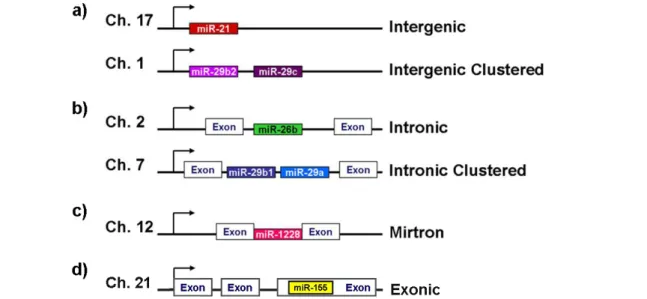
Figure 1.2. Genomic organization of miRNA genes. miRNA genes can reside (a) in-between
genes, named intergenic miRNAs (alone or clustered); (b) in the intron of ncRNA or protein-coding genes (alone or clustered), called intronic miRNAs; (c) in a short intron, the mirtrons; or (d) in the exon of ncRNAs, which are called exonic miRNAs. Adapted from Kapinas and Delany
(2011).
A few miRNA precursors, called ‘mirtrons’, comprise the full intron size and, when spliced, are able to bypass the first cleavage step in miRNA processing (Berezikov et al., 2007) (Fig. 1.2 c). Also, a small subset of miRNAs, approximately 10%, is located within exons of non-coding genes (Kim et al., 2009; Rodriguez et al., 2004) (Fig. 1.2 d). In addition, 36% to 47% of known miRNAs are found in clusters and might be transcribed as single polycistronic
primary transcripts in vertebrates (Griffiths-Jones et al., 2008; Olena and Patton, 2010; Thatcher et al., 2008) (Fig. 1.2 a, b). Clustered miRNAs are frequently related to each other in sequence, suggesting that miRNA clusters might be a consequence of gene duplication. Exceptionally, some clusters contain representatives of different miRNAs families without apparent sequence homology (Kim and Nam, 2006). A possible explanation for this relies on the ability for clustered miRNAs to target the same gene or different genes in the same pathway (Yuan et al., 2009b).
1.1.4. Identification of miRNAs
Nowadays, both biological and bioinformatics approaches for miRNA identification have yielded many thousands of miRNA sequences and novel miRNAs still appear almost on a daily basis. Identified miRNAs are deposited in miRBase (www.mirbase.org/) (Kozomara and Griffiths-Jones, 2011), a widely known public database for published miRNA sequences and respective annotation (miRBase database), and also for new miRNA genes prior to their publication (miRBase Registry). Each miRBase entry matches a predicted hairpin fraction of a miRNA transcript and contains information on the location and sequence of the mature miRNA, its genomic location, target prediction, conservation and experimental validation (Griffiths-Jones et al., 2008; Kozomara and Griffiths-Jones, 2011). The current version of miRBase (Release 20, updated in June 2013) contains 24521 entries, which express 30424 mature miRNAs in 206 different species.
The first miRNAs were identified by forward genetic screens (Lee et al., 1993; Reinhart et al., 2000). Directional cloning can be applied in every organism, which can be an advantage when the available genomic information is scarce or non-existent. Combined cloning and bioinformatics approaches proved to be particularly valuable in the first years of miRNA research. Nevertheless, with the recent advances in next-generation sequencing, deep sequencing has been applied to both miRNA discovery and quantification in several organisms (Bizuayehu et al., 2013; Castellano and Stebbing, 2013; Wei et al., 2012). In fact, this technology boosted the number of miRNAs identified in different species, further increasing the challenge of functional annotation and
driving a considerable advance in bioinformatics approaches for miRNA target prediction and systems-based analysis of miRNA function.
1.1.5. Biogenesis of miRNAs
1.1.5.1. miRNA Transcription
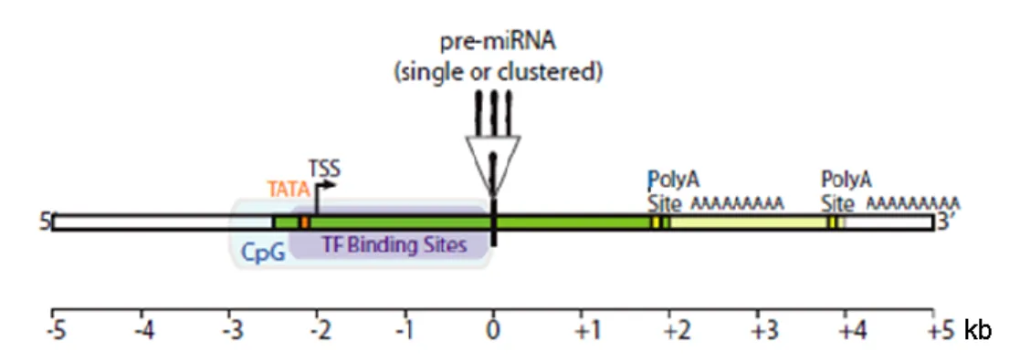
Characterization of miRNAs genomic organization, transcription and regulation is still ongoing. MiRNAs transcription is known to be mostly mediated by RNA polymerase II (Pol II), with primary transcripts (pri-miRNAs) bearing Pol II signatures such as a 7-methyl guanylate cap at the 5' end and poly (A) tail at the 3' end (Cai et al., 2004; Davis and Hata, 2009; Ozsolak et al., 2008). RNA Pol III has also been found to be involved in the transcription of some miRNAs, but this mechanism has been rarely observed (Borchert et al., 2006; Ozsolak et al., 2008). Supporting this, a large scale analysis of intergenic miRNAs structures and mapping of human miRNA promoters (involving chromatin immunoprecipitation) indicated that miRNA promoters display all features commonly associated with Pol II-mediated transcription, such as CpG islands, TATA boxes, transcription factor IIB recognition sites, initiator elements and histone modifications (Corcoran et al., 2009; Ozsolak et al., 2008; Saini et al., 2007). Besides having dedicated promoters, miRNAs were demonstrated to be first transcribed as long pri-miRNAs, with 3-4 kb (kilobase pairs) in length (Gu et al., 2006; Ozsolak et al., 2008) and containing transcript start sites (TSSs) and poly(A) signals located within approximately 2 kb upstream and downstream of the miRNA precursor (pre-miRNA), respectively (Fig. 1.3) (Saini et al., 2007). Ozsolak and colleagues also demonstrated that about one third of intronic miRNAs have their own promoter regions, enabling a different regulation from the host gene (Ozsolak et al., 2008). Furthermore, coupling between transcription and processing has been verified for both intergenic and intronic miRNAs. An important difference however, relies on the fact that intergenic miRNAs co-transcriptional processing is coupled with termination (Ballarino et al., 2009), while intronic miRNAs processing seems to occur co-transcriptionally in cooperation or preceding splicing of the primary transcript (Janas et al., 2011; Kim and Kim, 2007; Morlando et al., 2008).
Figure 1.3. Representation of a canonical intergenic pri-miRNA. Intergenic pri-miRNAs
(green) have dedicated promoters with TATA boxes, transcriptional start sites (TSS), CpG islands (CpG) and transcription factor (TF) binding sites. Pri-miRNAs transcripts can contain one or several pre-miRNAs and more then one polyadenylation (Poly A) sites. Adapted from
Saini et al. (2007).
Regarding transcriptional regulation, mRNAs and miRNAs also present important similarities. Indeed, the network of transcription factors (TFs) that control protein coding genes is also found to regulate miRNA transcription, orchestrating cell-fate decisions, cell differentiation and tissue and developmental stage-specificity (Schmeier et al., 2009). In addition, as in protein coding genes, TF-binding sites (TFBS) are 8-15 base pairs (bp) long and are generally located nearby TSSs, close to the pre-miRNA. Furthermore, 60% of human miRNAs have clustered TFBS preferentially located within a 1-kb region (Saini et al., 2007). Interestingly, emerging evidence indicates that miRNAs have a common tendency to regulate transcription factors that drive their expression, cooperating in complex regulatory networks through feedback loops to regulate cell decisions (Fazi et al., 2005).
In summary, regulation of pri-miRNA transcription is one of the most important features controlling miRNA abundance and a full understanding of that process requires a comprehensive characterization of the genomic location and extent of pri-miRNAs, including TSSs, promoters and TFBS.
1.1.5.2. miRNA Processing
As mentioned before, transcription of miRNA genes by RNA Pol II originates long, capped and polyadenylated pri-miRNAs which can contain one or more miRNAs (Cai et al., 2004). Once transcribed, pri-miRNAs fold into imperfectly base-paired stem-loop (also known as hairpin) structures that are
further processed. In animals, maturation of miRNAs occurs in two processing steps, each one catalyzed by a ribonuclease III (RNase III) endonuclease in cooperation with a double-stranded RNA-binding domain (DsRBD) protein. This process is summarized in Fig. 1.4 and will be described next.
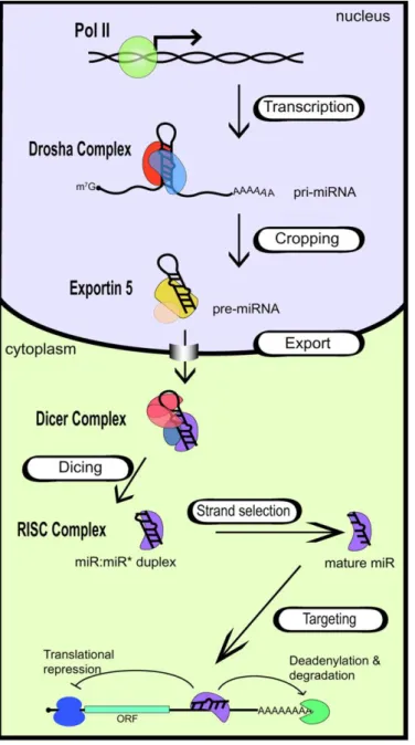
Figure 1.4. Biogenesis of miRNAs.
Transcription of miRNA genes by RNA Poll l originates capped and polyadenylated transcripts that fold into hairpin structures (pri-miRNAs). Cleavage by Drosha in the nucleus generates a smaller miRNA precursor (pre-miRNA) which is then exported into the cytoplasm and further processed by Dicer to generate a miRNA:miRNA* duplex. Once this duplex is assembled into RISC, the miRNA* is discarded, and the mature miRNA guides this complex to the target mRNA. Translation inhibition or mRNA degradation occurs by binding of the miRNA to the 3’UTR of target mRNA. In Davis and Hata (2009).
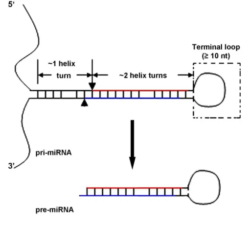
The first step of miRNAs processing is catalysed by the nuclear RNase III Drosha, which cleaves the stem-loop structures in pri-miRNAs to originate a ~70 nt pre-miRNA (Han et al., 2004; Lee et al., 2002, 2003, 2006). The accuracy and efficiency of this process is assured by the cooperation of DiGeorge syndrome critical region in gene 8 (DGCR8) (known as Pasha in
Drosophila and C. elegans), which interacts with Drosha forming a pri-miRNA processing complex named the Microprocessor complex (Faller et al., 2010; Han et al., 2004, 2006). Efficient processing by Drosha requires (i) an hairpin with a large terminal loop (≥ 10 nt); (ii) two helix turns (~22 nt) that encode the miRNA:miRNA* duplex plus (iii) one helix turn (~11 nt) of the lower stem (Han et al., 2006; Zeng et al., 2005b) (see Fig. 1.5). Cleavage by Drosha occurs approximately 11 nt away from the ssRNA-stem loop junction, defining one end of the mature RNA. The resulting pre-miRNA has a 5’ phosphate group and a 2-3 nt 2-3’ overhang characteristic of RNase II cleavage of dsRNA (Han et al., 2004, 2006; Zeng et al., 2005b).
Figure 1.5. Representation of pri-miRNAs structure and Drosha cleavage. Pri-miRNAs fold
into stem-loop structures with a central stem of approximately 33 nt (3 helix turns). The lower stem consists of one helix turn, flanked by ssRNA, and the upper stem (which encodes the miRNA duplex) comprises 2 helix turns, flanked by a terminal loop. Drosha cleavage sites are indicated in the pri-miRNA by verticals arrowhead and arrow. The miRNA duplex is represented by red and blue lines in the upper stem. Adapted from Zeng et al. (2005b).
After Drosha processing, Exportin-5 (XPO5), a nuclear export factor, recognizes the characteristic end structure of pre-miRNAs and exports it to cytoplasm in a Ran-GTP (RAs-related Nuclear protein) dependent manner,
through nuclear pore complexes (Bohnsack et al., 2004; Lund and Dahlberg, 2006; Lund et al., 2004). In the cytoplasm, the pre-miRNA is further processed by another highly conserved RNase III, Dicer, together with its dsRBD partners, which are apparently required for miRNA stability and effector complex formation (Lee et al., 2013; Pilotte et al., 2011). Then, Dicer cleaves the pre-miRNA at the terminal loop, liberating a ~22 nt-long RNA duplex (Bartel, 2004; Macrae et al., 2006; Pillai, 2005). This miRNA duplex contains the mature miRNA and the miRNA* (which is by definition the small RNA in the opposite side of the pre-miRNA stem loop), which are partially paired due to the 5’ and 3’ overhangs resulting from both Drosha and Dicer cleavages. Evidences collected in the last few years indicate that Dicer processing involves the binding of TRBP (trans-activator RNA (tar)-binding protein) to the miRNA duplex and, after cleavage, TRBP recruits Argonaute 2 (Ago2). Ago2 along with Dicer contribute to the assembly of RISC (RNA induced silencing complex) forming the RISC loading complex (RLC) (Chendrimada et al., 2005; Wahid et al., 2010). Once the miRNA duplex is loaded into Ago protein of RISC, the RNA strand with the lowest thermodynamic stability at its 5’-end (called guide strand) remains bounded to this complex while the miRNA* (passenger strand) is degraded (Schwarz et al., 2003). In some cases miRNAs* can be loaded into RISC and originate functional miRNAs. The precise mechanism through which the RNA loading into Ago occurs is still not understood. Concerning Ago2, this is the only one of the four Ago proteins in humans that is known to have endonucleolytic activity, being widely described as the RISC slicer (Song et al., 2004). However, Ago2 is also known to participate in the removal of miRNA passenger strand (Diederichs and Haber, 2007). Interestingly, all four Ago proteins are known to enhance production or stability of mature miRNAs (Diederichs and Haber, 2007). Finally, the mature miRNA guides the RISC to its target transcript, leading to its degradation or translation repression.
1.1.6.
miRNA Mechanism of action
The first identified miRNA, lin-4, was shown to down-regulate the protein levels of LIN-14 (Lee et al., 1993). Five years later, Mello’s group showed that dsRNA was far more effective in inhibiting the expression of specific mRNA
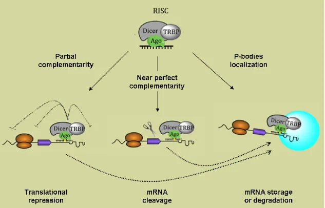
than ssRNA, uncovering the phenomena of RNAi (Fire et al., 1998). Although at that time these authors speculated that the inhibition process was based on a catalytic mechanism (Fire et al., 1998), miRNAs mode of action was later demonstrated to rely mostly on translation inhibition. However, the mechanism by which miRNAs regulate gene expression is still under debate. Indeed, different studies have demonstrated that down-regulation of protein levels can occur by either inhibition of translation initiation or elongation, premature termination of translation or co-translational inhibition (Eulalio et al., 2008). Additionally, miRNAs can induce target mRNA degradation (Schmitter et al., 2006) and also sequester mRNAs into cytoplasmatic foci called P-bodies, for storage or degradation (Castilla-Llorente et al., 2012) (Fig. 1.6). Recent mechanistic models proposed that miRNA-mediated gene silencing might occur by successive steps, combining translation inhibition and mRNA degradation (Béthune et al., 2012; Djuranovic et al., 2011).
Figure 1.6. Putative mechanisms for miRNA-mediated post-transcriptional regulation.
Translational repression occurs when miRNAs bind to their target mRNAs by partial complementarity. Protein production can be blocked by interference with the initiation, elongation or termination steps. A near perfect base pairing between miRNA and target mRNA can originate cleavage of the target mRNAs leading to mRNA decay. Both translation repression and mRNA cleavage can occur in P-bodies, where storage or degradation of mRNAs occurs. Adapted from Pedroza-Torres et al. (2014).
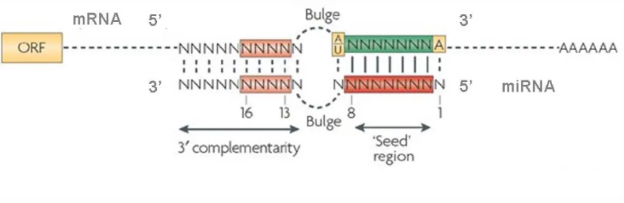
Despite the different mechanisms for miRNAs regulation of gene expression, a common feature was generally shown to be associated to this process, i.e. miRNAs binding to the 3’UTR of target mRNAs through imperfect complementarity. In few exceptional cases however, this regulation involves binding to the 5’UTR or to the coding sequencing of mRNA targets (Duursma et al., 2008; Ørom et al., 2008). The complementarity between miRNAs and mRNA targets is generally confined to the 5’ region (nucleotides 2-8) of the miRNA, which has been named the ‘seed region’, illustrating its contribution to target mRNA binding (Lewis et al., 2003a, 2005; Pillai, 2005) (Fig. 1.7).
Figure 1.7. Representation of miRNA:mRNA interaction in animals. Binding of miRNAs to
target mRNAs requires perfect complementarity between the seed region (nucleotides 2-8, red rectangle) and the 3’UTR of the mRNA (green rectangle). Possible base pairing involving the 3’ end of the miRNA might occur (pink rectangles), contributing for a best stabilization of the miRNA:mRNA duplex. The presence of a central bulge prevents the cleavage of the mRNA by Ago2. In Filipowicz et al. (2008).
This limited complementarity between miRNA and its target mRNA was proven to be an advantage in gene expression regulation, since the short length of miRNA seed region allows the simultaneous inhibition of hundreds of target mRNAs (Baek et al., 2008; Lewis et al., 2005; Selbach et al., 2008). Therefore, the biological effects of miRNAs can reflect synergistic effects through simultaneous regulation of different targets. The rules governing miRNA:mRNA Watson–Crick base pairing are quite complex, and though perfect pairing between seed regions and target 3’UTRs is crucial for gene regulation, 3′-end pairing might contribute to target recognition (Fig. 1.7), particularly when sites have weaker miRNA seed matches (Li et al., 2008). Imperfect miRNA:mRNA interactions with central bulges (nucleotides 9–12) facilitate translational
inhibition or exonucleolytic mRNA decay (Fig. 1.7), whereas the outcome of highly complementary binding sites is normally target regulation and slicing (Brodersen and Voinnet, 2009). Furthermore, the presence of multiple binding sites is though to enhance the degree of repression by miRNAs.
In the last few years, our knowledge about miRNA functions has greatly increased and some surprising modes of action have been identified. For instance, Zardo and colleagues demonstrated that miRNAs can regulate gene expression also at the transcriptional level, as was the case for miR-223 binding to the NFIA (nuclear factor I/A) promoter which repressed its transcription during granulopoiesis (Zardo et al., 2012).
Nevertheless, while miRNA-mediated gene regulation mechanisms are still under investigation, several other factors can influence miRNAs effects on their target miRNAs, but are not within the scope of this work and therefore will not be addressed here.
1.1.7. miRNA Functions
As key regulators of gene expression, miRNAs have been implicated in a variety of developmental, physiological and pathological processes, including cell proliferation, apoptosis, differentiation and cell fate decisions (Erson and Petty, 2008; Fatica et al., 2008; Ivey and Srivastava, 2010; Wang et al., 2013b; Wienholds and Plasterk, 2005; Wu et al., 2012; Xiong et al., 2010). Insights about the role of miRNAs in animals have been obtained through diverse approaches, including gain- and loss-of-function genetic screens in C. elegans and Drosophila (Brennecke et al., 2003; Lee et al., 1993; Reinhart et al., 2000; Slack et al., 2000), reverse genetic approaches by miRNA knockout or knockdown (Lee et al., 2005; Meister et al., 2004), miRNAs expression profiling (Bak et al., 2008; Chen et al., 2005a), mRNA target identification and validation (Jia et al., 2011; Wang et al., 2013a) and bioinformatics inference (Liu et al., 2012). Moreover, crucial evidences about miRNA essential roles resulted from approaches involving silencing of Dicer and consequent mature miRNAs depletion in several model systems. While Dicer-mutant mouse embryos failed to produce multipotent stem cells and ~50% died by day 7.5 (Bernstein et al., 2003), inactivation of zebrafish Dicer resulted in an inhibition of pre-miRNA
processing, loss of miRNA accumulation and abnormal morphogenesis, which was mainly attributed to miR-430 loss of function (Giraldez et al., 2005; Wienholds et al., 2003). Both studies evidenced the importance of miRNAs in vertebrate development. In particular organs and systems, conditional deletion of Dicer enabled to understand the role of miRNAs during embryonic stem cell proliferation (Murchison et al., 2005), formation of normal cartilage and bone (Gaur et al., 2010; Kobayashi et al., 2008), heart function (Roy et al., 2013) and neuronal function (Dorval et al., 2012).
Several studies have also implicated miRNAs in disease (Carissimi et al., 2009; Saito and Saito, 2012) and, in this field, cancer has been in the spotlight in the last years. In such pathological contexts, miRNAs can behave either as tumour suppressors or oncomiRs that become deregulated. Also, altered expression levels of Drosha, DGCR8, Dicer, XPO5, Ago2 and TRBP, which are crucial genes for the miRNA biogenesis machinery (see section 1.1.5), have been correlated with several cancer types including ovarian, lung, breast and prostate cancer (reviewed in Huang et al., 2014). This strongly suggests that the majority of miRNAs is implicated in cancer, supporting numerous studies that already demonstrated the tumorigenic effects of particular miRNAs (reviewed in Anwar and Lehmann, 2014; Bi and Chng, 2014; Calin and Croce, 2006; Christodoulatos and Dalamaga, 2014; Esquela-Kerscher and Slack, 2006; Pedroza-Torres et al., 2014).
Ultimately, miRNAs have emerged as biomarkers for cancer and other pathologies (Cao et al., 2014; Christodoulatos and Dalamaga, 2014), due to their ability to circulate in blood, either in blood cells or in a free state, transported by exosomes, lipoproteins or bound to proteins (Khalyfa and Gozal, 2014). These findings opened new doors for non-invasive miRNA-based detection strategies for disease diagnosis and prognosis, as well as for the development of new therapeutic tools (De Guire et al., 2013).
1.1.8. Investigating miRNA functions
1.1.8.1. miRNA Profiling
Characterization of miRNA temporal and spatial expression patterns is essential to understand miRNA function. Several studies have demonstrated that high expression of miRNAs in a specific cell type, tissue or developmental stage normally correlates with a regulatory function of that miRNAs in that system. In that sense, a range of techniques is currently available for profiling miRNAs according to their expression levels. Northern blotting was used in the first studies, where small RNAs from different samples were detected through labelled DNA probes complementary to miRNA sequences (Ambros et al., 2003; Lagos-Quintana et al., 2001). The use of highly sensitive and specific Locked Nucleic Acid (LNA) modified probes has improved this technique (Kloosterman et al., 2006; Válóczi et al., 2004) and further enabled to localize / detect miRNAs in tissues by in situ hybridization (ISH) (Kloosterman et al., 2006). None of these techniques however, allowed the detection of low abundant miRNAs and in fact, both require hard labouring when characterizing several miRNAs.
Recently, development of easy and high-throughput quantification methods has facilitated large-scale expression profiling of miRNAs. In this regard, microarray analysis was shown to be powerful method that was based on antisense oligonucleotides specifically binding to previously labelled mature miRNAs. This originated a signal which intensity can be quantified from scanned images using appropriate software (Baskerville and Bartel, 2005). This technique is still currently employed for miRNA profiling in different cell types, tissues, developmental stages and also diseases. Another currently used technique is the real time-PCR (qPCR), a highly sensitive method that allows the quantification of mature miRNAs with higher accuracy. Although this technique is widely used to validate microarray data, its major disadvantage concerns its inability to quantify miRNAs (and transcripts in general) in a high-throughput manner
The most recent advance in miRNA profiling is next-generation sequencing, a technique that allows quantification and sequencing of up to
million molecules. Its accuracy and sensitivity allows the detection of very low abundant miRNAs. The major drawback of this technique is the cost of each analysis. Even though, there are numerous examples of the successful use of this technique in miRNA profiling in different model organisms as reviewed by Cullum et al. (2011).
1.1.8.2. miRNA Targets: Identification and Validation
Functional characterization of miRNAs is largely based on the identification of their target genes. This can be a true challenge since a single miRNA can regulate hundreds of targets and one gene is normally modulated by more then one miRNA (Bartel, 2004; Filipowicz et al., 2008; Pillai, 2005; Rajewsky, 2006). Furthermore, it is now thought that most genes are regulated / controlled by miRNAs (Friedman et al., 2009). The majority of animal miRNAs pair imperfectly with their cognate targets and the identification of important biological targets is complex. The key element for identification of miRNA:mRNA interactions relies on the perfect pairing of the seed region (nucleotides 2-8) of the miRNA to the 3’UTR of the target mRNA. In the last years, several bioinformatics algorithms based on seed pairing and evolutionary conservation have been developed and became a powerful tool to identify miRNA targets (Bartel, 2009; Friedman et al., 2009; Lewis et al., 2003b, 2005). Among these are TargetScan, PicTar, miRanda, RNAhybrid, and many others. These algorithms use at least one of the following criteria to predict mRNA targets: (i) perfect or near perfect complementarity to the miRNA seed region; (ii) evolutionary conservation of the binding site (BS); (iii) free energy of the miRNA:mRNA duplex; (iv) multiple BSs in one single mRNA; (v) mRNA sequence features outside the target site (Chen and Rajewsky, 2006; Duursma et al., 2008; John et al., 2004; Lewis et al., 2003b, 2005; Thomas et al., 2010; Zhao et al., 2005). Algorithms such as TargetScan and PicTar, initially relied on the seed region in miRNA targeting. Thus, for example, TargetScan requires a perfect match to at least 7 nt of the seed sequence and evolutionary conservation is also considered (Friedman et al., 2009; Lewis et al., 2003b, 2005). In addition, a ‘context score’ is provided, based on features in the surrounding mRNA; targets with a high context score or multiple predicted BSs
are more prone to be truly regulated by a given miRNA (Grimson et al., 2007). Recently, this algorithm extended target prediction to zebrafish (TargetScanFish) allowing a deeper analysis on evolutional perspective (Garcia, et al. 2011; Grimson et al., 2007; Lewis et al., 2005; Ulitsky et al., 2012). Other algorithms, such as PITA, account for target site accessibility (Kertesz et al., 2007), estimating the free energy cost to unfold secondary structures surrounding the mRNA target site. An important advantage of PITA is the option for uploading both miRNAs and mRNAs of interest, allowing the study of new non-annotated miRNAs and genes unavailable in databases.
Robust comparisons of prediction algorithms are missing and, although many experimentally validated targets were found to be enriched in exact miRNA seed matches, high-throughput experimental analyses of Ago-bound miRNA-mRNA pairings (‘pull-down’) suggest that around 25%–45% of BSs lack a perfect seed match (Chi et al., 2009), indicating that filtering target genes by perfect seed mach criteria most likely eliminates bona fide targets. Regardless of some limitations, prediction algorithms are crucial starting points for the identification of putative miRNA targets. Nevertheless, posterior experimental validation using an in vitro and/or in vivo system has to be addressed in order to identify true miRNA targets. Experimental validation of targets is labour intensive and is normally based on: (i) validation of miRNA:miRNA interactions through reporter assays; (ii) confirmation of miRNA and target mRNA co-expression; (iii) miRNA effect on target protein and (iv) miRNA effects on target biological function (Kuhn et al., 2008). Outcome of prediction algorithms may result in multiple putative miRNA BSs, which can be tested using reporter assays. Briefly, the 3’UTR of the target gene is inserted in a plasmid downstream of the luciferase (Firefly or Renilla), green fluorescent protein (GFP) or another reporter open reading frame (ORF). Reporter construct and a mimic of the miRNA of interest are then transiently transfected into a host cell and the activity of the reported is measured. Alternatively, the reporter construct can be transfected into cells endogenously expressing the relevant miRNA. Binding of the miRNA to its target will repress reporter protein production, decreasing reporter activity, which is then normalized and compared to several controls (Kuhn et al., 2008). Further confirmation of bona fide BSs can be performed through point mutation approach or by specific knockdown of