Vol.48, n. 2 : pp. 227-233, March 2005
ISSN 1516-8913 Printed in Brazil BRAZILIAN ARCHIVES OF
BIOLOGY AND TECHNOLOGY
A N I N T E R N A T I O N A L J O U R N A L
Selenium Protective Activity Against Aflatoxin B
1Adverse
Affects on
Drosophila
melanogaster
Handan Uysal* and Güleray Agar
Department of Biology; Faculty of Science and Arts; Atatürk University; 25240/Erzurum; TURKEY
ABSTRACT
The aim of this work was to investigate the effects of AFB1 and AFB1+Se4+ on various developmental stages of
Drosophila melanogaster were investigated. Both different concentrations of AFB1 and Se4+ applied with AFB1 were
fed during the flies developmental period (egg, larva and pupae). When F1 progeny of control and application
groups were compared with each other, AFB1 was found to have extending the process of metamorphosis and
decreasing the total number of offsprings. But, these negative effects were inhibited with selenium treatment at different concentrations (4.0 and 8.0 ppm). These results suggested that selenium could effectively inhibit AFB1-
induced abnormalities of the developmental stages of D.melanogaster.
Key words: Selenium, Aflatoxin B1, Developmental stages, Protective effect, D.melanogaster
INTRODUCTION
Aflatoxins are a group of naturally occurring, highly toxic mycotoxins that contain a characteristic dihydrobisfuran moiety in their molecular structures.These fungal metabolites are produced by specific strains of Aspergillus flavus
and Aspergillus parasiticus, which are commonly found as contaminants of a variety of foodstuffs.The most common form of aflatoxins, aflatoxin B1 (AFB1), has been shown to be a potent
mutagen ,hepatocarcinogen and teratogen in several species of experimental animals (Wogan, 1973; Garner and Martin, 1979; Garner,1980; Busby and Wogan, 1985).
These adverse biological effects of AFB1 are
manifested after its metabolic activation and subsequent interaction with cellular macromolecules (Wogan, 1973; Garner and Martin, 1979; Garner, 1980; Essigman et al., 1982). Several factors naturally present in foods of
common consumption have been shown to modify these critical reaction of AFB1, i.e. its microsomal
activation andinteraction with DNA (Bhattacharya et al., 1984). These include vitamin, trace metals, fatty acids, flavonoids, phenolic acids and other compounds. It is expected that these substances will also counteract the adverse biological effects of AFB1. Several studies have demonstrated that
certain selenium dietary provide a protective effect against AFB1 toxicity in several animal species.
Newberne and Conner (1974) first reported that selenium supplemantion up to a dietary level 1.00 ppm progressively reduced the acute toxicity of AFB1 in rat, while greater levels enhanced the
observed mortality. Studies that have been initiated to test this concept recorded that selenium and certain trace metals have exceptional ability to modify microsome mediated mutagenic activation of AFB1 in Salmonella typhimurium strains
Brazilian Archives of Biology and Technology Selenium is an essential micronutrient in the
mammalian diet and deficiency of this trace element can cause a variety of severe pathological conditions (Diplock, 1981). There is increasing evidence that selenium can act as an anticarcinogen and inhibit tumor inititation and progression (Horvath and Ip,1983; Darodo et al., 1985; Milner, 1985). Epidemiological studies have revealed an inverse relation between cancer risk and dietary intake or geographic levels of selenium content. Studies in laboratory animals suggested that selenium was also effective in inhibiting AFB1
induced hepatocarcinogenesis (Baldwin and Parker, 1987; Yu et al., 1988; Lei et al., 1990). This protective effect was confirmed in several later studies with swine and turkeys (Burguera et al., 1983; Davila et al., 1983). In view of the demonstrated protective effect of selenium against the acute toxicity of AFB1 and the postulated
anticarcinogenic effect of selenium studies were conducted by Chen and associates to investigate the possible influence of selenium and vitamin E on certain aspects of the metabolism of AFB1 in
rats and chicks ( Chen et al., 1982a; Chen et al.,
1982b). Their results suggested that combined
vitamin E-selenium deficiency enhanced aflatoxin binding to hepatic DNA and RNA in rat.
Howewer, the protective effect of selenium against AFB1 induced teratogenic effects and influence on
some development stages of Drosophila melonogaster has not been elucidated. The main aim of present study was to see whether selenium has any protective effect against the adverse effects of AFB1 on Drosophila melonogaster.
MATERIALS AND METHODS
The flies used in experiments were Oregon-R wild type (w.t.) strain of Drosophila melanogaster
Meigen (Diptera: Drosophilidae). This stock had been maintained for many years in culture vials and was, therefore, highly inbred with little genetic variation. The experimental medium was a yeast-agar-sugar medium (Standard Drosophila Medium:SDM). Flies were grown and aged in culture bottles containing SDM. All experiments were carried out at 25oC and 40-60% relative humidity.
The tested substance, crystalline aflatoxin B1 (
AFB1, Acros Organics, No:227340100, New Jersey,
USA), was dissolved in a 10% solution of dimethyl sulfoxide (DMSO, Sigma-Aldrich Laborchemikalies GmbH). A sodium selenite solution (4.0 and 8.0 ppm) was prepared by adding of Na2SeO3 (Sigma
Chemical Co., St. Louis, MO) to distilled water. In our experiments, parental generations of
D.melanogaster were treated with various concentrations of both AFB1 (0.2, 0.5 and 0.8 ppm
AFB1/ ml SDM) and sodium selenite (4.0 and 8.0
ppm Se4+/ ml SDM). To test the effects of the AFB1
and AFB1+Se4+ on the growth of D.melanogaster,
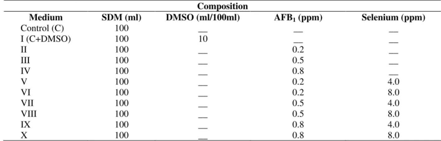
this species was cultivated in 9 different media, shown in Table 1. All the females used in the experiments were virgins. The flies with the same age were used for experiments and seven pairs were mated. Then, the developmental stages were followed daily. Offsprings were counted everyday from the first day of eclosion and phenotypic abnormalites of F1 individuals examined under
microscope were observed. Statistical analysis of data was done using Duncan’s one-way range test.
Table 1 - The composition of different media used for this study
Composition
Medium SDM (ml) DMSO (ml/100ml) AFB1 (ppm) Selenium (ppm)
Control (C) 100 __ __ __
I (C+DMSO) 100 10 __ __
II 100 __ 0.2 __
III 100 __ 0.5 __
IV 100 __ 0.8 __
V 100 __ 0.2 4.0
VI 100 __ 0.2 8.0
VII 100 __ 0.5 4.0
VIII 100 __ 0.5 8.0
IX 100 __ 0.8 4.0
RESULTS AND DISCUSSION
The effects of the AFB1 and AFB1+Se4+ on the
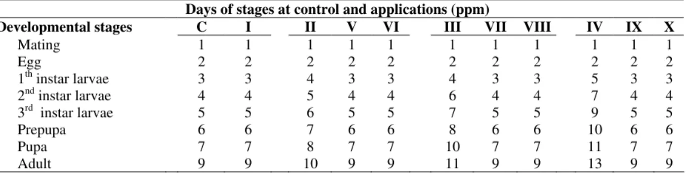
development stages of D. melanogaster are shown in Table 2. In the laboratory at 25±1oC, the life cycle (egg-adult) was 9 days (Uysal and Bahçeci, 1995; Uysal et al., 2002). In control (C), C+DMSO (Medium I) and all experimental groups laid eggs were observed at the second day of
mating.In control and Medium I, first adult was emerged from the pupa at 9th days of mating. But in the presence of 0.2, 0.5 and 0.8 ppm of AFB1
(Medium II, III and IV), various development stages were retarded. At these concentrations of AFB1, first offsprings of F1 progeny were seen at
10th,11th and 13th days, respectively (Table 2).
Table 2 - Occurrence of egg- adult developmental stages after application of different doses of AFB1 and AFB1+ Se4+ Days of stages at control and applications (ppm)
Developmental stages C I II V VI III VII VIII IV IX X
Mating 1 1 1 1 1 1 1 1 1 1 1
Egg 2 2 2 2 2 2 2 2 2 2 2
1th instar larvae 3 3 4 3 3 4 3 3 5 3 3
2nd instar larvae 4 4 5 4 4 6 4 4 7 4 4
3rd instar larvae 5 5 6 5 5 7 5 5 9 5 5
Prepupa 6 6 7 6 6 8 6 6 10 6 6
Pupa 7 7 8 7 7 10 7 7 11 7 7
Adult 9 9 10 9 9 11 9 9 13 9 9
On the other hand, one of the most interesting results obtained was that the developmental stages of D. melanogaster were observed at the same days with control (C) with shown that Se4+ applications (Medium. V-X). Table 2, both 4.0 and 8.0 ppm Se applications prevented the negative effects of AFB1 on developmental stages and these
stages were completed within their normal developmental phases (9 days).
Similar findings have been observed in previous studies also. For example, Lalor et al., (1976) found that growth on AFB1 media of
D.melanogaster caused significant increases in egg-to- adult developmental time. Larval and pupal toxic effects caused by AFB1 have also been
demonstrated by various authors in Musca domestica (Beard and Walton, 1971) and
D.melanogaster (Kirk et al., 1971). Besides, according to Chinnici et al.,(1979), second and third larvae of strain A-9 showed significant mortality rates when grown at 0.88 ppm AFB1. At
the 0.88 ppm concentration, both A-9 and A-11 strains showed significant mortality rates for first instar larvae, but the A-9 larvae died at higher
affected by all application groups ( Medium II, III and IV) of toxin ( Table 3). The maximum inhibition effect was produced with higher toxin concentration (0.8 ppm/ ml, Medium IV). As seen in Table 3 too, the difference between the number of the offsprings of control (2305) and control+DMSO (Medium I, 2218) was statistically unimportant (P> 0.05). But, as 2305 F1 individuals
were in control group, the number of offsprings decreased in the application groups (Medium II, III and IV), depending on the dosage increase. Statistical analysis showed that this decreasing was significant (P< 0.01).
In our unpublished study, besides the above mentioned dosages of AFB1, three different
dosages have also been examined (1.1, 1.4 and 1.7 ppm/ ml), but, no offsprings were obtained. Furthermore, metamorphosis stopped in 1st, 2nd and 3rd instar larvae according to the increasing concentrations and was not completed.
For many researchers, the most important reason of the decrease of offsprings in D. melanogaster
exposed AFB1 was the decrease of fertility, both in
Brazilian Archives of Biology and Technology
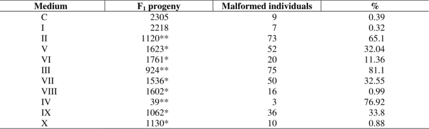
Table 3 - The effect of different concentrations of selenium together with AFB1 on the total number of F1
individuals and formation of malformed individuals (%)
Medium F1 progeny Malformed individuals %
C 2305 9 0.39
I 2218 7 0.32
II 1120** 73 65.1
V 1623* 52 32.04
VI 1761* 20 11.36
III 924** 75 81.1
VII 1536* 50 32.55
VIII 1602* 16 0.99
IV 39** 3 76.92
IX 1062* 36 33.8
X 1130* 10 0.88
Duncan’s one-way range test: * P<0.05 ; ** P<0.01
Besides, the extreme sensitivity of larvae and pupae and also their mortality in high concentrations (1.4 ppm) were one of the most important reasons of the decrease in the number of offsprings ( Kirk et al., 1971; Llwellyn and Chinnici, 1978).
Furthermore, when F1 individuals were examined
as phenotypic, malformed individuals were also observed and malformation was concentrated mainly on wing, leg and thorax and neither the formation of extremities nor their lackness have been found. While the rate of the malformed individuals in the control was 0.39%, they changed between 65.17-81.16% in Medium II, III and IV, respectively. This ratio of change was statistically important (P<0.01). Depending on the increase of concentration in Medium II, III and IV, both the number of individuals decreased and malformation increased (Table 3).
In previous studies, similar results have also been obtained. For example, teratogenic effects of AFB1
were observed on fetuses of hamster, mice, rat and cow by Aleksandrowicz and Smyk (1973). These teratogenic effects were also found as follows: decreases in body size (Chinnici et al., 1976) and wing length (Lalor et al., 1976), formation of tumor on different body parts (Sidorov et al., 2001), hepatocarcinogen effects at vertebrates (Pier, 1981). These findings are in accordance with our results. According to our data, the most important reason for the formation of phenotypic abnormalities was delaying of metamorphosis, because AFB1 caused some faults during
transcription of developmental genes and defects
in homeotic genes which affected the final condition of imaginal discs (Wallace et al., 1991). It has also been reported that AFB1 is metabolically
activated by the microsomal mixed-function monooxygenase system to the 8, 9-epoxide, which readily binds to the nucleophilic sites in DNA to form DNA adducts (Stark,1986). AFB1
preferentially attacks guanine residues in DNA and the major adduct for is 8,9- dihydro-9-hydroxy-(N7-guanyl) AFB1, which accounts for over 90% of
the total adducts (Essigmann et al., 1982). The formation of AFB1- DNA adducts in the target cell
gives rise to promutagenic sites in DNA.
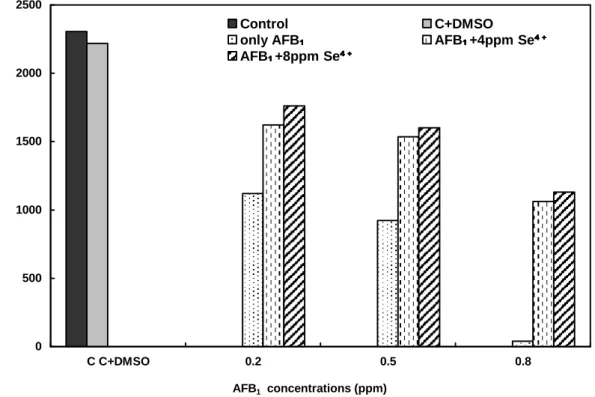
The addition of different concentrations of Se4+ to aflatoxin containing media (Medium V-X), was found to be interesting for the increase of F1
individuals and the decrease of the rate of malformation. The individuals number belonging to the F1 was 1120 and the rate of malformed
individuals was 65.17 % in the medium containing 0.2 ppm AFB1 (Medium II). In the media
containing 4.0 and 8.0 ppm Se4+ (Medium V and VI), F1 individuals were 1623 and 1761,
respectively. Furthermore, the number of malformation decreased from 65.17% to 11.36%. Similarly, while the number of F1 individuals was
924 in the medium of 0.5 ppm AFB1 (Medium III),
0 500 1000 1500 2000 2500
C C+DMSO 0.2 0.5 0.8
AFB1 concentrations (ppm)
T
h
e
t
o
ta
l
n
u
m
b
e
r
o
f
o
ff
s
p
ri
n
g
s
(
fo
r
F
1
p
ro
g
e
n
y
)
Control C+DMSO
only AFB₁₁₁₁ AFB₁₁₁₁+4ppm Se⁴ ⁺⁴ ⁺⁴ ⁺⁴ ⁺
AFB₁₁₁₁+8ppm Se⁴ ⁺⁴ ⁺⁴ ⁺⁴ ⁺
Figure 1 - Comparison of the total number of offsprings obtained from D.melanogaster treated with different concentrations of AFB1 and AFB1+Se4+.
As only 39 individuals were counted at the 0.8 ppm AFB1, the individuals number with the
increasing Se4+ concentrations was found to be important. This was statistically important (P<0.05), (Fig. 1).
Although protective effects of selenium on teratogenic disorders has not been identified, some studies demonstrated that it has protective effect against the acute toxicity of AFB1 and
carcinogenesis in animal species (Chen et al., 1982a).Carcinogen-induced cellular oxidative damage and its role in the cytotoxicity and carcinogenesis have attracted much attention (Imlay and Linn,1988; Farber et al., 1990). Oxidative damage usually refers to the impairment of the function of cellular components, e.g., enzymes, nucleic acids, membranes and proteins, by reactive oxygen species such as superoxide radicals (O2-), hydroxyl free radical (OH), and
hydrogen peroxide (H2O2). Oxidative damage
substances with antioxidant properties such as selenium, vitamin A, β karotene etc. Selenium has antimutagenic and anticarcinogenic activity. Previous studies on the protective effects of antioxidants such as selenium and vitamin A against the cytotoxicity and genotoxicity of AFB1
mostly focused on the metabolism and detoxification of AFB1 or the formation of AFB1
-DNA adduct (Chen et al., 1982b; Mandel et al., 1987; Decoudu et al., 1992). It was also suggested that the protective effect of selenium could be mediated through a cellular mechanism related to glutathione detoxification pathways (Shen et al., 1994). Glutathione has been shown to play an important role in the detoxification of AFB1
Brazilian Archives of Biology and Technology Our results have also showed that selenium has a
protective effect on the development disorders of
D. melanogaster. It could be concluded that more investigations should be carried on in order to understand whether or not the mechanism of the effect of selenium on D. melanogaster is as shown in the present experiments.
RESUMO
O objetivo deste trabalho foi investigar os efeitos da AFB1 e AFB1 + Se 4 em vários estágios de
desenvolvimento da Drosophila melanogaster.
Ambos diferentes concentrações da ABF1 e Se4+
aplicado com AFB1 foram alimentados durante a
fase de desenvolvimento da mosca (ovo, larva e pulpa). Quando a progenese F1 do controle e
aplicações foram comparadas com outros grupos, ABF1 ampliou o processo de metamórfose e na
redução do número total de ovos. Porém esses efeitos negativos foram inibidos com o tratamento com selênio em diferentes concentrações (4.0 e 8.0 ppm). Esses resultados sugerem que o selênio pode efetivamente inibir AFB1 que induz
anomalias nos estágios do desenvolvimento da
Drosophila melanogaster
REFERENCES
Aleksandrowicz, J. and Smyk, B. (1973), The association of neoplasmic diseases and mycotoxins in the environment. Tex. Rep. Biol. Med., 31, 715-726. Baldwin, S. and Parker, R. S. (1987), Influence of
dietary fat and selenium in initiation and promotion of aflatoxin B1-induced preneoplastic foci in rat liver.
Carcinogen.,8,191-207.
Bashandy, S. A. E.; Galil, M. A. E.; Bashandy, M. A. and Morsy, F. (1994), Effect of dietary aflatoxin and vitamin C on gonadal activity in male rats. Al-Azhar Bull. Sci., 5, 697-707.
Beard, R. L. and Walton, G. S. (1971), Insecticidal mycotoxins produced by Aspergillus flavus var. colunaris. Bull. Conn. Agr. Exp. Sta., 725, 1-26.
Bhattacharya, R. K.; Firozi, P. F. and Aboobaker, V. S. (1984), Factors modulating the formation of DNA adduct by aflatoxin B1 in vitro. Carcinogen., 5, 1359-1362.
Bronzetti, G.; Cini, M.; Andreoli, E.; Caltavuturo, L.; Panunzio, M. and Croce, C. D. (2001), Protective effects of vitamin and selenium compound in yeast. Mutat. Res. Gen. Toxicol. Enviroment. Mutagen.,
429, 105-115.
Burguera, J. A.; Edds, G. T. and Osuna, O. (1983), Influence of selenium on aflatoxin B1 or crotolaria
toxicity in turkey poults. Am. J. Vet. Res., 44, 1714-1719.
Busby Jr., W. F. and Wogan, G. N. (1985), Aflatoxins. In: Searle, G. (Ed.). Chemical Carcinogen. Washington : American Chemical Society. pp. 945-1136.
Chen, J.; Goetchius, M. P.; Campbell, T. C. and Combs, G. F. (1982a), Effects of dietary selenium and vitamin E on hepatic mixed-function oxidase activities and in vivo covalent binding of aflatoxin B1
in rat. J. Nutr., 112, 324-328.
Chen, J.; Goetchius, M. P.; Combs, G. F. and Campbell, T. C. (1982b), Effects of dietary selenium and vitamin E on covalent binding of aflatoxin to chick liver cell macromolecules. J. Nutr., 112, 350-357. Chinnici, J. P.; Booker, M. A. and Llewellyn, G. C.
(1976), Effect of aflatoxin B1 on viability, growth,
fertility and crossing over in D. melanogaster (Diptera). J. Invertebr. Pathol., 27, 255-258.
Chinnici, P. J.; Erlanger, L.; Charnock, M.; Jones, M. and Stein, J. (1979), Sensitivity differences displayed by Drosophila melanogaster larvae of different ages to the toxic effects of growth on media containing aflatoxin B1. Chem. Biol. Interact., 24 : (3), 373-380.
Darodo, R. D.; Porta, E. A. and Aquino, T. M. (1985), Effects of dietary selenium on hepatic and renal tumorogenesis induced in rat by diethylnitrosamine. Hepatol., 5, 1201-1208.
Davila, J. C.; Edds, G. T.; Osuna, O. and Simpson, C. F. (1983), Modification of effects aflatoxin B1 and
warfarin in young pigs given selenium. Am. J. Vet. Res., 44,1877.
Decoudu, S.; Cassand, P.; Daubeze, M.; Frayssinet, C.; and Narbonne, J. F. (1992), Effect of vitamin A dietary intake in vitro and in vivo activation of aflatoxin B1. Mutat. Res., 269, 269-278.
Diplock, A. T. (1981), Metabolic and functional defects in selenium deficiency. Philos. Trans. R. Soc. London. Ser. B. Biol Sci., 294 : (1071), 105-117. Essigman, J. M.; Croy, R. G.; Bennet, R. A. and
Wogan, G. N. (1982), Metabolic activation of aflatoxin B1: pattern of DNA adduct formation,
removal and excretion in relation to carcinogenesis, Drug Metabol. Rev., 13, 581-602.
Farber, J. L.; Kyle, M. E. and Coleman, J. B. (1990), Biology of disease: Mechanisms of cell injury by activated oxygen species. Lab. Invest., 62, 670-679. Francis, A. R.; Shetty, T. K. and Bhattacharya, R. K.
(1988), Modifying role of dietary factors on mutagenenicity of aflatoxin B1: In vitro effect of trace
elements, Mutat. Res., 199, 85-93.
Garner, R. C. and Martin, C. N. (1979), Fungal toxins, aflatoxins and nucleic acids. In: Grover, P. L. (Ed.). Chemical Carcinogens and DNA. Florida : West Palm Beach. pp. 187-225.
Gracy, R W.; Talent, J. M.; Kong, Y. and Conrad, C. C. (1999), Reactive oxygen species: the unavoidable environmental insult. Mutat. Res., 428 : (1/2), 17-22. Horvath, P. M and Ip, C. (1983), Synergistic effect of
vitamin E and selenium in the chemoprevention of mammary carcinogenesis in rat. Cancer Res., 43, 5335-5341.
Imlay, J. A. and Linn, S. (1988), DNA damage and oxygen radical toxicity. Science, 240, 1302-1309. Kirk, H. D.; Ewen, A. B.; Emson, H. E. and Blair, D. G.
(1971), Effect of aflatoxin B1 on development of
D. melanogaster (Diptera). J. Invertebr. Pathol., 18, 313-315.
Lalor, J. H.; Chinnici, J. P. and Llewellyn, G. C. (1976), Effect of a fungal metabolite, aflatoxin B1, on larval
viability and gross morphology in D. melanogaster. Dev. Ind. Microbiol., 17, 443-449.
Lei, D. N.; Wang, L. Q.; Ruebner, B. H.; Hsieh, D. P.; Wu, B. F.; Zhu, C. R. and Du, M. J. (1990), Effect of selenium an aflatoxin hepatocarcinogenesis in the rat. Biomed.Environ.Sci., 3, 65-80.
Llwellyn, G. C. and Chinnici, J. P. (1978), Variation in sensitivity of aflatoxin B1 among several strains
of D. melanogaster (Diptera). J. Invertebr. Pathol.,
31, 37-40.
Mandel, H. G.; Manson, M. M.; Judah, D. J.; Simpson, J. L.; Green, J. A.; Forrester, L. M.; Wolf, C. R. and Neal, G. E. (1987), Metabolic basis for the protective effect of the antioxidant ethoxyquin on aflatoxin B1 hepatocarcinogenesis in rat. Cancer Res., 47, 5218-5223.
Matsumura, F. and Knight, S. G. (1967), Toxicity and chemosterilizing activity of aflatoxin against insects. J. Econ. Entomol., 60, 871-872.
Milner, J. A. (1985), Effect of selenium on virally induced and transplantable tumor models. Fed. Proc.,
44, 2568-2572.
Nair, A. and Verma, R. J. (2000), Effect of aflatoxin on testis of mouse and amelioration by vitamin E. Indian J. Toxicol., 7, 109-116.
Newberne, P. M. and Conner, M. W. (1974), Effect of selenium on acute response to aflatoxin B1. In:
Hemphill, D. D. (Ed.). Trace Substances in Environmental Health. Columbia : University of Missouri. pp.323-327.
Pier, A. C. (1981), Mycotoxins and animal health. Adv. Vet. Sci. Comp. Med., 25, 185-243.
Reis, J. (1975), Insecticidal and larvicidal activities of the mycotoxins aflatoxin B1, rubratoxin B,
Patulin and diacetoxyscirpenol towards Drosophila melanogaster.Chem. Biol. Interact., 10 : (5), 339-342. Stark, A. A. (1986), Molecular aspects of aflatoxin B
Sidorov, R. A.; Ugnivenko, E. G.; Khovanova, E. M. and Belitsky, G. A. (2001), Induction of tumor clones in D. melanogaster wts/+ heterozygotes with chemical carcinogens. Mut. Res., 498, 181-191. Uysal, H. and Bahçeci, Z. (1995), Effect of mercury
chloride on the durations of developmental stages of D. melanogaster. Dros. Infor. Serv., 76, 162-163. Uysal, H.; Aydoğan,. N. and Algur,O. F. (2002), Effect
of single cell protein as a protein source in Drosophila culture. Braz. J. Microbiol., 33, 314-317. Verma, R. J. and Nair, A. (2001), Vitamin E
ameliorates aflatoxin-induced biochemical changes in the testis of mice. Asian J. Androl., 3, 305-309. Wallace, R. A.; Sanders, G. P. and Ferl, R. J. (1991),
The Science of Life. New York : Harper Collins Publishers Inc.
Wogan, G. N. (1973), Aflatoxin carcinogenesis. In: Bush, H. (Ed.). Methods in Cancer Research. New York : Academic Press. pp. 309-344.
Yu, S. Y.; Chu, Y. J. and Li, W. G. (1988), Selenium chemoprevention of liver cancer in animals and possible human applications. Biol. Trace Element Res., 15, 231-241.