1. Mestrado Integrado em Medicina da Faculdade de Medicina da Universidade do Porto.
2. Licenciatura em Medicina. Unidade de Reumatologia Pediátrica do Hospital de São João do Porto.
manifestations, diagnosis and treatment of juvenile NPSLE.
Keywords: Juvenile Systemic Lupus Erythematosus; Neuropsychiatric Manifestations; Autoantibodies; Treatment.
IntroductIon
Systemic Lupus Erythematosus (SLE) is an autoimmune, chronic and multisystemmic autoimmune disea -se1-3, characterized by a broad clinical spectrum, an ar-ray of immunological abnormalities and a variable course and prognosis. In its pathogenesis are involved environmental, hormonal, immunologic and genetic factors, although its etiology is still unknown4,5.
The American College of Rheumatology classifica-tion criteria are used to diagnose juvenile SLE (jSLE)3, considering its appearance before sixteen years old a very important criterium6. The median age of onset of disease is 10-12 years7, being rare in children under five years old. The age of onset may influence the course of disease in terms of clinical presentation, organ in-volvement, and serological findings8,9. jSLE represents 15-20% of all SLE cases. The estimated incidence of jSLE ranges from 10 to 20 per 100000 children, de-pending on the ethnic population3,10.
The clinical spectrum of jSLE can be quite variable3. As it can assume characteristics of other pathologies, its diagnosis in pediatric age is very difficult. Systemic constitutional symptoms such as fatigue, weight loss and fever3 are considered to be the most common pre-sentation symptoms, followed by skin involvement, in-cluding malar rash and fotossensibility, musculoskele-tal disease including arthralgia/arthritis11, renal and neuropsychiatric disease7.
Neuropsychiatric involvement in SLE (NPSLE) in-cludes the neurological syndromes of the central ner-vous system (CNS), of the peripheral nerner-vous system
Juvenile Systemic Lupus Erythematosus:
neuropsychiatric manifestations
ACTA REUMATOL PORT. 2012;37:117-125 Helena Fernandes1, Iva Brito2
AbstrAct
Juvenile Systemic Lupus Erythematosus (jSLE) is a chronic and multisystemmic autoimmune disease, which appears before 16 years old with an incidence of 10 to 20 cases per 100000 children. The clinical spec-trum of jSLE can be quite variable. The most common symptoms are constitutional, followed by the cuta-neous, musculoskeletal, renal, and neuropsychiatric in-volvement.
Neuropsychiatric involvement in jSLE has a preva-lence ranging from 20 to 50.9% and results in signifi-cant morbidity and mortality.
The most common clinical manifestations of juvenile neuropsychiatric SLE (NPSLE) are headache, cognitive dysfunction, mood disturbances and seizures.
The pathophysiology of juvenile NPSLE is not yet fully known, but immunological and inflammatory fac-tors, such as autoantibodies, cytokines and prothrom-botic states are widely described. The role of autoanti-bodies in the onset of specific clinical manifestations has also been recognized.
Juvenile NPSLE manifestations are often difficult to diagnose. In addition to semiological aspects, the study and validation of neuropsychological testing and neu-rocognitive assessment for the juvenile SLE population are essential. The role of advanced imaging techniques should be explored.
The treatment of juvenile NPSLE must be individua lized according to the type and severity of clinical mani festations, relying on symptomatic therapy, anticoagu lants or steroids. New therapeutic approaches, inclu -ding biotherapies need controlled randomized trials for further validation.
(PNS) and of the autonomic nervous system (ANP) and psychiatric disorders12, in the absence of se -condary causes6,13,14.
NPSLE can precede the onset of lupus15 or occur at any time during its course, mainly within the first year from the time of diagnosis4,11,16.
Juvenile NPSLE is a major cause of morbidity and less so of mortality in jSLE patients16, and its prevalence ranges from 22 to 50, 9% among several stu -dies6,11,13,15-20. Some studies suggest that jSLE has a more serious course and a more frequent neuropsychiatric involvement than adult SLE7,9.
There are no guidelines for the treatment of NPSLE, thus, it should be individualized, according to the type and severity of clinical manifestations and other system or organs involvement21.
Based on up-to-date references, this article aims to review the main clinical manifestations, pathogenesis, diagnosis and treatment of juvenile NPSLE.
MAterIAl And Methods
Forty-four scientific articles published between 1999 and 2010 in journals with an impact factor of 3 were researched. Those articles were obtained in the PubMed database.
The keywords used were “systemic lupus erythe-matosus”, “juvenile systemic lupus erytheerythe-matosus”, “neuropsychiatric”, “autoantibodies”, “pathogenesis” and “treatment”.
neuropsychIAtrIc MAnIfestAtIons of juvenIle sle
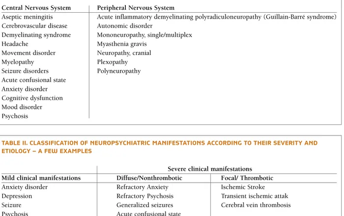
The prevalence of neuropsychiatric manifestations of jSLE ranges from 22 to 50,9%6,11,13,15-20. A major source of variance in the rates of occurrence has to be at-tributed to the lack of standard definitions for the pe-diatric lupus population and for the NPSLE 15,16. In or-der to develop a standardized nomenclature system for the NPSLE in 1999 “The American College Rheumatology nomenclature and case definitions for Neuropsychiatric Manifestations in Systemic Lupus Erythematosus” has been published22, which describes twelve syndromes concerning CNS and seven syn-dromes concerning PNS (Table I).
Neuropsychiatric manifestations can be classified according to their etiology and severity (Table II). The majority of thrombotic mechanisms cause focal mani -festations and diffuse mani-festations are described as
being due to non-thrombotic mechanisms. However, some studies mention that diffuse manifestations can also be consequent to thrombotic mechanisms21.
Headache is the most common neuropsychiatric manifestation, and its prevalence ranges from 39.6 to 75% among several studies6,11,13,15-20. Headache is sub-divided into five categories: migraine, tension headache, cluster headache, headache from intracra-nial hypertension and non-specific intractable headache16. Headache is frequently seen in associa-tion with more severe CNS involvement, including psychosis, acute confusional state11,16and seizures6.
Cognitive dysfunction is one of the most common neuropsychiatric manifestations, which prevalence ranges from 16.9 to 70.8%6,11,13,15-20. There is no specific clinical spectrum, and the degree of impairment can range from mild or subclinical findings to severe de-mentia11. The most common reported areas of impair-ment include verbal and nonverbal learning and memo ry, verbal fluency, complex problem solving, psychomotor speed and executive functioning23. Availa ble data do not suggest that demographic fac-tors such as race and ethnicity are predictive of cogni-tive impairment in SLE19.
Cognitive development, which critical cognitive maturation period from late childhood through ado-lescence and into young adulthood coincides with the pediatric age spike for SLE onset, is a complex pro-cess. Thus, patients who develop juvenile SLE are at a period of exceptional vulnerability of the CNS, which may result in a particular risk for delays and impair-ments in cognitive development19.
Mood disorders, with a prevalence which ranges from 11.4 to 57%6,11,13,15-20, include depression, the most common of the mood disorders and mania and bipolar disorder that are rare. A reactive depression secondary to chronic disease and a depression se -condary to corticosteroid use must be excluded16,25.
Seizures have a prevalence that ranges from 11.4 to 51%6,11,13,15-20and are associated with other CNS in-volvement in most cases, such as headaches, cere-brovascular disease and cognitive dysfunction, with isolated seizures occurring in less than 25% of cases. Generalized seizures are more common than focal seizures11.
Psychosis, which prevalence ranges from 5 to 37.1%6,11,13,15-20, has visual hallucinations as its main characteristic, which in turn is not present in idiopa -tic schizophrenia of childhood, what helps to exclude this diagnosis16. Tactile and auditory hallucinations
and suicidal ideation might be present. Steroid-in-duced psychosis may be differentiated from NPSLE by the presence of uncommon features of NPSLE, such as mania and excessive crying11.
Cerebrovascular disease occurs in 11.3 to 30% of cases6,11,13,15-20and includes a spectrum of SLE-associated cerebral blood vessel abnormalities, ranging from small arteries to large arteries, such as cerebral vein thrombo-sis, which is seen in about 25% of juvenile NPSLE pa-tients16. Microthrombotic and microembo lic events in the form of ischemic stroke and transient ischemic attack are common, while cerebral bleeding is rare.
Headaches and seizures are the most common clini -cal signs and symptoms of cerebrovascular disease11 but hemiparesis, quadriparesis, spastic diplegia or spastic quadriplegia should also be considered6.
Anxiety disorders occur in 1.8 to 14% cases6,11,13,15-20 and possible psychological reactions related to the pos-sibility of having a major chronic systemic illness might be excluded22.
Movement disorders have a prevalence which ranges from 5.6 to 20%6,11,13,15-20. Chorea is the most common movement disorder and it is more common in juvenile SLE thanin adult-onset SLE11. Most patients only have one episode of chorea, and unilateral chorea, which is seen more commonly than bilateral chorea16. Chorea also affects more frequently the upper limbs6. PNS involvement occurs in 5.6 to 15%6,11,13, 15-20of all patients with juvenile SLE. It may occur with or without concomitant CNS involvement. Optic neu-ropathy and oculomotor palsy are the most common cranial neuropathies and facial palsy and trigeminal neuropathy are less frequent. Unlike in adults with SLE, autonomic dysfunction has been only rarely re-ported in juvenile patients16.
Less common CNS symptoms include: Transverse myelitis, which may present with acute paraplegia or quadriplegia and may be the presenting sign of SLE6,11,16, diabetes insipidus, Parkinson`s syndrome, leukoencephalopathy, as well as retinal vascular disea -se, consisting of arterial or venous occlusion, cotton-tAble I. neuropsychIAtrIc systeMIc lupus erytheMAtosus syndroMes
Central Nervous System Peripheral Nervous System
Aseptic meningitis Acute inflammatory demyelinating polyradiculoneuropathy (Guillain-Barré syndrome) Cerebrovascular disease Autonomic disorder
Demyelinating syndrome Mononeuropathy, single/multiplex
Headache Myasthenia gravis
Movement disorder Neuropathy, cranial
Myelopathy Plexopathy
Seizure disorders Polyneuropathy Acute confusional state
Anxiety disorder Cognitive dysfunction Mood disorder Psychosis
tAble II. clAssIfIcAtIon of neuropsychIAtrIc MAnIfestAtIons AccordIng to theIr severIty And etIology – A few exAMples
Severe clinical manifestations Mild clinical manifestations Diffuse/Nonthrombotic Focal/ Thrombotic Anxiety disorder Refractory Anxiety Ischemic Stroke Depression Refractory Psychosis Transient ischemic attak
Seizure Generalized seizures Cerebral vein thrombosis
Psychosis Acute confusional state
wool spots, optic disc edema, retinal hemorrhages or ischemic optic neuropathy, which can be found in up to 10% of patients16.
pAthogenesIs of juvenIle npsle
The exact mechanisms underlying the pathogenesis of NPSLE remain unknown19. However, it is acknow -ledged that NPSLE pathogenesis is a complex process incorporating immunologic factors such as the pro-duction of autoantibodies against brain structures and the deposition of immune complexes in the central nervous system.
A mechanism proposed by some studies27,28, but not yet confirmed, suggests that the binding of certain au-toantibodies, such as the Anti-endothelial cell anti-bodies (AECA) and the Anti-phospholipid autoanti-bodies (aPL) or the immune complexes to the endothelium of bloodbrain barrier (BBB) leads to stimu -lation of signaling pathways, with activation of transcription factors such as nuclear factor kappa B27. Thus, there will be an induction of synthesis of cy-tokines and chemokines and expression of adhesion molecules, which leads to an increased permeability of the BBB. Access of antibodies to the central nervous system may occur through a disrupted blood brain barrier or again through synthesis in the nervous sys-tem. Autoantibodies, binding molecules exposed on the surface of neurons, lead to a neurotoxic effect28.
Some cytokines and chemokines were shown to be elevated intrathecally, such as interleukin-6 (IL-6), IL-1, IL-8, IL-10, tumor necrosis factor (TNF)-α, in-terferon (IFN)-γ, monocyte chemotactic protein 1 (MCP-1)/CCL2, Interferon-gamma inducible protein--10 (IPprotein--10)/CXCL10 and Fractalkine/CX3CL119.
During the systemic inflammation inherent to SLE, homeostatic balance shifts toward coagulation that fa-vors pro-thrombotic states, which might play a role in the pathogenesis of NPSLE26.
Autoantibodies are potential immunological mar -kers for the diagnosis and prognosis of NPSLE. Ac-cording to their specificity (neurons, endothelium, ubiquitous cellular components) these autoantibodies are divided into three groups (Tables III, IV)21. AUTOAnTIbODIES
A significant number of reports confirmed the associa -tion between neuropsychiatric manifesta-tions in SLE and the presence of autoantibodies (Table V)4,19,28-30. The studies included patients with jSLE and adult SLE. However, only a few studies have focused in children
so the conclusions in SLE adult patients were extrapo lated to jSLE. There is a high variability among diffe -rent studies because of the differences in the popula-tions of patients studied and the laboratory tests used to detect serum antibodies28.
Some studies show an association between Anti--ganglioside antibodies (AGA) and migraine, de-mentia and peripheral neuropathy, depression27,28and cognitive disfunction, suggesting that the detection of AGA has a predictive value for the onset of a neu-ropsychiatric flare29.
In comparison to SLE patients without neuropsy-chiatric symptoms, an increased incidence of Antineurofilament antibodies (ANFA) has been repor -ted in patients with NPSLE. Anti-microtubule-asso-ciated protein 2 antibodies (AMAP2) have been de-tected in sera from patients with NPSLE, but, like the Anti-glial fibrillary acidic protein antibodies (AGFPA), there is no conclusive evidence about its role in NPSLE pathogenesis28.
Anti-DNA antibodies cross-reactive with NMDA receptor (ARNMDA) are found in 25 to 50% of pa-tients with SLE and brain dysfunctions seem clearly to be correlated with the presence of antibodies in CSF, and symptom severity correlates with antibody titles. In a murine model, these antibodies induce non-in-flammatory, excitatory and apoptotic19 neuronal dama -ge in the hippocampus and amygdala, when a break-down of the BBB is caused by a bacterial lipopolysac-charide and epinephrine, respectively28. The neuronal damage in the hippocampus results in cognitive dys-function and the damage in the amygdala leads to be-havioral disorders.
Anti-endothelial cell antibodies (AECA) have a clinical association with disease activity and neu-ropsychiatric manifestations in SLE31,32. Anti-Nedd5 antibodies (ANedd5) titles are higher on the group of patients with neuropsychiatric manifestations than in patients without these disorders. However, there is no conclusive evidence concerning their pathogenic role. The serum Anti-triosephosphate isomerase anti-bodies IgG index is higher in the NPSLE than in other autoimmune diseases, demonstrating a high specificity for the diagnosis of NPSLE. The anti-his-tone antibodies are prevalent in approximately 50% of NPSLE patients28.
Anti-ribosomal P antibodies (ARP) are highly spe-cific for SLE, since they do not occur in healthy indi-viduals or in patients with other diseases4. Several stu -dies show a strong association between elevated
plas-ma ARP titles and neuropsychiatric plas-manifestations, specially psychosis and severe depression19,28. Other studies report a possible relationship between changes in ARP titles and clinical changes in psychosis, sugges -ting its periodic monitoring16. However, 15 to 25% of patients without NPSLE have these autoantibodies pre-sent in their serum and less than one third of NPSLE patients have positive ARP33,34.
Anti-phospholipid antibodies (APL) lead to mi-crothrombotic events, which are responsible for focal
manifestations, such as ischemic stroke, transient is-chemic attacks and cerebral vein thrombosis35,21.
The association between aPL and etiopathogenesis is not yet known, such as headache, seizures, cognitive dysfunction, psychosis and depression is suggested in some studies, but not yet fully established25. Chorea is universally associated with aPL in several pediatric studies7,11and in most patients with chorea and trans-verse myelitis these antibodies are present16. A recent study concerns an association between aPL and mul-tAble III. AntI-neuronAl AutoAntIbodIes AssocIAted to neuropsychIAtrIc systeMIc lupus
erytheMAtosus
Autoantibodies Target
AGA Component of the cell plasma membrane which modulates cell signal transduction events ; predominantly in the nervous system
ANFA Intermediate filament occurring with neurotubules in the neurons and having cytoskeletal and transport functions
AMAP2 Restricted to neurons; important in the control of cytoskeletal integrity and other neuronal functions
AGFAP Involved in cell structure and movement, in cell communication and in the functioning of the blood brain barrier.
ARNMDA NR2 receptors bind the neurotransmitter glutamate; play a role in learning and memory
Anti-endothelial ANedd5 Cytoskeletal GTPase that play an essential role in cytokinesis cell antibodies
AGA – Anti-ganglioside antibodies; ANFA- Anti-neurofilament antibodies; AMAP2 – Anti-microtubule-associated protein 2 antibodies; AGFAP – Anti-glial fibrillary acidic protein antibodies; ARNMDA – Anti-DNA antibodies cross-reactive with NMDA receptor; ANedd5 – Anti-Nedd5 antibodies
Anti-neuronal Autoantibodies
tAble Iv. AutoAntIbodIes specIfIc to ubIquItous cellulAr coMponents AssocIAted to neuropsychIAtrIc systeMIc lupus erytheMAtosus
Autoantibodies Target
ATPI Plays an important role in glycolysis and energy production. ASSA/Ro Nuclear and cytoplasmic polypeptid
ASm Ribonucleoproteins that plays a central role in the processing of pre-mRNA in the nucleus
AHT Chief proteic components of chromatin
ARP Makes up the ribosomal subunits in conjunction with rRNA; ARP recognizes a new integral membrane protein of the neuronal cell surface preferentially distributed in areas involved in memory, cognition, and emotion
APL Anionic phospholipids or protein-phospholipid complexes; includes ACLA and LAC
ATPI – Anti-triosephosphate isomerase antibodies; ASSA/Ro – Anti-SSA/Ro antibodies; ASm – Anti-Smith antibodies; AHT – Anti-histone antibodies; ARP – Anti-ribosomal P antibodies; APL – Anti-phospholipid antibodies; ACLA – Anti-cardiolipin antibodies;
LAC – Lupus anti-coagulant antibodies.
Autoantibodies specific to ubiquitous cellular components
tiple small high-density lesions on the brain Magnetic resonance imaging (MRI), which in turn are compati-ble with subclinical cognitive manifestations, that are only detected through formal neuropsychometric as-sessment36.
Anti-cardiolipin antibodies (ACLA) are reported in a study as the only type of aPL with statistically sig-nificant association with ischemic stroke18. Several studies show an association between ACLA and seizure, chorea6 and PNS involvement16.
Lupus anti-coagulant antibodies (LAC) are uni-versally associated with the risk of cerebral thrombo-sis13,16. One study refers a positive association between LAC and cerebrovascular disease at diagnosis and an association between persistently high LAC titles and chorea37.
dIAgnosIs of juvenIle npsle
Making the diagnosis of juvenile NPSLE is not an easy task29, and it should include a comprehensive clinical history and a physical examination19.
Secondary causes of NP manifestations such as in-fection, hypertensive encephalopaty, metabolic ab-normalities13, drug side effects, including corticos-teroids14and congenital or acquired CNS disease not related to SLE6, should be excluded. Complete blood cell count, inflammatory markers, serum autoanti-bodies, lumbar puncture and cerebrospinal fluid (CSF) analysis, electroencephalography (EEG) and CNS imaging6,11,19,25,38must be performed in order to exclude secondary causes and to confirm the diagnosis of NPSLE.
Inflammatory markers such as erythrocyte sedi-mentation rate and C-protein rate may be normal in one third of patients with psychosis and cognitive dis-function and they are often elevated in patients with cerebrovascular disease and seizures11,19,25. One study did not detect any association between serum com-plement and neuropsychiatric manifestations6.
Some authors argue that a child with SLE should do an APL test at the time of diagnosis and then an-other at least once a year as part of routine screening38. EEG is reported in a study as a valuable mean of diag nosis of cognitive dysfunction39, however, ano ther study found that EEG findings are usually abnormal only when seizures are present16.
One study showed that microembolic signals on transcranial Doppler were more frequent in the cases with NPSLE when compared with the cases without NPSLE and were higher for all SLE patients than for healthy controls40.
Ideally, computorized tomographic (CT) or MRI should be performed in all patients with NPSLE15to as-sess for structural and functional abnormalities19. However they rarely play a role in the assessment of non-thrombotic manifestations.
CT imaging of the brain can be useful in emergen-cy settings to exclude cerebral haemorrhage, large in-farct, intracranial hypertension and cerebral vein throm bosis, when MRI is unavailable19. In contrast MRI scans are more effective in detecting early lesions and lesions secondary to small vessel involvement. Le-sions seen on MRI are frequently small, multifocal, and bilateral with high signal intensity affecting more com-monly the white matter16, suggestive of cerebral vas-culopathy or thought to be due to multiple small in-farcts36.
According to one study, MRI with fluid attenuated inversion recovery seems to be more sensitive in NPSLE than conventional MRI, especially for lesions that are close to CSF-tissue interfaces40.
Vasculopathy, if present, involves small vessels not detectable by MR angiography. If a vasculopathy is strongly suspected, conventional arteriography may be the best option. However, arteriography may also fail to detect changes in the smallest vessels19.
Positron emission tomography (PET) and Single photon emission computerized tomography (SPECT) tAble v. AutoAntIbodIes AssocIAted wIth clInIcAl MAnIfestAtIons
Autoantibodies Clinical Manifestation
AGA Cognitive dysfunction; Depression; Migraine; Peripheral neuropathy ARNMDA Cognitive dysfunction; Behavioral disorders
ARP Psychosis; Severe depression
APL Ischemic stroke; Transient ischemic attacks; Cerebral vein thrombosis; Headache, Seizures, Cognitive dysfunction, Psychosis and Depression; Chorea; Transverse myelitis
have been shown to be highly sensitive for the diag-nosis of diffuse manifestations in NPSLE. PET fre-quently identifies areas of hypometabolism, most of-ten in the parietal or frontal lobes19and SPECT reveals, more commonly, multiple brain areas with hypoper-fusion, most often in the frontal lobe.
SPECT may be used to monitor disease severity and guide therapy41, although it has little or no value in monitoring CNS disease activity16.
Despite their high sensitivity, PET and SPECT do not have a clear specificity. These modalities require further study and results should be interpreted cau-tiously19.
Promising imaging modalities, including Proton magnetic resonance spectroscopy (MRS), Magnetiza-tion transfer imaging (MTI) and Diffusion weighted imaging (DWI), may reveal important information in the future, assessing biochemical aspects, quantifica-tion of brain activity and differentiaquantifica-tion inflammatory lesions from ischemic damage, respectively. Further studies are necessary to provide a true understanding of the clinical usefulness of these techniques14,19,40,42.
A multidisciplinary assessment is needed and it may include self, teacher and parent reports of cognitive difficulties or recent behaviour disorders19.
A psychiatric consultation may be required for diag -no sis of depression and for the exclusion of reactive depression secondary to chronic disease, psychosis and anxiety disorder22.
Formal neuropsychological assessment is recom-mended whenever cognitive decline is suspected, and some authors argue that all children with SLE should undergo formal neurocognitive assessment at diagno-sis to establish a baseline level of cognitive function, given the challenge in detecting cognitive abnormali-ties19.
A wide array of instruments that can provide com-prehensive and useful information is available for for-mal neurocognitive testing, such as Mini Mental Status Exam22, a standardized core set of neuropsychological tests for assessment of pediatric cognitive function pro-posed by Childhood Arthritis and Research Alliance and the computer-administered neurocognitive assessment Pediatric-Automated Neuropsychological Assesssment Metrics19. However, no standardized battery has al-ready been validated for the pediatric SLE population. Changes inherent to age and development, and the limited information about premorbid functioning dif-ficult the effectiveness and interpretation of neu-rocognitive tests in pediatric population. Moreover,
they take several hours to be performed and the lan-guage and cultural factors and the potential for score inflation via “learning effects” after repetitive testing are limitations of all neurocognitive tests19.
treAtMent of juvenIle npsle
The clinical diversity of NPSLE, the difficulty in defi -ning outcome measures and the lack of diagnostic cri-teria have been considered the biggest obstacles to the performance of controlled clinical trials related to study treatment options which difficult the compari-son between different therapeutic regimens. Once there are no guidelines for the treatment of juvenile NPSLE, it should be individualized and based upon clinical experience6.
The management of patients with juvenile NPSLE has to overcome these challenges: trying to control the disease and preventing the damages induced by medications7.
It is important to differentiate between mild and se-vere manifestations and between thrombotic and non-thrombotic disease.
Patients with mild neuropsychiatric disease such as anxiety, depression, seizures, psychosis and headache or migraine may be treated conservatively with anxio lytics, antidepressants, anticonvulsants, antipsycho -tics, analgesics / anti-inflammatory drugs / calcium an-tagonists and ergotamine, respectively. Long-term antiepileptic treatment is indicated in the presence of concomitant cerebrovascular disease and / or persistent EEG abnormalities. Anticoagulation may be conside -red in a selected group of patients with seizures and persistent aPL positivity. Avoiding corticosteroids in these particular cases has also helped in the reduction of complications such as secondary infection, osteo-porosis or aseptic necrosis21. Patients with cognitive disfunction should receive psychological support and educational interventions to maximize memory and function19.
The recognition of the role of APL in the appearan -ce of severe thrombotic/focal events on NPSLE con-tributed to a more assertive therapeutic approach. Due to the high risk of recurrence, in the case of previous thrombosis or high titers of APL, including ACLA, long-term anticoagulation with warfarin is mandatory, with an International Normalized Ratio values between 2.5 to 3.5. For patients with low or moderate levels of ACL, without any clinical symptom, and no previous episode of thrombosis, a prophylaxis with low-dose aspirin may be sufficient, even though its application
is controversial7.
There is no evidence whether patients with subcli -nical cognitive manifestations and small lesions with high signal intensity in the MRI, which in turn are as-sociated with the presence of APL, would benefit from antiagreggrant therapy based on aspirin. Patients who fail to respond to antiaggregant therapy, show progres-sion on the brain MRI leprogres-sions or develop cognitive dys-function and should receive anticoagulant therapy with warfarin28.
Acute severe diffuse CNS manifestations generally require high doses of intravenous corticosteroids and chronic severe diffuse CNS manifestations generally re-quire oral corticosteroids, preferably for short pe riods21.
The use of intrathecal corticosteroids43or cy-clophosphamide may be used in refractory cases to cor-ticosteroids. There are no randomized controlled trials comparing the effectiveness and safety of cyclophos-phamide and corticosteroids21. A combination between cyclophosphamide and corticosteroids has been des -cribed in some pediatric studies as useful for the treat-ment of severe NPSLE6,11,16.
Combination therapy with syncronized plasma-pheresis and subsequent cyclophosphamide in severe NPSLE has been proposed by some authors and it seems that patients who respond better to plasma-pheresis are those with more severe illness44.
In selected patients with APL positivity and whose neuropsychiatric manifestations are difficult to be dif-ferentiated between thrombotic and non-thrombotic ones, such as chorea and transverse myelitis, can be treated with a combination and monitored closely the -rapy with corticosteroids, cyclophosphamide and an-ticoagulation. Chorea may also respond to an addi-tional anticonvulsant therapy21.
Intrathecal methotrexate or corticosteroids, hyper-baric oxygen, azathioprine, mycophenolate mofetil, memantine, plasmapheresis and biotherapies as ritu -ximab16,19 require further controlled clinical trials to confirm their usefulness in the NPSLE.
conclusIons
Neuropsychiatric manifestations in jSLE represent an important diagnostic challenge because of its diversity and complexity. The most common neuropyschiatric manifestations are headache, cognitive dysfunction, mood disturbances and seizures.
Although its true pathogenesis is still unknown, it
seems that some autoantibodies have a positive pre-dictive value concerning to some manifestations, such as Anti-ganglioside antibodies, Anti-DNA antibodies cross-reactive with NMDA receptor, Anti-ribosomal P antibodies and Anti-phospholipid antibodies.
However, many areas remain unclear. Formal neuropsychological tests should be validated for the pedia -tric population with SLE and new imaging techniques should be explored. In order to improve the prognosis of children with NPSLE and to clarify pathophysio -logy, diagnosis and treatment aspects, further con-trolled clinical trials are needed.
correspondence to
Helena Fernandes Rua Monte de Cima, 372 4765-445 Guardizela E-mail: med05199@med.up.pt
references
1. Smith PP, Gordon C. Systemic lupus erythematosus: Clinical Presentations. Autoimmun Rev 2010; 10:43-45.
2. Haji Muhammad Ismail Hussain I, Loh WF, Sofiah A. Child-hood cerebral lupus in an Oriental population. Brain Dev 1999; 21: 229-235.
3. Thucker LB. Making the diagnosis of systemic lupus erythema-tosus in children and adolescents. Lupus 2007; 16:546-549. 4. Abdel-Nasser AM, Ghaleb RM, Mahmoud JA, Khairy W,
Mah-moud RM. Association of anti ribosomal P protein antibodies with neuropsychiatric and other manifestations of systemic lu-pus erythematosus. Clin Rheumatol 2008; 27:1377-1385. 5. Brito I, Silva H, Ferreira A, et al. Nefrite lúpica na criança. Acta
Reumatol Port 2000; 96:13-19.
6. Olfat MO, Al-Mayouf SM, Muzaffer MA. Pattern of neuropsy-chiatric manifestations and outcome in juvenile systemic lupus erythematosus. Clin Rheumatol 2004; 23:395-399.
7. Kone-Paut I, Piram M, Guillaume S, Tran TA. Lupus in adoles-cence. Lupus 2007; 16:606-612.
8. Hedrich CM, Zappel H, Straub S, et al. Early onset systemic lu-pus erythematosus: differential diagnoses, clinical presentation, and treatment options. Clin Rheumatol 2010; 30:275-283. 9. Pluchinotta FR, Schiavo B, Vittadello F, Martini G, Perilongo G,
Zulian F. Distinctive clinical features of pediatric systemic lu-pus erythematosus in three different age classes. Lulu-pus 2007; 16:550-555.
10. Macdermott EJ, Adams A, Lehman TJ. Systemic lupus erythe-matosus in children: current and emerging therapies. Lupus 2007; 16:677-683.
11. Benseler SM, Silverman ED. Systemic lupus erythematosus. Pe-diatr Clin N Am 2005; 52:443– 467.
12. Pego-Reigosa JM, Isenberg DA. Psychosis due to systemic lupus erythematosus: characteristics and long-term outcome of this rare manifestation of the disease. Rheumatology 2008; 47:1498–1502.
13. Avcin T, Benseler SM, Tyrrell PN, Cucnik s, Silverman ED. A fol-low up study of antiphospholipid antibodies and associated neuropsychiatric manifestations in 137 children with systemic lupus erythematosus. Arthritis Rheum 2008; 59:206–213.
14. Bosma GP, Rood MJ, Huizinga TW, Jong BA, Bollen EL, van Bu-chem MA. Detection of cerebral involvement in patients with ac-tive neuropsychiatric systemic lupus erythematosus by the use of volumetric magnetization transfer imaging. Arthritis Rheum 2000; 43: 2428–2436.
15. Singh S, Gupta MK, Ahluwalia J, Singh P, Malhi P. Neuropsy-chiatric manifestations and antiphospholipid antibodies in pe-diatric onset lupus: 14 years of experience from a tertiary cen-ter of North India. Rheumatol Int 2009; 29:1455-1461. 16. Benseler SM, Silverman ED. Neuropsychiatric involvement in
pediatric systemic lupus erythematosus. Lupus 2007; 16:564--571.
17. Sibbitt WL, Brandt JR, Johnson CR, et al. The incidence and prevalence of neuropsychiatric syndromes in pediatric onset systemic lupus erythematosus. J Rheumatol 2002; 29:1536--1542.
18. Harel L, Sandborg C, Lee T, von Scheven E. Neuropsychiatric manifestations in pediatric systemic lupus erythematosus and association with antiphospholipid antibodies. J Rheumatol 2006; 33:1873-1877.
19. Levy DM, Ardoin SP, Schanberg LE. Neurocognitive impairment in children and adolescents with systemic lupus erythemato-sus. Nat Clin Pract Rheumatol 2009; 5:106-114.
20. Muscal E, DR Bloom DR, Hunter JV and Myones BL. Neuro-cognitive deficits and neuroimaging abnormalities are preva-lent in children with lupus: clinical and research experiences at a US pediatric institution. Lupus 2010; 19: 268–279. 21. Sanna G, Bertolaccini ML, Mathieu A. Central nervous system
lupus: a clinical approach to therapy. Lupus 2003; 12:935-942. 22. ACR Ad Hoc Committee on Neuropsychiatric Lupus
Nomen-clature. The American College of Rheumatology nomenclature and case definitions for neuropsychiatric lupus syndromes. Arthritis Rheum 1999; 42:599–608.
23. Roebuck-spencer TM, Yarboro C, Nowak M, et al. Use of com-puterized assessment to predict neuropsychological functioning and emotional distress in patients with systemic lupus erythe-matosus. Arthritis Rheum 2006; 55: 434–441.
24. Denburg SD, Denburg JA. Cognitive dysfunction and anti-phospholipid antibodies in systemic lupus erythematosus. Lu-pus 2003; 12:883–890.
25. Demirkaya E, Bilginer Y, Aktay-Ayaz N, et al. Neuropsychiatric involvement in juvenile systemic lupus erythematosus. Turk J Pediatr 2008; 50: 126-131.
26. Kwiecin ski J, Kłak M, Trysberg E, Blennow K, Tarkowski A, Jin T. Relationship between elevated cerebrospinal fluid levels of plasminogen activator inhibitor 1 and neuronal destruction in patients with neuropsychiatric systemic lupus erythematosus. Arthritis Rheum 2009; 60: 2094–2101.
27. Santer DM, Yoshio T, Minota S, Möller T, Elkon KB. Potent in-duction of IFN-{alpha} and chemokines by autoantibodies in the cerebrospinal fluid of patients with neuropsychiatric lupus. J Immunol 2009; 182:1192-1201.
28. Colasanti T, Delunardo F, Margutti P, et al. Autoantibodies in-volved in neuropsychiatric manifestations associated with sys-temic lupus erythematosus. J Neuroimmunol 2009; 212:3-9. 29. Mostafa GA, Ibrahim DH, Shehab AA, Mohammed AK. The role
of measurement of serum autoantibodies in prediction of pe-diatric neuropsychiatric systemic lupus erythematosus. Neu-roimmunol 2010; 227:1-2.
30. Huizinga TWJ, Diamond B. Lupus and the central nervous sys-tem. Lupus 2008; 17:376-379.
31. Meroni PL, Tincani A, Sepp N, et al. Endothelium and the brain in CNS lupus.Lupus 2003; 12:919-928.
32. Conti F, Alessandri C, Bompane D, et al. Autoantibody profile in systemic lupus erythematosus with psychiatric manifesta-tions: a role for anti-endothelial-cell antibodies. Arthritis Res Ther 2004; 6:366-372.
33. Reichlin M. Ribosomal P antibodies and CNS lupus. Lupus 2003; 12:916-918.
34. Karassa FB, Afeltra A, Ambrozic A, et al. Accuracy of anti–ribo-somal p protein antibody testing for the diagnosis of neuropsy-chiatric systemic lupus erythematosus. Arthritis Rheum 2006; 54: 312–324.
35. Muscal E, Myones BL. The role of autoantibodies in pediatric neuropsychiatric systemic lupus erythematosus. Autoimmun Rev 2007; 6:215-217.
36. Sanna G, Bertolaccinni ML, Cuadrado MJ, Laing H, Mathieu A, Hughes GR. Neuropsychiatric manifestations in systemic lupus erythematosus: prevalence and association with antiphospho-lipid antibodies. J Rheumatol 2003; 30:985-992.
37. Avcin T, Benseler SM, Tyrrell PN, Cucnik S, Silverman ED. A fol-low-up study of antiphospholipid antibodies and associated neuropsychiatric manifestations in 137 children with systemic lupus erythematosus. Arthritis Rheum 2008; 59:206-213. 38. Av in T, Silverman ED. Antiphospholipid antibodies in
pedia-tric systemic lupus erythematosus and the antiphospholipid syndrome. Lupus 2007; 16:627-633.
39. Stojanovich L, Stojanovich R, Kostich V, Dzjolich E. Neuropsy-chiatric lupus favourable response to low dose i.v. cyclophos-phamide and prednisolone (pilot study). Lupus 2003; 12:3-7. 40. Nived O, Sturfelt G, Liang MH, De Pablo P. The ACR
nomen-clature for CNS lupus revisited. Lupus 2003; 12:872-876. 41. Driver CB, Wallace DJ, Lee JC, et al. Clinical validation of the
watershed sign as a marker for neuropsychiatric systemic lupus erythematosus. Arthritis Rheum 2008; 59:332-337.
42. Peterson PL, Howe FA, Clark CA, Axford JS. Quantitative mag-netic resonance imaging in neuropsychiatric systemic lupus erythematosus. Lupus 2003; 12:897-902.
43. Funauchi M, Ohno M, Nozaki Y, Sugiyama M, Kinoshita K, Ka-namaru A. Intrathecal corticosteroids for systemic lupus eryt-hematosus with central nervous system involvement. J Rheu-matol 2003; 30:207-208.
44. Sanna G, Khamashta MA. Low-dose pulse cyclophosphamide in the treatment of neuropsychiatric lupus. Lupus 2003; 12:1-2.