i
iii
Copyright Sara Raquel Marques Correia, FCT/UNL, UNL
A Faculdade de Ciências e Tecnologia e a Universidade Nova de Lisboa têm o direito, perpétuo e sem limites geográficos, de arquivar e publicar esta dissertação através de exemplares impressos reproduzidos em papel ou de forma digital, ou por qualquer outro meio conhecido ou que venha a ser inventado, e de a divulgar através de repositórios científicos e de admitir a sua cópia e distribuição com objectivos educacionais ou de investigação, não comerciais, desde que seja dado crédito ao autor e editor.
v Quero agradecer, primeiramente, ao Professor Peter Whitton, orientador desta dissertação. Agradeço a oportunidade de realizar o meu projecto de mestrado no seu laboratório, por me ter aceitado, abrindo-me as portas ao mundo científico fascinante que é a neurobiologia.
Ao Professor Doutor Bryan Thompson, chefe do Departamento de Farmacologia da School of Pharmacy, um obrigado cheio de gratidão por todos os conselhos e sugestões acerca desta dissertação.
À Professora Doutora Margarida Castro Caldas, orientadora interna desta dissertação, ao Professor Doutor José Paulo Sampaio e à Professora Doutora Paula Gonçalves, coordenadores do Mestrado em Genética Molecular e Biomedicina, agradeço a prontidão e a eficiência com que sempre esclareceram as minhas dúvidas.
Á Emma, aluna de doutoramento e colega de laboratório, a holandesa mais amorosa que eu conheço, agradeço a tua alegria, o teu apoio, os teus conselhos, a tua maturidade. Como te disse quando nos despedimos, se não fosse com a tua ajuda, sugestões e mensagens de ânimo, nunca teria feito metade do trabalho desta dissertação. Thank you for everything sweetie!
A todos os habitantes do gabinete 632 do piso 6 da School of Pharmacy, Martin, Jessica, Archie, Adam e Laura, obrigado por me terem acolhido de forma tão alegre. Agradeço o apoio e disponibilidade que sempre tiveram para os meus desabafos. É um orgulho ter-vos ensinado a dizer “pastel de nata”! Desejo-vos o maior sucesso ao longo deste vosso caminho como “PhD student”. Tentem não chorar debaixo da secretária ou ter pesadelos com eppendorfs e elisas!
A minha experiência em Londres não teria sido a mesma sem as minhas duas colegas de quarto. Primeiro, completas desconhecidas, e depois, amigas para uma vida. Elisabeth e Christiane, as duas austríacas que irão sempre ter um lugar no meu coração. Com vocês, sabia que tinha uma confidente do outro lado do quarto. Obrigado pela vossa amizade, apoio, abraços, sorrisos e cumplicidade. Juntas, tirámos o melhor desta experiencia. Tenho a certeza que nos iremos ver em breve, em Lisboa ou em Viena.
Todos os passeios de sightseeing, todas as idas a pubs comer um hambúrguer e beber um pint, todas as horas de conversa com um cappuccino ao lado em qualquer coffee shop, todas as caminhadas de windowshopping pela Oxford Street, todas as compras por Camden e por Notting Hill, todas as noites de clubbing, todos os picnics e, pessoalmente, a minha favorita, todas as quintas-feiras de karaoke, não teriam tido qualquer significado sem o meu grupo de London’s Friends. Vocês foram a família que escolhi para partilhar estes sete meses. A vocês: thank you, gracias, merci, danke, grazie, OBRIGADO! Quero ainda escrever umas palavras especificamente para o Alex e o Miguel, os
vi Durante estes setes meses não estivemos sempre juntas mas não foi por isso que nos afastámos. As minhas miúdas: Diana, Joana e Sara. Tive muitas saudades vossas. Tinha receio de que quando regressasse algo tivesse mudado, que não houvesse mais tema de conversa, que pairasse um ambiente mudo. Mas não. Quando se tem amigas verdadeiras, do coração, uma pausa não muda nada.
Ao meu grupinho de mestrado, Joana, Pedro, Raimundo e Diana agradeço a vossa companhia durantes os almoços no Spot e os sorrisos vindos das conversas mais disparatadas. Vocês fizeram-me rir durante meses a fio, o que ajudou a enfrentar os dias longos no Departamental. Desejo-vos a maior das sortes nesta fase que termina e que o vosso futuro seja brilhante.
E agora o ultimo agradecimento. Um agradecimento que não quero fazer nesta tese mas sim ao longo do resto da minha vida. Aos meus pais. Á Bela e ao Carlos. Quem me proporcionou a melhor experiência da minha vida. As pessoas que não me deixavam sair de casa sem passar (no mínimo) uma hora no Skype. O meu maior suporte e o meu porto de abrigo. Tenho imenso orgulho em ser vossa filha. Um obrigado pelo vosso amor incondicional, pela vossa paciência, pelo vosso carinho. Uma saudades que sentia todos os dias. Não consigo deixar de sorrir ao pensar na típica pergunta do meu pai “Então filha, este muito frio hoje?”. Pai, apesar de me chatear mais do que deveria com a tua preocupação, és o MEU “pai-galinha” e vais ser sempre o meu super-herói.
Mãe, a minha mommy. És a minha melhor amiga. E depois és a minha mãe. A minha melhor conselheira, quem me dá os abraços mais apertados e os beijinhos mais repenicados. Quem me dá colo quando choro e um puxão de orelhas quando preciso. Muita da minha personalidade vem de ti, a alegria, a sensibilidade, a amabilidade, o sorriso fácil. Todas as vezes que te ia esperar ao aeroporto dava-me um friozinho na barriga, até te ver chegar e puder dar-te um abraço. És a mãe mais cool do mundo! Mãe, ouvir-te dizer que tens orgulho em mim faz os meus olhos brilhar. Esta tese é para ti.
vii Research on Parkinson’s disease (PD) has mainly focused on the degeneration of the dopaminergic neurons of nigro-striatal (NS) pathway; also, post-mortem studies have demonstrated that the noradrenergic and the serotonergic transmitter systems are also affected (Jellinger, 1999). Degeneration of these neuronal cell bodies is generally thought to start prior to the loss of dopaminergic neurons in the NS pathway and precedes the appearance of the motor symptoms that are the “hallmark” of PD.
Gastrointestinal (GI) motility is often disturbed in PD, manifesting chiefly as impaired gastric emptying and constipation. These GI dysfunction symptoms may be the result of a loss in noradrenergic and serotonergic innervation. GI deficits were evaluated using an organ bath technique. Groups treated with different combinations of neurotoxins (OHDA alone, OHDA + pCA or 6-OHDA + DSP-4) presented significant differences in gut contractility compared to control groups. Since a substantial body of literature suggests the presence of an inflammatory process in parkinsonian state (Whitton, 2007), changes in pro-inflammatory cytokines in the gut were assessed using a cytokine microarray. It has been found in this work that groups with a combined dopaminergic and noradrenergic lesion have a significant increase in both expressions of IL-13 and VEGF. IL-6 also shows a decrease in treatment groups; however this decrease did not reach statistical significance.
The therapeutic value of Exendin-4 (EX-4) was evaluated. It has been previously demonstrated that EX-4, a glucagon-like peptide-1 receptor (GLP-1R) agonist, is neuroprotective in rodent models of PD (Harkavyi et al., 2008). In this thesis it has been found that EX-4 was able to reverse a decrease in gut contractility obtained through intracerebral bilateral 6-OHDA injection. Although more studies are required, EX-4 could be used as a possible therapy for the GI symptoms prominent in the early stages of PD.
Keywords: Parkinson’s Disease, EX-4, Inflammation, Gastrointestinal Dysfunction, Enteric Nervous System.
ix A investigação sobre a doença de Parkinson (DP) é centrada, principalmente, na degeneração dos neurónios dopaminérgicos do eixo nigro-estriado; no entanto, estudos post-mortem demonstraram que os sistemas de neurotransmissores noradrenérgico e serotonérgico são, também, afetados (Jellinger, 1999). A degeneração destes corpos celulares neuronais ocorre, geralmente, previamente à perda de neurónios dopaminérgicos do eixo nigro-estriado e precede o aparecimento dos sintomas motores, que são a "marca" da DP.
A motilidade gastrointestinal (GI) é, frequentemente, afetada na DP manifestando-se, principalmente, através de esvaziamento gástrico lento e obstipação. Estes sintomas de disfunção gastrointestinal podem ser o resultado de uma perda quer na inervação noradrenérgica, quer na inervação serotonérgica. A contractilidade GI foi avaliada por meio de uma técnica de banho de órgãos. Os grupos tratados com diferentes combinações de neurotoxinas (6-OHDA sozinho, 6-OHDA + pCA ou 6-OHDA + DSP-4) apresentaram diferenças significativas na contractilidade intestinal em comparação com grupos controlo.
Uma vez que um corpo substancial de literatura sugere a presença de um processo inflamatório no estado parkinsoniano (Whitton., 2007), alterações na expressão de citocinas pró-inflamatórias no intestino foram avaliadas através de um microarray. Observou-se que, os grupos com uma lesão dopaminérgica combinada com uma lesão noradrenérgica têm um aumento significativo na expressão de IL-13 e VEGF. Apesar de não apresentar significância estatística, a expressão de IL-6 também diminui.
O valor terapêutico de Exendina-4 (4) foi avaliado. Foi previamente demonstrado que EX-4, um agonista de GLP-1R’s (do inglês: glucagon-like peptide-1 receptor), é neuroprotetor em modelos da PD (Harkavyi et al., 2008). Neste trabalho, sugere-se que EX-4 foi capaz de reverter a diminuição da contractilidade do intestino a partir de injeções bilaterais intracerebrais de 6-OHDA. Com maior quantidade de estudos, a EX-4 poderá ser utilizada como um possível tratamento para os sintomas gastrointestinais proeminentes nas fases iniciais da DP.
Palavras-chave: Doença de Parkinson, EX-4, Inflamação, Disfunção Gastrointestinal, Sistema Nervoso Entérico.
xi Acknowledgements ... v Abstract ... vii Resumo ... ix Index of Contents ... xi Index of Figures ... xv
Index of Tables ... xvii
List of Abbreviations ... xviii
1. Introduction ... 1
1.1.History of PD ... 2
1.2. General Introduction... 2
1.3.Neuroanatomy in PD ... 4
1.4.Motor and Non-motor Symptoms ... 5
1.5. Physiology ... 6 1.5.1. Dopamine ... 6 1.5.2. Noradrenaline ... 7 1.5.3. Serotonin ... 8 1.6. Gastrointestinal Dysfunction in PD ... 9 1.6.1. Neuroanatomy of GI Dysfunction in PD ... 10 1.6.2. Gastrointestinal Complications ... 11
1.6.3. Catecholamines in the ENS ... 12
1.6.4. Serotonin in the ENS ... 13
1.6.5. Diagnostic and Therapy ... 14
1.7. Neuroinflammation ... 14
1.7.1. Cytokines as inflammatory mediators ... 16
1.8. Causative Factors ... 17
1.8.1. Genetic Factors ... 17
1.8.2. Environmental Factors ... 18
1.9. Current Treatments for Motor Symptoms ... 18
1.9.1. Pharmacological Treatments ... 18
xii
1.10.2. 5-HT4 Receptor Agonists ... 21
1.11. Glucagon Family and Exenatide ... 21
1.11.1. Introduction to the GLP-1 family peptides ... 21
1.11.2. Glucagon-Like Peptide 1 Receptor ... 22
1.11.3. Exenatide ... 23
1.11.4. Neuroprotective Effects ... 24
1.12. Aims of the project ... 25
2. Materials and Methods ... 27
2.1. Reagents ... 28
2.2. Equipment ... 29
2.3. Solutions ... 30
2.4. Methods ... 31
2.4.1 Animals and Husbandry ... 31
2.4.2 Stereotaxic surgery ... 31
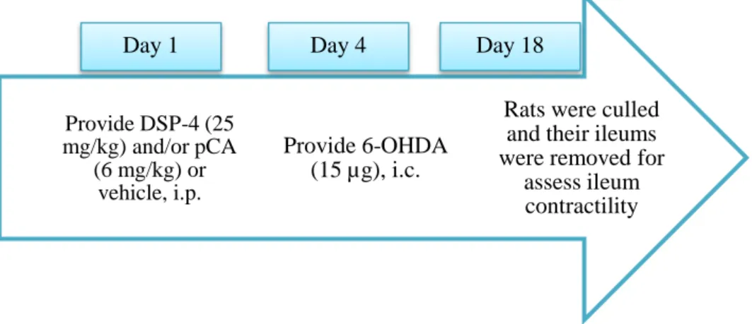
2.4.3. 6-OHDA Injection ... 31
2.4.4 DSP-4 Intraperitoneal Injection ... 32
2.4.5. pCA Intraperitoneal Injection ... 32
2.4.6. Removing the gut ... 38
2.4.7. Mounting the gut in the organ bath ... 38
2.4.8. Effect of acetylcholine chloride (AchC) concentration in gut contractility... 39
2.4.9. Effect of Exendin-4 (EX-4) in gut contractility... 39
2.4.10. Effect of Exendin-(9,39) in gut contractility ... 40
2.5. Experimental Protocol and Animal Usage ... 32
2.6. Tissue dissection and homogenization ... 41
2.7. Biorad analysis for a 96 well-plate assay ... 41
2.8. Rat Cytokine Array ... 42
2.9. Measurements and Statistical Analysis ... 43
3. Results ... 41
3.1. Introduction and Experimental Details ... 42
3.2. Dose-Response Curve Sham vs. 6-OHDA treated ... 43
xiii
3.6. Effect of EX-(9, 39) in ileum contractility ... 46
3.7. Gut Cytokine Array ... 47
4. Discussion ... 49
4.1. Introduction and Project Details ... 50
4.2. SHAM vs. 6-OHDA treated rats ... 51
4.3. SHAM vs. 6-OHDA + DSP-4 treated rats... 52
4.4. SHAM vs. 6-OHDA + pCA treated rats ... 53
4.6. Effect of EX-(9, 39) in ileum contractility ... 55
4.7. Cytokines expression in the GI tract ... 56
4.8. Future Work ... 61
4.9. Final Remarks ... 64
5. References ... 67
Appendix ... xxiii
xv
Figure 1.1 – Brain regions affected by Parkinson’s disease.. ... 3
Figure 1.2 - Progression of PD-related intraneural pathology ... 5
Figure 1.3 - Schematic representation of some of the connections involved in local enteric reflexes .. 11
Figure 1.4 - A simplified schematic of the interaction between microglia and astrocytes. ... 16
Figure 1.5 – Structure of proglucagon gene fragment contains sequences coding for several biologically active peptides. ... 22
Figure 2.2 – Scheme of a gut segment mounted in the organ chamber for recording. ... 39
Figure 2.3 – Diagram representing the addition of EX-4 to the organ bath. ... 40
Figure 2.4 – Diagram representing the addition of EX-(9, 39) to the organ bath. ... 40
Table 2.5 – Bradford assay to quantify protein amount. ... 42
Figure 3.1 - Effect of acetylcholine chloride (AchC, 10 µM) in the isolated ileum of Sham and 6-OHDA treated rats with a selective dopaminergic lesion... 43
Figure 3.2 - Effect of acetylcholine chloride (AchC, 10 µM) in the isolated ileum of Sham and 6-OHDA + DSP-4 treated rats with a combined noradrenergic and dopaminergic lesion ... 44
Figure 3.3 - Effect of acetylcholine chloride (AchC, 10 µM) in the isolated ileum of Sham and 6-OHDA treated rats with a combined serotonergic and dopaminergic lesion. . ... 45
Figure 3.4 - Effect of EX-4 (EX-4, 1 µg/mL) in the isolated ileum of 6-OHDA and 6-OHDA+EX-4 treated rats with a selective dopaminergic lesion. ... 46
Figure 3.5 - Effect of EX-(9,39) (1 µg/mL) in the isolated ileum of vehicle (saline) injected rats. ... 47
Figure 3.6 – Cytokine array with ileum tissue from Sham, 6-OHDA, 6-OHDA + pCA and 6-OHDA + DSP-4 lesioned rats. . ... 48
Figure 4.1 - Proposed mechanism of action of GLP-1R dependent signal transduction pathways in the intestinal cell.. ... 55
xvii
Table 1.1 – GI symptoms associated with PD ... 12
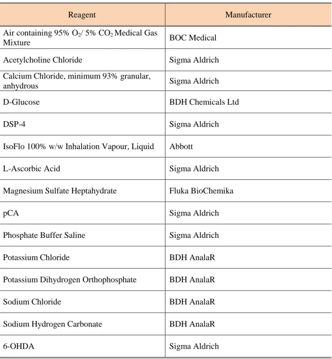
Table 2.1 - List of reagents and supplementary materials used in the course of work. ... 28
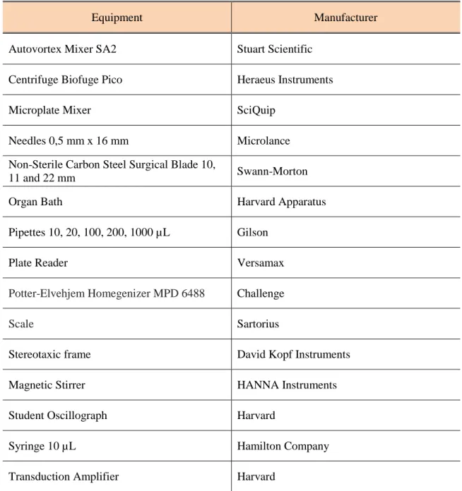
Table 2.2 - List of equipment used in the course of work. ... 29
Table 2.3 – Composition of Krebs Solution used in the course of work.. ... 30
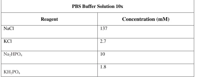
Table 2.4 – Composition of PBS Buffer Solution used in the course of work.. ... 30
xviii 5-HIAA - 5-Hydroxyindoleacetic acid
5-HT - 5-hydroxytryptamine, serotonin 5-HTP - 5-Hydroxytryptophan 6-OHDA - 6- Hydroxydopamine
AADC - Aromatic L-Amino Acid Decarboxylase AC - Adenylate cyclase
Ach – Acetycholine
AchC – Acetycholine Chloride AIF – Apoptosis-Inducing Factor ANS - Autonomic Nervous System AS – α – synuclein
ATP – Adenosine Triphosphate ATP13A2 – ATPase Type 13A2 BBB- Blood Brain Barrier
cAMP - Cyclic adenosine monophosphate c-cas-3 – cleaved caspase-3
CREB - cAMP Response Element Binding Protein CNS – Central Nervous System
COMT - Catechol-O-methyl transferase
DA - Dopamine (3, 4-dihydroxyphenethylamine) DAT - Dopamine transporter
DMV – Dorsal Motor Nucleus of the Vagus nerve DPP-4 - Dipeptidyl Peptidase-4
xix EC50 – Half maximal Effective Concentration
ENS – Enteric Nervous System
Epac2 - Exchange Proteins directly Activated by cAMP 2 EX-4 - Exendin-4
EX-(9, 39) – Exendin-(9, 39)
GDNF - Glial cell line-Derived Neurotrophic Factor GI - Gastrointestinal
GLP-1- Glucagon-like peptide-1
GLP-1R - Glucagon-like peptide-1 receptor GLP-2- Glucagon-like peptide-2
GRPP – Glicentin-Related Pancreatic Polypeptide i.c. - Intracerebral (injection)
i.p. – Intraperitoneal IFN-γ – Interferon-gamma IL-6 – Interleukin-6 IL-1β – Interleukin-1β IL-13 – Interleukin-13
IL-13R1 – Interleukin-13 Receptor 1 IP-1 – Intervening Peptide-1
IP3 – Inositol Triphosphate
IPAN – Intrinsic Primay Afferent Neuron KO- Knockout
xx LRRK2 - Leucine-Rich Repeat Kinase 2
LPS - Lipopolysaccharide
MAPK - Mitogen Activated Protein Kinase MAO - Monoamine oxidase
MPTP - 1-methyl-4-phenyl-1, 2,3,6-tetrahydropyridine NA - Noradrenaline, norepinephrine
NAT - Norepinephrine (noradrenaline) transporter NFκB - Nuclear Factor kappa B
NO – Nitric Oxide OXM - Oxyntomodulin PARK7 – Parkinson Disease 7 PBS - Phosphate buffered saline pCA - Para-chloroamphetamine PD - Parkinson’s disease
PINK1 - PTEN-Induced Putative Kinase 1 PKA - Protein kinase A
PKC - Protein kinase C PLC – Phospholipase C
ROS - Reactive Oxygen Species SERT - Serotonin Transporter SN – Substantia Nigra
SNpc - Substantia nigra pars compacta TH - Tyrosine hydroxylase
xxi TPH1 - Tryptophan hydroxylase 1
TPH2 - Tryptophan hydroxylase 2 TNF-α – Tumor Necrosis Factor-alpha VEGF – Vascular Endothelial Growth Factor VIP – Vasoactive Intestinal Peptide
1
1. Introduction
2
1.1. History of PD
The pathology now referred to as Parkinson’s disease (PD) has been in existence since medieval times and has afflicted all global populations. The ancient Indian medical system of Ayurveda described some symptomatic features of PD under the name Kampavata (Manyam and Sanchez-Ramos, 1999). Traditional therapies in the form of herbal preparations containing anticholinergics, levodopa, and monoamine oxidase inhibitors were used in the treatment of PD in India, China, and the Amazon region of South America (Gourie-Devi et al., 1991). Galen of Pergamum (AD 138-201), a prominent Roman physician and philosopher, also described several features of PD and characterized it as the “shaking palsy”. However, PD was not formally recognized and its symptoms were not documented until 1817 in An Essay on the Shaking Palsy by the British physician James Parkinson (Parkinson, 2002). PD was then known as paralysis agitans, the term "Parkinson's disease" being coined later by Jean-Martin Charcot. The underlying biochemical changes in the brain were identified in the 1950s largely due to the work of Swedish scientist Arvid Carlsson, who later went on to win a Nobel Prize in Physiology or Medicine for his research on dopamine (DA) (Carlsson et al., 1957). PD is a progressive disorder and the motor symptoms manifest only when approximately 70% of the DA neurons in the substantia nigra have already degenerated (Fearnley and Lees, 1991). Although there is no current cure for the disease, there are a number of effective symptomatic therapies available (Poewe, 2006). The first specific treatment to be used for PD was L-dihydroxyphenylalanine (L-DOPA), which entered clinical practice in 1967 (Hornykiewicz, 2002). The first large study reporting the efficacy of this drug in patients with PD was published in 1968 (Cotzias, 1968).
1.2. General Introduction
The characteristic symptoms of PD are the result of a selective degeneration of dopaminergic neurons of the nigro - striatal axis ( substance nigra - striatum ) in the central nervous system (CNS), a highly reduced DA synthesis capacity and a consequent inability to activate DA receptors in the striatum (Whitton, 2007; Iarlori, 2009) .
Despite being strongly related to age PD approximately affects 3 % of the population aged 65 years and 4-5 % of the population aged over 85 years old, 5-10 % of patients are aged less than 40 years old (Iarlori, 2009) . The symptoms are visible only after a "silent" loss of approximately 80 % of dopaminergic neurons in the substantia nigra pars compacta (SNpc), possibly due to an ongoing apoptotic cascade, free radicals formation and neuroinflammation (Perry, 2012) (Figure 1.1).
3
Figure 1.1 – Brain regions affected by Parkinson’s disease. Adapted from Dauer and Pzedborski, 2003.
However, due to the growing number of aging population - a result of increased quality of public health service - and, as the average life expectancy increases, it is expected that the percentage of patients with PD will increase. Epidemiological studies have shown that sporadic PD cases with late onset occurs in 95% of patients while the remaining 5 % are cases of familial PD with early onset (Tanner and Ben- Shlomo, 1999).
Although most cases of PD may not be associated with a specific cause, familial PD has been associated to mutations in several autosomal dominant and autosomal recessive genes encoding proteins such as parkin , PINK1 (PTEN-Induced Putative Kinase 1) or α - synuclein (Pilsl and Winklhofer, 2012) .
After four decades of scientific research, a therapeutic strategy that provides a cure or at least the possibility of preventing the progress of the disease remains elusive. Treatments are based on drugs that provide symptomatic relief and do not show efficacy in all patients. Additionally, the side effects of most drugs used in PD patients are associated with a high mortality rate, especially in cases of chronic use. Likewise, the treatment effectiveness inevitably decreases as cell death progresses. For this reason, there is a need and a lack of an innovative therapy that is both affordable and, most importantly, has the potential to prevent disease progression and mitigate symptoms (Harkavyi et al., 2008; Hurtig, 1997).
4
1.3. Neuroanatomy in PD
The extended observation of Lewy bodies - the main location of α - synuclein (AS) - is a major feature of the pathology. AS is a small and relatively unstructured protein, with sticky elements that make it suitable to aggregate. Lewy bodies - eosinophilic inclusions with a diameter of approximately 8-30 µm and consisting of abnormal, insoluble aggregates of AS - are usually circular with a eosinophilic core and surrounded by a pale halo. Researchers believe that when a small cluster of AS is formed, it tends to attract proteins in the same neighborhood and eventually triggers the formation of long, insoluble fibrils which may be broken into fragments of shorter length . AS clusters of smaller size and formed near the site where the aggregation process starts are the most toxic forms of the protein and its toxicity will destroy neighboring neurons. If these structures are symptomatic or causative of PD is still unclear.
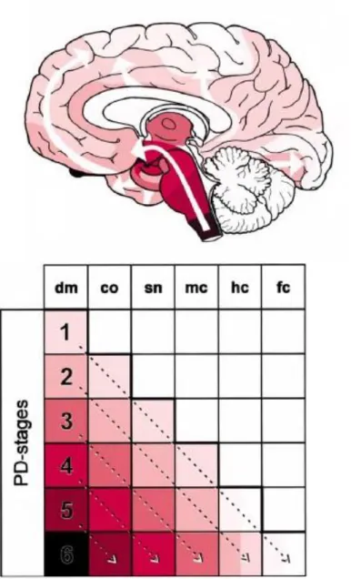
Braak and colleagues found that, in samples with mild pathology, which Braak called Stage 1, the Lewy bodies are typically confined to the olfactory bulb and the dorsal motor nucleus of the vagus nerve (DMV). In Stage 2, Lewy bodies continue to ascend into the brainstem, reaching the medulla oblongata and pontine tegmentum, parts of the brainstem that control swallowing, sleep, and other autonomic functions sometimes affected in PD. By Stage 3, pathology starts to show up in the amygdala (a mass of neurons involved in processing fear and other emotions, but also the sense of smell) and in the substantia nigra; this is the stage when the motor phase of the disorder begins. In Stage 4, pathology in areas affected in earlier stages worsens, and Lewy bodies progress to the forebrain and invade a portion of the cerebral cortex (the temporal mesocortex), whereas the neocortex, the part of the brain involved in higher functions, remains unaffected. In Stages 5 and 6, the pathology is full blown, appearing initially in the anterior association and prefrontal areas of the neocortex and then spreading to the posterior association areas, which are involved with memory and learning, and planning movement (Figure 1.2). Defects in these areas could explain many of the cognitive problems associated with advanced PD (Braak et al., 2003a; Braak et al., 2004).
5
Figure 1.2 - Progression of PD-related intraneural pathology (Adapted from Braak et al., 2003a). Upper – Diagram showing the ascending pathological process (white arrows). The increasing intensity of the colored areas indicates the growing severity of the pathology in vulnerable brain regions. Lower – Simplified diagram showing the topographic expansion of the lesions (from left to right: dm to fc) and, simultaneously, the growing severity on the part of the overall pathology (from top to bottom: stages 1–6). List of abreviations: dm, dorsal motor nucleus of the vagal nerves; co, coeruleus complex; sn, substantia nigra; mc, mesocortex; hc, high order sensory association areas; fc, first order sensory association areas.
1.4.Motor and Non-motor Symptoms
The best motor symptoms associated with PD include tremors, rigidity in movement, language problems, cognitive problems, bradykinesia - slowness of coordinated voluntary movements - and
6 hypokinesia - loss of coordinated voluntary movements. Postural instability, which is characterized by loss of postural reflexes, leads to balance difficulties and a higher propensity for falls.
Other motor symptoms include dystonia - abnormal and painful muscle contractions - mostly on the feet but may include other skeletal muscles. Patients with PD have difficulty in swallowing; the facial muscles are also affected.
In the last decade, the so-called non-motor symptoms have attracted considerable scientific attention and, particularly, due to the appearance of new hypotheses concerning the beginning and progress of the disease (Woitalla and Goetze, 2011). The non-motor symptoms experienced by patients with PD have been increasingly recognized as part of the degenerative process associated with the pathology and contribute significantly to the morbidity and mortality related to the disease (Salat - Foix and Suchowersky, 2012).
Some of the non-motor symptoms include neuropsychiatric and cognitive disorders, anxiety, fatigue, apathy and dementia (visual and auditory hallucinations) and complications in the gastrointestinal (GI) system - constipation, fecal incontinence and abnormalities in intestinal transit. Although some of these symptoms are manifested before diagnosis of the disease (PD diagnosis requires identification of parkinsonian motor symptoms), its prevalence, severity and impact on quality of life of patients increases its progression.
A comprehensive survey of PD patients in the 1980’s found that problems with saliva occurred in 70% of PD patients, dysphagia (trouble swallowing) in 52%, nausea in 24%, and constipation in 29%. In addition, defecatory dysfunction was reported by two thirds of PD patients – twice the control prevalence (Edwards et al., 1994). Subsequent studies have confirmed the high frequency of GI abnormalities in PD. Abnormal gastric emptying has been described in 43%-88% of PD patients and can worsen as PD progresses (Goetze et al., 2006; Goetze et al., 2005). The incidence rate of constipation has be estimated to be anywhere from 29%-89% and problems with defecation are also much more common in PD (~60%) than in age-matched controls (Siddiqui et al., 2002; Singer et al., 1992). Total GI tract transit time is also significantly prolonged in PD (Davies et al., 1996).
1.5. Physiology
1.5.1. Dopamine
DA was first discovered in 1952 by the Swedish scientist Arvid Carlsson. Arvid Carlsson was awarded the Nobel Prize for demonstrating that DA was a neurotransmitter in its own right and not just a precursor of noradrenaline and adrenaline. DA is the predominant neurotransmitter belonging to the group of catecholamines in the mammalian brain. The name “dopamine” is derived from its
7 precursor L- DOPA (Dihydroxy L- phenylalanine). DA belongs to the group of the catecholamine neurotransmitters – a family that possesses a catechol functional group in its structure.
Three of the major four dopaminergic pathways originate in the substantia nigra (SN) of the midbrain. The nigro - striatal axis is involved in the control of movement and is affected in PD and in other pathologies responsible for perturbations in the movement. The remaining two pathways originating from the SN, via the mesolimbic and mesocortical pathways, have a central role in the control of emotion and motivation. The fourth route of DA synthesis starts in the hypothalamus and is projected to the pituitary gland, which regulates the secretion of hormones.
The rate limiting step in the synthesis of DA is catalyzed by the enzyme tyrosine hydroxylase (TH), which is an oxidase that converts tyrosine to L- DOPA. TH is present in all cells with the ability of producing catecholamine neurotransmitters. L- DOPA is then decarboxylated by aromatic amino acid decarboxylase (AADC), forming DA and CO2.
Neurons that use DA as their neurotransmitter only possess TH and AADC unlike, for example, noradrenergic neurons that also possess β-hydroxylase which in turn adds a hydroxyl group to DA to form noradrenaline. During development, the expression of genes encoding enzymes responsible for the synthesis of catecholamines can be regulated independently.
There are several mechanisms for the inactivation of DA after release into the synaptic cleft . The main mechanism is the reuptake of DA in the synaptic cleft back into the presynaptic dopaminergic neuron through membrane transporters. Released DA into the synaptic cleft also undergoes enzymatic degradation by monoamine oxidase (MAO) – found on the outer membrane of mitochondria – and by catechol -O-methyl transferase (COMT).
There are, at least, five dopamine receptors - D1 to D5. The five receptors have similarities among them since all of them present metabotropic properties – meaning they are G-protein-linked. The D1 and D5 receptors stimulate the production of cyclic adenosine monopohosphate (cAMP), while the D2, D3 and D4 receptors inhibit the production of cAMP (Kebabian and Calne, 1979). D1 and D2 were the first to be discovered and are the most common in nigro-striatal axis. The D4 receptor is expressed in the hypothalamus and limbic areas associated with emotions.
1.5.2. Noradrenaline
Noradreanaline (NA) was isolated by Polish physiologist Napoleon Cybulski in 1895. NA shares the first two reactions in its biosynthetic pathway with DA in a series of enzymatic steps from the amino acid tyrosine. However, cells producing NA possess dopamine- β -hydroxylase enzyme, which adds a hydroxyl group to DA to form NA. Unlike other enzymes in the biosynthetic pathways of small molecules, DA - β -hydroxylase is a membrane associated protein strongly attached to the
8 inner surface of aminergic vesicles. Consequently, NA is synthesized within vesicles, and thus synthesized as a single transmitter.
In the CNS, NA is used as a neurotransmitter by neurons whose cell bodies are present in the locus coeruleus (LC). Although these noradrenergic neurons are present in small numbers, their projections are widespread across the cortex, cerebellum, striatum, hypothalamus, hippocampus and spinal cord. NA is usually excitatory in nature and has been implicated in governing arousal, motivation, emotional state and alertness.
NA exerts its effects via activation of noradrenergic G-protein coupled receptors, which are clustered into two main groups - α (α1 and α2) and β (β1 , β2 and β3) . Upon activation of α1-adrenergic receptors, a G-protein (Gq) activates phospholipase C (PLC), which causes an increase in
inositol triphophate (IP3) and Ca2+. This triggers further effects, primarily through the activation of Protein Kinase C (PKC). The activation of α2-adrenergic receptors causes the inactivation of adenylate cyclase (AC), resulting in a decrease of the second messenger cAMP produced from adenosine triphosphate (ATP). The activation of β-adrenergic receptors causes an increase in the intracellular concentration of cAMP. The effector of cAMP is the protein kinase A (PKA), which will enhance protein phosphorylation.
There are several mechanisms of NA inactivation after its release in the synaptic cleft. The major mechanism is reuptake back into the presynaptic terminal via NA transporters (NAT) present in the membrane. Released NA is also subject to enzymatic cleavage by MAO and by COMT.
In addition to acting as neurotransmitter, NA also functions as a hormone. As a hormone, NA is released through the adrenal medulla, along with adrenaline in the blood in conditions of sympathetic nervous system activation (control under stress). This activation usually takes the form of the “fight or flight response”, hereafter NA has the ability to increase heart rate, blood pressure, glycogenolysis in the liver and adipose tissue lipolysis.
1.5.3. Serotonin
Vittorio Erspamer was the first scientist to isolate serotonin (5–HT), in 1935 from enterochromaffin cells - a type of endocrine cells in the epithelium of the digestive tract and respiratory system. These cells contain approximately 90 % of the 5-HT stored in the body.
Two enzymes are necessary to synthesize serotonin from tryptophan - tryptophan hydroxylase (TPH) and 5- hydroxytryptophan (5-HTP) decarboxylase. The rate limiting step of the reaction is catalyzed by the first enzyme in the biosynthetic pathway - TPH. In the CNS, the neuronal cell bodies of serotoninergic neurons are found in and around the nucleus of Raphe and in the brain stem, which are
9 involved in regulating the level of attention and other cognitive functions. The projections of these neurons are widely distributed in various areas of the brain and spinal cord.
In the peripheral nervous system, 5-HT functions to regulate GI motility, blood clotting, vasoconstriction, and cell growth.
There are seven families of 5-HT receptors (5-HT1 - 5HT7) responsible for both inhibitory and
excitatory signals. With the exception of 5- HT3 receptor - an ion channel regulated by binding of its
ligand -, the other 5-HT receptors are coupled to G proteins, thereby activating an intracellular second messenger cascade. 5-HT receptors modulate the release of many neurotransmitters, including glutamate, GABA, DA, adrenaline / NA, and acetylcholine (Ach).
There are several mechanisms of 5-HT inactivation after its release in the synaptic cleft. The major mechanism is reuptake back into the presynaptic terminal through 5-HT transporter (SERT) present in the membrane. The released 5-HT is also subject to enzymatic cleavage and is converted to 5- hidroxindoleacetic acid (5- HIAA) by MAO.
The 5-HT receptors influence various biological and neurological processes such as aggressive behavior, anxiety, appetite, cognition, learning, memory, sleep and thermoregulation. The 5 -HT receptors are subject to a wide variety of drugs and illegal drugs such as antidepressants, antipsychotics, hallucinogens and amphetamines.
1.6. Gastrointestinal Dysfunction in PD
The most prominent clinical features associated with PD are the motor symptoms, mainly associated with the loss of dopaminergic neurons in the SNpc. Symptoms associated with GI dysfunction had previously attracted a minority of attention but now a new focus of interest and an increasing number of references are addressing the GI system in the pathogenesis of PD. The GI tract has a special importance because it can be the starting point of the disease (Woitalla and Goetze, 2011).
The GI tract is the largest interface between neural tissue and the environment and has a sufficiently high number of neuronal cells to be known as the "second brain". The lumen of the GI tract holds the largest and most diverse resident microbial community in the human body, with the ability to induce inflammation and pro-oxidative pathways. In the various regions of the gastrointestinal (GI) tract, muscle layers of the gut wall and their innervation are adapted and organized to subserve the motor functions of that region. Along the GI tract, the gut interacts with the CNS through autonomic neurons. Various types of GI dysmotility have been documented repeatedly in PD, mainly delayed gastric emptying and obstipation, and most likely reflect dysfunction at one or more levels of the brain-gut axis (Kellow et al., 1999).
10 Korczyn described that the gastrointestinal symptoms precede motor symptoms in Parkinson patients (Korczyn and Gurevich, 2010). The DMV is always involved in the pathology of the disease in Stage 1 (Braak et al., 2004). Once the vagus nerve connects the brain to the enteric nervous system (ENS), the authors proposed that the Lewy bodies could form in the gut and move along the vagus nerve in an upstream, or retrograde, direction toward the brain.
1.6.1. Neuroanatomy of GI Dysfunction in PD
The basis of an appropriate GI transport is a coordinated contraction of smooth muscle cells (most of the GI tract is composed of smooth muscle cells). The contractions and local reflections are coordinated by the ENS located in the intestinal wall. The ENS besides being responsible for bowel movements also integrates impulses from the CNS (through extrinsic innervation from the vagal nerve).
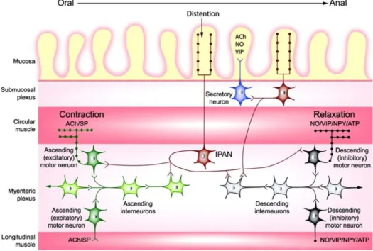
The ENS is a subdivision of the autonomic nervous system (ANS). The ENS comprises two neuronal plexus - the submucosal (or Meissner’s) plexus, which controls mucosal secretion and blood flow, and the myenteric (or Auerbach’s) plexus, which controls peristaltic movements. From a functional point of view the Auerbach’s plexus operates as a multisynaptic reflex, in which local factors, such as wall distention or the chemical composition of intraluminal contents, are sensed by the so-called intrinsic primary afferent neurons (IPANs). IPANs subsequently transmit this information to specific neurons capable of triggering motor, secretomotor or vasomotor responses that ultimately reach the smooth muscle cells (Figure 1.3) (Salat-Foix and Suchowersky, 2012). The ENS has approximately one hundred million neurons (one thousandth of the number of neurons in the brain) and a number of neurotransmitters, including DA and Ach. The main excitatory neurotransmitter in the ENS and in the parasympathetic nerves is Ach, and in the sympathetic nerves the main inhibitory neurotransmitter is NE. In all the other systems nitric oxide (NO), vasoactive intestinal peptide (VIP) and ATP are the main inhibitory neurotransmitters (Figure 1.3). Despite the ability of the ENS to run autonomously, it interacts closely with the CNS. In the stomach, the myenteric plexus connects directly to the vagus nerve, providing a direct link between the CNS and stomach.
The mobility in the small intestine as well as in all parts of the GI system is modulated predominantly by excitatory or inhibitory signals from the ENS. However, GI mobility is also regulated by signals originating in the CNS. GI hormones (gastrin, GLP1, GLP2) also appear to affect the GI mobility in some way.
11
Figure 1.3 - Schematic representation of some of the connections involved in local enteric reflexes. (Adapted from
Benarroch, 2007). List of abreviations: IPAN, intrinsic primary afferent neurons; Ach, acetylcholine; NO, nitric oxide; VIP, vasoactive intestinal peptide; SP, substance P; NPY, neuropeptide Y; ATP, adenosine triphosphate.
Phosphorylated AS in Lewy bodies and Lewy neurites - one of the pathological hallmarks of idiopathic PD - are present in the myenteric plexus of patients with PD and the first observation of AS in the ENS coincides with the appearance of the same in the DMV. These inclusions have been detected in patients with advanced PD, but also in non- symptomatic patients with lesions limited to the brain stem, thus supporting the hypothesis that changes in the GI tract may be an initial characteristic of pathology. Although animal models have inherent limitations and the findings related to the accumulation of AS and its dissemination are not wholly consistent, there is a growing body of evidence to suggest that the GI system may be critical to the pathogenesis of PD.
Drolet et al.. exposed healthy rats to low doses of rotenone for six weeks, a mitochondrial complex I inhibitor. Animals lost some of their ability to coordinate digestion and showed evidence of Lewy bodies in enteric neurons (Drolet et al., 2009).
1.6.2. Gastrointestinal Complications
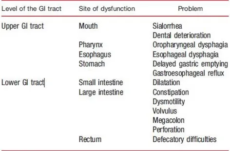
GI disorders are common in all stages of PD patients. Approximately 30% of PD patients report structural and functional abnormalities in the GI tract, and practically, vulnerabilities have been observed throughout the entire GI system (Table 1.1).
12 Swallowing difficulties and constipation symptoms were included in the first description of PD in 1817 (Parkinson, 2002). Constipation is the most traditional symptom of GI dysfunction in PD patients. Constipation was observed in 90% of patients and it has been described as a feature of the pre-motor stages of the disease.
The stomach is one of the first places where deposition of AS is observed in parkinsonian patients, suggesting that the stomach, although not receiving as much attention as abnormalities of gut motility, is also involved in the pathogenesis of PD.
Gastrointestinal disorders affect the quality of life of many patients and an altered gastric function may cause inadequate absorption of drugs, exacerbating the classic motor symptoms of PD. The worsening of GI dysfunction in PD is repeatedly associated with the progression of PD state
.
Table 1.1 – GI symptoms associated with PD (adapted from Marrinan et al., 2013)
Except for dysphagia, which may lead to weight loss and respiratory tract infections (aspiration pneumonia is a leading cause of death among PD patients), GI symptoms are mainly a source of discomfort and social embarrassment, and their overall impact on the patient’s life expectancy is considered marginal.
1.6.3. Catecholamines in the ENS
Catecholamines modulate GI motility. Nerve endings of the sympathetic nervous system release NA, which inhibits the release of ACh from parasympathetic motor neurons (by α2-adrenoceptors) and decreases the contraction of intestinal smooth muscle cells. Moreover, the release
13 of ACh from parasympathetic nerve terminals enhances the contraction of smooth muscle through activation of M receptors.
DA was recently hailed as an enteric neurotransmitter as DA, TH and DA transporter (DAT) are co-localized in neurons of the ENS.
DA acts as a negative regulator of motility in the GI tract to inhibit the release of ACh from cholinergic neurons expressing dopamine D2 receptor.
Tian et al.. recently demonstrated an increased expression of DA, TH and DAT in the GI tract of mice subjected to a bilateral injury induced by a bilateral injection of 6- hydroxydopamine (6-OHDA), a neurotoxin with selective toxicity to dopaminergic neurons. Given the inhibitory nature of DA in ENS, an overexpression of this neurotransmitter would reduce the mobility and contractions of the GI tract, especially gastric emptying (Tian et al., 2008).
In fact, in patients with PD, administration of the D2 antagonist, domperidone receptor, accelerates gastric emptying delayed by administration of therapy with L- DOPA (Tonini et al.., 2004).
1.6.4. Serotonin in the ENS
For decades, it has been extensively described in the literature the role of 5-HT in regulating GI function. The majority of 5-HT is synthesized and stored in the intestine and different types of serotonergic receptors are found in the intestinal wall. However, despite many evidences, the actual functions of this neurotransmitter in the GI tract have proven difficult to identify. The reasons for this gap are related to the existence of 5-HT prevenient from neurons in the CNS and 5-HT prevenient from neurons in the ENS and to the widespread and overlapping distribution of specific subtypes of serotonergic receptors.
In 2011, Li et al. (Li et al., 2011) used knockouts (KO) of tryptophan hydroxylase 1 (TPH1), the rate-limiting enzyme in the synthesis of enteric 5–HT, and KO of tryptophan hydroxylase 2 (TPH2), the rate-limiting enzyme in the synthesis of neural 5–HT, to selectively delete the production of 5-HT from the two possible sources. The TPH1 KO did not differ from controls in any studied function - GI emptying, intestinal transit and colonic motility. By contrast, TPH2 KO showed major changes in each of the examined functions; KO mice for both enzymes were indistinguishable from TPH2 KO animals. The presence of a mediator to replace 5- HT in KO TPH1 is plausible. However, without a likely candidate for a compensatory mediator, the most obvious conclusion is that 5-HT in the intestinal mucosa has a very minor role in the regulation of GI motility in the mouse whereas, neural 5-HT may have a more substantial role than previously thought.
Perhaps the 5-HT from the intestinal mucosa plays a significant role only after some pathophysiological insult, for example, inflammation. Moreover, neuronal 5-HT is clearly necessary
14 for normal functioning of the GI tract. The role of enteric 5-HT remains unclear and requires further study, especially, being the source of all circulating 5–HT.
1.6.5. Diagnostic and Therapy
The identification of gastrointestinal symptoms in parkinsonian patients is relatively direct and straighforward through appropriate questions directly to the patient. Gl symptoms may be related to the administration of antiparkinsonian drug therapies, either as a side effect, or as a manifestation of improper treatment.
The management of these symptoms in PD patients should be performed identically to the management of the same in the general population - anticholinergics and botulinum toxin to prevent the frequency of drooling, proper diet and laxatives if constipation . The treatment of dysphagia within the PD patients is usually ineffective (Menezes and Melo, 2009). The adjustment of dopaminergic treatment is rarely effective and usually maintaining a proper diet with intake of homogeneous food with thick texture is the best solution. Patients are advised to eat small portions and chew their food several times before swallowing (Baijens and Speyer, 2009). If the above measures prove ineffective, feeding through a gastric tube may be necessary.
The management of constipation in PD is carried out along the general lines that are observed in any other setting. Nonpharmacological measures, such as increasing dietary fiber and fluid intake, and avoiding sedentarism, are recommended, although their effectiveness has not been proven in well-designed clinical trials. Pharmacologic treatment, if needed, can be attempted with various laxatives (Salat-Foix and Suchowersky, 2012).
1.7. Neuroinflammation
Several hypotheses have been postulated regarding the possible causes of neuronal neurodegeneration observed in PD patients. These include genetic factors, environmental toxins, mitochondrial dysfunction and cell death mediated by free radicals (Schapira, 1994; Rosenberg, 2002). Although there is a smaller body of evidence to suggest that neuroinflammation is the main stimulus to the onset of neurodegeneration, preclinical and epidemiological data suggest that chronic, slow and steady, neuroinflammation may be a reason for neuronal dysfunction during the asymptomatic phase of PD (Lee et al., 2009).
Neuroinflammation induced by exposure to either infectious agents or toxic agents with proinflammatory characteristics is currently recognized as a major contributor to the pathogenesis of PD (Whitton, 2007).
15 The traditional features of neuroinflammation include the presence of activated microglia cells and reactive astrocytes, direct participation of the adaptive immune system and overproduction of cytokines, chemokines, prostaglandins and reactive oxygen species (ROS 's), which have the ability, in certain cases, to penetrate the blood-brain barrier (BBB) (Ransohoff and Perry , 2009).
Over a long period of time, the CNS has been considered a prime location of the immunological point of view because of an absent classic immune response (Di Filippo et al., 2008). Thus, it is also thought that the brain would not be greatly affected by systemic immune or inflammatory reactions (Lucas et al., 2006).
It is now widely accepted that there is an interaction between the nervous and immune systems involving a two-way cross-talk. Indeed, the CNS is stocked with a classical active immune surveillance and can be the starting point of an inflammatory responses caused by different types of lesions (Di Filippo et al., 2008).
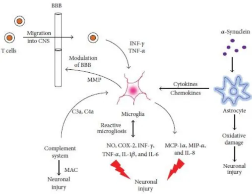
The secreted inflammatory mediators are responsible for the expression of cytokines and adhesion molecules, activation of the glial cells and stimulating astrogliosis (Figure 1.4). Therefore, neuroinflammation is seen as a complex cellular and molecular response of neuronal cells to harmful stimuli such as injury, stress or sepsis (Semmler et al., 2008; Whitney et al., 2009). The main objectives of neuroinflammation are defending against these damaging insults, removal of damaged neurons and maintain homeostasis of the CNS.
16
Figure 1.4 - A simplified schematic of the interaction between microglia and astrocytes (Adapted from More et al., 2013).
Abbreviations: NO: nitric oxide, COX-2: cyclooxygenase, INF-𝛾: interferon-𝛾, TNF-𝛼: tumor necrosis factor 𝛼, IL-1𝛽: interleukin-1𝛽, 6: interleukin-6, MCP-1𝛼: monocyte chemotactic protein-1, MIP-𝛼: microphage inflammatory protein, IL-8: interleukin-8, MAC: membrane attack complex, 𝛼-syn: 𝛼-synuclein, MMP: matrix metalloprotein, BBB: blood brain barrier, C3a: complement component 3a, and C4a: complement component 4a.
However, when neuroinflammation is not controlled, its beneficial effects are subdued and may contribute to progression of the damage. In this case, the recruited cells release more inflammatory mediators, and establish a positive feedback leading to neuronal damage with changes in neurogenesis (Swarup et al., 2008, Whitney et al., 2009). Additionally, oligodendrocytes are extremely vulnerable to inflammatory molecules, which can result in damage to white matter in the appearance of neuromotor, cognitive and behavioral limitations in neurodegenerative disorders such as PD or Alzheimer’s Disease.
1.7.1. Cytokines as inflammatory mediators
Cytokines are small signaling molecules - peptides, proteins or glycoproteins - involved in cell-to-cell communication, immune responses and movement of cells towards sites of inflammation, infection and trauma. Previously, cytokines were thought to only act in the peripheral system.
Nowadays, an increasing number of observations reveal that these molecules exert different actions in the CNS and they are involved in neuronal development. Proinflammatory cytokines such as Tumor
17 Necrosis Factor–alpha (TNF-α) and interleukin-1beta (IL- β) are synthesized by neural cells and participate in standard intercellular communication, assuming an important role in maintaining cellular homeostasis (Giulian et al., 1988, Zhao and Schwartz, 1998).
Typically, the levels of these cytokines are decreased (in order of picomolar), but their expression increases rapidly in the event of neuroinflammation and this increase may be 1000 fold. When this increased expression is unsustainable, proinflammatory cytokines can become pathological, deregulating cytokine release and causing the death of neurons and oligodendrocytes (Donnelly and Popovich, 2008).
Yet, the detrimental effect of cytokines may rely on the type and level of cytokines produced. In other words, the low physiological rates of cytokine expression may be important for the cross-talk between the neural cells during development, but the overexpression observed during the neuroinflammatory stage may compromise neuronal survival and plasticity.
Therefore, regulating the production of neuroinflammatory mediators or their action on their receptors would be an effective approach to mitigate the inflammatory processes in PD. This knowledge may be helpful in developing pharmacologic strategies for treating neuroinflammation in PD (Whitton, 2007).
1.8. Causative Factors
Most cases of PD are not inherited and although aging is a clear risk factor, the disease affects only a fraction of the elderly population. This indicates the involvement of external factors - genetic and environmental - as causatives of the disease.
1.8.1. Genetic Factors
PD is a multifactorial disease in which 5% of cases are hereditary in nature, caused by mutations in autosomal dominant and recessive genes. Autosomal dominant mutations in genes encoding proteins such as α-synuclein and Leucine-Rich Repeat Kinase 2 (LRRK2), and autosomal recessive mutations in genes encoding parkin, PTEN-Induced Putative Kinase 1 (PINK1), ATPase Type 13A2 (ATP13A2) and Parkinson Disease 7 (PARK7) are involved in the degeneration associated the pathogenesis of PD.
Mutations in the proteins encoded by these genes have resulted in the formation of aggregates of misfolded proteins, changes in the ubiquitination pathway and changes and defects in the mitochondrial respiratory chain.
18
1.8.2. Environmental Factors
Sporadic PD (95% of cases have a sporadic nature) is caused by environmental factors and toxins. Herbicides such as paraquat and rotenone, or the toxin MPTP (1-methyl-4-phenyl-1, 2,3,6-tetrahydropyridine) appear to induce parkinsonism in humans and animal models. Either MPTP or rotenone are inhibitors of complex I of the mitochondrial respiratory chain, which will cause a decrease in ATP production and an increase in the synthesis of ROS's. Activation of apoptotic cascades (members of the Bcl-2 and caspase family) will trigger the death of neurons in the nigro-striatal axis.
These findings were observed when analyzing post mortem brain tissue of patients with PD who presented elevated levels of lipid peroxidation markers in the CNS, suggesting high levels of oxidative stress (Andersen, 2004).
1.9. Current Treatments for Motor Symptoms
1.9.1. Pharmacological Treatments
Currently, there are several sites of action for pharmacological therapies related to PD, however, existing drug therapies on the market have the potential to only attenuate or delay the progression of symptoms and do not prevent or reverse the motor and non-motor symptoms of the disease.
Administration of Levodopa will increase DA levels.
In PD, DA depletion is accompanied by an increased activity of the striatal cholinergic system, subsequent rearrangement of the striatal circuitry, and appearance of the motor symptoms. Ach inhibitors block the action of this neurotransmitter in the striatum, attenuating the stiffness and gear-wheel movements, typical of patients with PD. Antagonists of muscarinic cholinergic receptors were the first drugs approved and introduced in the market for the treatment of PD (Hobson et al., 2002), being the first demonstration of the functional antagonism between DA and Ach - activation of inhibitory dopamine D2 receptors reduces the release of Ach (Polymeropoulos et al. 1997). Although this class of drugs has shown beneficial effects, it is also associated with adverse neuropsychiatric effects (Katzenschlager et al., 2003) .
The use of Amantadine stimulates the release of DA in the synaptic cleft and inhibits the reuptake of the released DA in the synaptic cleft back into the presynaptic neuron, increasing the period of time during which DA remains bound to its receptors.
The use of MAO inhibitors - present in the outer mitochondrial membrane which plays a major role in oxidative deamination of monoamines, such as DA - is another recurring
19 pharmacotherapeutic strategy in the treatment of PD. There are two isoforms of the enzyme, MAO-A and MAO-B. The B-isoform is present predominantly in the brain and is found in astrocytes but not neuronal cells (Westlund et al., 1985). The most well-known MAO inhibitor in the treatment of PD is selegiline, which was introduced in 1989, followed by rasagiline - a selective compound for MAO-B . The administration of selegiline will prevent degradation of DA, which will be available for release into the synaptic cleft (Deane et al., 2004).
Another pharmacotherapy strategy for the treatment of PD is the administration of COMT inhibitors. COMT is an intracellular enzyme located in postsynaptic neurons whose substrate is any compound having a catechol structure, including L -DOPA, DA and NA (Deane et al. 2004). COMT inhibitors act peripherally to prevent L-DOPA and DA degradation. COMT inhibitors cause a prolonged stay of DA in the synaptic cleft and enhance its effects.
There are two drugs in this class which have been approved for PD treatment. Tolcapone, introduced in 1997, followed by entacapone, in 1999. Both drugs increase the bioavailability of L- DOPA , but also causes a large number of side effects, as diarrhea, hypotension, acute liver failure (Rinne et al., 1998; Obeso et al., 2000).
One of the most common therapeutic strategies is to stimulate DA receptors in the striatum directly through the administration of DA receptors agonists. Several DA are available clinically, including rotigotine, pramipexole and piribedil.. Some of the side effects observed in patients under administration of DA agonists include excessive sleepiness and sleep attacks (Richard and Kurlan, 1997), pathological gambling, and related impulse control disorders where there is an inability to resist an impulse desire despite negative consequences.
1.9.2. Experimental Treatments
The possibility of developing cell transplantation therapies for PD is based on the premise that it is the degeneration of nigro-striatal axis the main cause of the onset of symptoms associated with the disorder.
Neurotransplantation is a recently studied experimental strategy for the treatment of PD involving the implantation of cells producing AD in the brain of patients. However, information regarding the effectiveness of this treatment is limited and is not, to date, an accepted treatment throughout the medical community. Promising results were obtained in studies involving rats undergoing treatment with 6-OHDA, where improvements were observed in the progression of motor symptoms (Hurtig et al., 2000).
Chemical infusion directly into the basal ganglia was also performed in some patients with PD. This technique aims an infusion of growth factors into the brain, preventing cell death and
20 simultaneously stimulating the growth of DA-producing neurons. This procedure is still in clinical trials and 10 to 20 years will be needed until its effects are visible.
For many years research has been done in order to deepen the possibility of replacing damaged DA producing cells by brain tissue from human fetuses, hoping that fetal tissue has the ability to produce DA and possibly fix the problems caused by the failure of the same. To date, results have proved ambiguous and treatment is only experimental. Scientists believe that this type of treatment will probably remain experimental in the next 5-10 years.
Stem cells, especially human embryonic stem cells have the ability to differentiate into dopaminergic neurons , although behaviors and cell survival are limited and the potential risk of teratoma formation is high (Goya et al., 2008).
1.10. Current Treatments for GI Dysfunction
Although GI symptoms significantly impair quality of life for PD patients, their treatment is made more difficult by ignorance of the complex enteric regulating mechanisms and lack of medical treatment options. Established treatment options for GI dysfunction differ according to the individual clinical picture and the symptoms to be treated.
Despite growing knowledge of the factors influencing GI motility, therapeutic options are still limited, and only a small number of studies are being performed to examine drug effects on GI function in humans. Limitations of medical knowledge are even more striking, especially concerning studies on GI dysmotility as a condition in PD (Woitalla and Goetze, 2011).
1.10.1. Dopamine Receptors
The clinical effects of levodopa on gastric emptying are controversial. By stimulating dopaminergic chemoreceptors, levodopa causes nausea. In its peripheral effects, levodopa normalizes partially myoelectric activity in the stomach, probably due to its dopaminergic activity in the stomach itself (Lu et al., 2004). Other reports describe the inhibition of ileum coordination, an effect that can be antagonized by blocking peripheral DA receptors (Schuurkes and Van Neuten., 1984).
Domperidone, a potent, mainly peripheral-acting DA antagonist relieves symptoms of nausea, vomiting, anorexia, abdominal bloating and regurgitation but not constipation in PD patients (Soikan et al., 1997). Domperidone increases the amplitude of oesophageal motor function and accelerates gastric emptying (Weihrauch and Ehl, 1981). Considering the effects of domperidone on a cellular level, its action is probably due to blocking D2 receptors (Reddymasu et al., 2007).
21
1.10.2. 5-HT
4Receptor Agonists
Of the 5-HT4 receptor agonists, cisapride has the widest use. Studies in PD patients have been
conducted with cisapride, showing beneficial effects on motor fluctuations (Djaldetti et al., 1995)
.
Cisapride is a peripherally acting substance with positive effects on GI function in PD (Jost, 1997), improving levodopa absorption and motor fluctuations in PD patients (Djaldetti et al., 1995).
Tegaserod, a 5-HT4 agonist was approved for constipation. Studies in PD revealed a tendency for
improvement of abdominal discomfort and symptoms (Sullivan et al., 2006).
Prucalopride is a 5-HT4 agonist approved for colonic constipation. The stimulating effects of
prucalopride in gastric and colonic transit have been demonstrated (Bouras et al., 2001).
1.11. Glucagon Family and Exenatide
1.11.1. Introduction to the GLP-1 family peptides
Glucagon-Like Peptide 1 (GLP-1) is a newly discovered molecule, derived from the L cells of the intestine. This peptide has insulinotropic properties and this is the predominant feature that generated interest in its research. GLP-1 has been described for the first time in 1985 (Schmidt et al., 1985) after cloning the gene encoding proglucagon. GLP-1 is largely responsible for increasing the amount of insulin released by pancreatic β cells after ingestion of nutrients, and for decreasing the secretion of glycogen. GLP-1 acts on a transmembrane G-coupled receptor, the Glucagon-Like Peptide 1 Receptor (GLP-1R) thus stimulating AC and cAMP formation, with downstream effects (Yada et al., 1993) in gene expression.
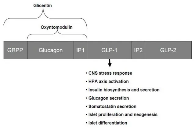
In addition to its insulinotropic properties, GLP-1 is involved in neogenesis, differentiation and proliferation of β cells in islets of Langerhans of the pancreas, in the activation of the hypothalamic-pituitary-adrenal axis and neuroprotection and response to stress in the CNS. GLP-1 functions are summarized in Figure 1.5.
Processing of the proglucagon gene gives rise to 29 amino acid glucagon itself and a number of biologically active peptides including glicentin, oxyntomodulin (OXM), GLP-1 and glucagon-like peptide 2 (GLP-2). Both OXM and glicentin contain the whole 29 amino acid sequence of glucagon and a C-terminal 8-amino acid extension called intervening peptide-1 (IP-1). Compared to OXM (37 amino acids), glicentin (69 amino acids) also contains the N-terminal extension called glicentin-related pancreatic polypeptide (GRPP). OXM has recently been found to suppress appetite and a recent clinical study found that it could be used as a treatment for obesity (Wynne et al., 2006b). The mechanism of action of OXM is poorly understood. It has been shown to bind to both the
glucagon-22 like peptide 1 receptor (GLP-1R) and the glucagon receptor, but it is likely that its effects are mediated by a novel receptor (Wynne and Bloom, 2006a). Effects of glicentin are not well understood, but it is thought to be implicated in the growth of intestinal mucosa by mechanisms involving the GLP-1R (Ayachi et al., 2005). On the other hand, a lot more is known about functions of GLP-1 and its mechanisms of action
Figure 1.5 – Structure of proglucagon gene fragment contains sequences coding for several biologically active peptides:
Glicentin Related Pancreatic Polypeptide (GRPP), glucagon, Intervening Peptide 1 (IP1), Glucagon-Like Peptide 1 (GLP1), Intervening Peptide 2 (IP2) and Glucagon-Like Peptide 2 (GLP-2). Figure also summarizes the known functions of GLP-1 (Adapted from Rampersaud, 2010).
1.11.2. Glucagon-Like Peptide 1 Receptor
GLP-1R is a classic seven transmembrane domain G-protein coupled receptor. The cDNA s of GLP-1R of rat and human have been cloned and sequenced, for the first time in the 90’s . The rat and the human GLP-1Rs show 95 % homology in amino acid sequence, differing only at position 42 (Tibaduiza et al. 2001). The human GLP-1R gene was mapped on the long arm of chromosome 6, band p21.1 and encodes a protein of 64 kDa (Stoffel et al. 1993).
GLP-1R has been identified and expressed in a wide range of tissues and cells including α, β and δ cells of the islets of Langerhans, intestines, lungs, heart, kidneys and various CNS regions,