• Nenhum resultado encontrado

Arq. Bras. Cardiol. vol.82 número1 en a08v82n1

N/A
N/A
Protected

Academic year: 2018

Share "Arq. Bras. Cardiol. vol.82 número1 en a08v82n1"

Copied!
6
0
0

Texto

(1)

8 8 8 8 8 8 8 8 8 8

Fundação Cardiovascular São Francisco de Assis - Serviço do Coração Mailing address: Nilcéa Leal Moraes Assis - Rua Barão do Rio Branco, 737 Cep 35010-030 - Governador Valadares, MG - Brazil - E-mail: nilcealm@cardiol.br Received 11/19/02

Accepted 5/5/03

Arq Bras Cardiol, volume 82 (nº 1), 88-93, 2003

Nilcéa Leal de Moraes Assis, Otoni Moreira Gomes, Silvério Leonardo Macedo Garcia, Guilherme Gustavo do Valle

Belo Horizonte, MG - Brazil

Effects of Propafenone Associated with Propofol on

Myocardial Contractility, Heart Rate, Coronary Flow, and the

Incidence of Arrhythmia in Isolated Hearts of Rats

Cardiac rhythm disturbances are a frequent finding in patients with heart diseases, and are responsible for the de-velopment of increasing clinical and experimental research. To recognize and treat arrhythmias correctly is of utmost im-portance because they are risk factors for sudden cardiac death. Since the study by Wenckeback 1, at the beginning

of the 20th century, about the efficiency of antiarrhythmic therapy, several studies have been conducted demonstra-ting that the control of rhythm disturbances is essential in the prevention of sudden cardiac death and in the reduction of mortality because of acute myocardial infarction 2-5.

Maintenance of arrhythmia control usually demands additional care, and chronic treatment is still a challenge, es-pecially in light of evidence of the arrhythmogenic effects of most known medications 6-9. The clinical decision to treat

patients with cardiac arrhythmias involves careful analysis and knowledge of the elected antiarrhythmic medication.

Among the most commonly used antiarrhythmic medi-cations in our country for the treatment of ventricular and supraventricular arrhythmias, propafenone stands out, classified as an IC drug according to Vaughan Willians 10,11.

2-3 hydrochloride (hydroxy-2 propylamine) 3 propoxy phe-nyl (propafenone) was synthesized in 1970 and has been widely used in the suppression of supraventricular and ven-tricular arrhythmias. The medication contributes to the re-duction of the permeability of the cellular membrane to so-dium and to a lesser extent to calcium, disrupting the action potential of myocardial fibers 12,13. It also has an

antiadrener-gic effect to block nonselective beta-adrenerantiadrener-gic receptors. These mechanisms suggest negative inotropic and chrono-tropic effects especially if the dose exceeds that predicted. Because of specific studies 14-17 that demonstrated the

antiarrhythmic efficiency of propafenone, it has been used in patients with heart diseases that can be surgically treated.

Regarding anesthesia in cardiovascular surgery, the se-verity of heart diseases leads to concern about the level of myo-cardial depression caused by most anesthetic medications.

Propofol (2,6-diisopropyphenol) is an intravenous

Objective - To study the influence of propafenone

as-sociated with propofol on myocardial contractility (dP/dt and heart rate), coronary flow, and the incidence of ar-rhythmia in isolated rat hearts.

Methods - Forty albino rats were anesthetized with

sulfuric ether, a modified Langendorff method was perfor-med, and the rats were fed with Krebs-Henseleit (K-H) so-lution, (95% O2, 5% CO2, pH 7.4±0.1, perfusion pressure between 90 and 100cm of water, and temperature 37± 0.5oC). Control records were obtained after a

stabiliza-tion period and rats were distributed into the following 4 groups: I (control), II (100mcg propafenone), III (25mcg propofol), and IV (propafenone-propofol).

Results - A decrease (P<0.05) in the heart rate in

groups II and IV was observed, with a greater decrease in group II. A decrease was noted in the dP/dt ratio (P< 0.05) in groups II and IV, during all periods. Group III experien-ced depression from the 1st to the 3rd minute. Coronary flow had a decrease (P<0.05) in all groups, compared with the control group, especially in group IV with a decrease from 14mL/min to 11mL/min. Arrhythmogenic effects of propafenone (pro-arrhythmia) were verified in 50% of group II. In the association with propofol (group IV), no significant difference occurred, and arrhythmias (pro-arrhythmic effect) were observed in 40% of the hearts.

Conclusion - The association propafenone-propofol

was not harmful to the use of propafenone solely, regar-ding the effects observed in myocardial contractility, coro-nary flow, and in the incidence of arrhythmias.

(2)

8 9 8 9 8 9 8 9 8 9

anesthetic agent for use in the induction and maintenance of an anesthetized state with rapid action and elimination and is widely used in cardiovascular surgeries. Intravenous in-jection of a therapeutic dose produces hypnosis rapidly with minimal excitation. Propofol binds to plasma proteins above 95% with elimination through hepatic metabolism, forming inactive compounds that are eliminated in the uri-ne18. Studies of propofol 19-20 have demonstrated decreases

in systolic and diastolic blood pressure and in the cardiac index, in addition to the decrease in peripheral vascular re-sistance without a significant alteration in heart rate. The negative inotropic effect has been presented both in clinical studies and in experimental models in animals 21-24.

A special concern is that patients with heart diseases who use propafenone may undergo cardiac surgery and re-ceive propofol, because both medications are myocardial depressors with the possibility of an enhancing effect con-cerning myocardial contractility. As studies in the literature about the possible enhancing effect of interaction of these medications were not found, the present experimental in-vestigation was conducted aiming at studying the heart’s performance, assessing the variables: myocardial contracti-lity, heart rate, coronary flow, and incidence of arrhythmias.

Methods

Forty adult albino Wistar rats housed in the vivarium at the Medical School of Barbacena – UNIPAC, were studied without regard to sex, with weights ranging from 210 to 350g (mean, 280g). All animals were treated according to the “Ethical Principles in Animal Research” adopted by the Bra-zilian College of Animal Experimentation (COBEA) 25, and by

the “Guide for the Care and Use of Laboratory Animals” pu-blished by the National Institutes of Health - USA 26.

After anesthesia with inhalation of sulfuric ether, the animals underwent a wide thoracotomy. The anterior thora-cic wall was reflected, and the heart and the great vessels were exposed. After that, 500 UI of sodium heparin were ad-ministered in the posterior vena cava to prevent thrombosis. The animal’s aorta was isolated and repaired with a 2.0 cot-ton thread, and cannulated with a 20G-caliber plastic cathe-ter, making sure that the integrity of the aortic valve was maintained during introduction of the catheter. The catheter was then fixed to the aorta with another 2.0 cotton thread. Next, the left ventricle was drained with the introduction of a fenestrated 18G-caliber plastic cannula through the left atrium and was taken out through the cardiac apex, avoiding greater coronary vessel lesions. The heart was released by cutting the vena cava, pulmonary artery, and the aorta abo-ve the cannulation region and was immediately connected to the coronary perfusion system.

The method used was the modified Langendorff 27,28,

using Krebs-Henseleit solution for perfusion (composition:

NaCl 126mM/l, NaHCo325mM/l, KH2PO4 1.2mM/l, KCl

4.8mM/l, MgSO4 1.2mM/l, CaCl2 2.5mM/l, and glucosin

11.5mM/l), gassed with a mixture containing 95% O2 and 5% CO2, obtaining 7.4±0.1 pH and 37±0.5oC temperature with

the use of a heating unit for thermal permutation (COMEX Ind. Com. Ltda, Belo Horizonte, MG). The solution was filte-red with a CardioPro monofilament filter (American BioTech Corporation, New York, NY, USA) with holes of 3 micra, pla-ced right after the deposit of the Krebs-Henseleit (K-H) so-lution. Perfusion pressure was kept constant, between 90 and 100cm H2O.

After heart perfusion for 15 minutes, for the purpose of recovering and stabilizing cardiac activity, an empty plastic balloon was introduced into the left ventricle through a hole in the left atrium. The mitral valve was preserved, and the balloon was connected to a monitor (model DH 073, BESE-Bioengenharia, Belo Horizonte, MG) with the help of a metal cannula. Systolic and diastolic pressure, heart rate, and an electrocardiogram were recorded (printer Epson LX-810). Diastolic pressure was adjusted to 5+2 mmHg, through mo-vements of introduction and withdrawal of redistilled water in the latex balloon in all periods of group I; in groups II, III, and IV, the diastolic pressure was not adjusted except in t1 period (fig. 1). Coronary flow was determined by the asses-sment of the drained volume of the right and left cavities in 1 minute, collected in a graduated glass bottle. The incidence of arrhythmia was assessed by observing cardiac rhythm, recorded on the electrocardiogram in D2 derivation by 2 elec-trodes placed on the heart.

The 40 hearts were distributed into 4 groups and pre-pared according to the method described. The groups were perfused with a Krebs-Henseleit solution at 37oC

tempera-ture for 30 minutes, and the parameters were recorded in the control (t0), after the 1st (t1), 3rd (t2), 5th (t3), 10th (t4), and 15th (t5) minute. The groups can be characterized as follows: group I control: 10 hearts without medication; group II -propafenone: 10 hearts differing from the control group due to the administration of 100mcg of propafenone diluted in 0.1 mL of the K-H solution via a proper lateral route above the bulb of the cannula inserted into the aorta; 10 hearts dif-fering from the control group because of the administration of 25mcg of propofol diluted in 0.1 mL of the K-H solution via a proper lateral route above the bulb of the cannula inserted into the aorta; group IV - propafenone: 10 hearts dif-fering from the previous groups by the administration of 0.1 mL of the K-H solution containing 100mcg of propafenone in an appropriate lateral vein above the cannular bulb inser-ted in the aorta followed by 0.1 mL of the solution contai-ning 25 mcg of propofol by the same route.

The variations of heart rate in bpm, of the velocity of circumferential cardiac fiber shortening (dP/dt in mmHg.s-1),

of coronary flow (mL/min), and the incidence of arrhythmias were studied. For statistical analysis, we used the Student t test for paired data (significance level: P<0.05) and single-factor analysis of variance with the same significance level.

Results

(3)

9 0 9 0 9 0 9 0 9 0

In group II, a decrease was recorded from 100% (control) to 71% (1st min), 68% (3rd min), 71% (5th min), 83% (10th min), and 78% (15th min); in group IV decreases occurred from 100% to 77% (1st min), 69% (3rd min), 70% (5th min), 76% (10th min), and 80% (15th min), on average. In the comparison between groups, groups II and IV demonstrated significant variation compared with the control group; however, they did not have significant variation when compared with each other. Heart rate had a significant variation (P<0.05) only in the first minute in the comparison between groups II and III (fig. 2 and tab. I). The dP/ dt decreased in group II (from the 1st to the 15th min), in group III (1st and 3rd min), and in group IV (from the 1st to the 15th min). In percentage values, in groupII, a decrease was recorded from 100% (control) to 62% (1st min), 75% (3rd min), 80% (5th min), 78% (10th min), and 76% (15th min). In group III, a per-centage decrease occurred from 100% (initial) to 77% (1st min), and 86% (3rd min). In group IV, a percentage decrease occurred from 100% (initial) to 54% (1st min), 62% (3rd min), 68% (5th min), 73% (10th min), and 76% (15th min). All these decreases demonstrated in mean percentile values were statistically

sig-nificant. In the comparison between groups, the dP/dt ratio was smaller (P<0.05) in group II when compared with that in the control group in all periods. Group III had a significant de-crease (P<0.05) only in the first minute regarding the control group. Group IV had a significant decrease (P<0.05) from the first to the fifth minute compared with the control group. In the comparison between groups II and III, the dP/dT ratio did not have a significant decrease. Variations occurred between groups II and IV, and II and IV were not significant (fig. 3 and tab. II).

Coronary flow decreased in groups II and IV (1st to 15th min), and in group III (3rd, 5th, 10th, and 15th min). In terms of percentages, in group II a decreases from 100% (control), to 88% (1st min), 86% (3rd min), 80% (5th min), 75% (10th min), and 72% (15th min) was recorded. In group III, a percentage decrease from 100% (control), to 99% (1st min), 92% (3rd min), 89% (5th min), 84% (10th min), and 77% (15th min) was recorded. In group III, a percentage decrease from 100% (control), to 91% (1st min), 88% (3rd min), 85% (5th min), 79% (10th min), and 74% (15th min) was recorded.

Fig. 1 - Diagram of the isolated heart preparation used in the Langendorff @ method.

Reservoir with

K-H solution

Water column

regulating

pressure

Filter

Heat

permuter

Water reservoir

at 37°C

Heater

Pulser pump

Biomonitor

Syringe

Printer

Carbogeny

records

Carbogeny

Syringe for

drug injection

Balloon inside

left ventricle

Isolated

heart

(4)

9 1 9 1 9 1 9 1 9 1

Discussion

Despite the great advances in clinical and experimental electrophysiology in the last 20 years, sudden cardiac death is still a complex physiopathologic event. Patients with co-ronary disease as well as those with congestive heart failure are an important risk group for sudden death 29-31.

Around 75% of sudden deaths are due to episodes of ventricular tachycardia or ventricular fibrillation 32-35.

Pa-tients with congestive heart failure experience severe ven-tricular arrhythmias and sudden death in up to 50% of ca-ses36. Aiming at preventing these severe complications,

an-tiarrhythmic therapeutics are indicated. However, the pro-per treatment of a cardiac arrhythmia is a challenge in clini-cal practice due to the adverse effects of antiarrhythmic me-dications, which may even worsen the congestive heart fai-lure due to the negative effect of myocardial contractility.

With these data, several studies have searched for the ideal antiarrhythmic medication to prevent and treat these arrhythmias. However, despite the studies conducted, so-me of the available antiarrhythmic drugs, have pro-arrhyth-mic effects, and may cause myocardial depression and in-creased mortality 37,38.

The Cardiac Arrhythmia Suppression Trial (CASH) 39, a

randomized multicenter study, demonstrated that patients treated with IC antiarrhythmic medications have a greater risk of sudden death compared with the placebo group. Other studies 40,41 demonstrated similar results with the use of IC

medications, which led to the use of these antiarrhythmics only in potentially lethal cases, avoiding their chronic use.

The difficulties in approaching clinical treatment of a patient are frequently due to concomitant diseases, which re-quires the administration of several medications at the same

Table I - Heart rate variation (bpm) in the groups studied

Períods

Groups t0 t1 t2 t3 t4 t5

I 225±18 231±15 226±24 226±28 224±29 221±25 II 226±28 155±56 152±63 163±50* 187±28* 175±31* III 204±19 196±34 192±26* 193±27 195±23* 196±18 IV 236±29 182±57* 162±64* 167±56* 178±52* 188±54*

Group I: control; group II: propafenone; group III: propofol; group IV: propo-fol+propafenone; control(t0), 1st min (t1), 3rd min (t2), 5th min (t3), 10th min (t

4), 15th min(t5).*P<0.05 - Student t test.

Table II - ST/Dt variation (mmHg/s) in the groups studied

Períods

Groups t

0 t1 t2 t3 t4 t5

I 1876±273 1840±318 1854±269 1983±334 1965±327 1935±285

II 2218±578 1339±350* 1569±30* 1676±333* 1650±397* 1593±410*

III 1800±232 1399±381* 1564±400* 1700±358 1811±255 1840±254

IV 2354±736 1305±601* 1405±404* 1518±498* 1666±411* 1687±455*

Group I: control; group II: propafenone; group III: propofol; group IV: propofol+propafenone; control(t0), 1st min (t1), 3rd min (t2), 5th min (t3), 10th min (t4), 15th min(t

5).*P<0.05 - Student t test.

All these decreases where statistically significant, demons-trated in mean percentage values (P<0.05). In the compari-son between groups, coronary flow did not decrease in groups II and IV as compared with that in the control group. A significant decrease occurred (P<0.05) in the 10th and 15th minutes of group III, as compared with the control group. No significant difference occurred (P<0.05) between groups II and IV, between groups II and IV, and when group II was compared with group III (fig. 4 and tab. III).

During the study, arrhythmias were not present in groups I (control) and III (propofol). In group II (propafeno-ne), arrhythmias were observed in 5 of the 10 hearts, and in the propofol-propafenone association (group IV) an increase in the arrhythmias were observed in 4 of the 10 hearts studied.

Fig. 2 - Group I: control; group II: propofol; group IV: propofol+propafenone. Va-lues obtained in table I (mean and standard deviation). *P<0.05 - Student t test.

Heart Rate

Groups

Fig. 3 - Group I: control; group III: propofol; group IV: propofol+propafenone. Va-lues obtained in table II (mean and standard deviation). *P<0.05 - Student t test.

(5)

9 2 9 2 9 2 9 2 9 2

Table III - Coronary flow variation in the groups studied (mL/min)

Períods Groups t

0 t1 t2 t3 t4 t5

I 13±4 14±4 13±3 13±4 13±4 12±3*

II 15±3* 13±3* 13±3* 12±4* 12±4* 11±3* III 13±2 13±2 12±2* 11±2* 10±2* 9±2* IV 14±3 13±3* 13±4* 12±4* 11±4* 11±4*

Group I: control, group ± I: propafenone; group III: propofol; group IV: pro-pofol+propafenone; control(t

0), 1st min (t1), 3rd min (t2), 5th min (t3), 10th min

(t4), 15th min(t5).*P<0.05 -Student t test.

time. The interaction of these medications may generate unex-pected consequences in the initial approach to the patient. Several possibilities of interaction exist, and, therefore, they are an extensive research field, extremely important in clinical practice. The study of the interaction of these medications in clinical trials is difficult, and the use of experimental animal models is essential. In addition to that, the effects of an in vivo medication may be difficult to assess, due to the metabolic interferences of the autonomous nervous system and due to the changes in the heart rate, preload, and afterload.

Among the most commonly used medications in clini-cal practice, propafenone stands out because of its efficacy in suppressing ventricular and supraventricular arrhyth-mias. The use of this medication is not rare during surgeries where anesthesia is used, and therefore the possibility of an interactive effect is present.

Propofol is a medication that has been used frequently in cardiovascular surgeries with the possibility of interac-tion with propafenone. However, studies on the add-on ef-fect of the propafenone-propofol association were not found. Taking into account that the 2 medications have a suppressor myocardial effect, a study is necessary.

Salerno et al 42 reported a decrease in left ventricular

function in patients receiving propafenone. Baker et al 43

re-ported similar results. Meneghin 44 observed that even in

healthy hearts, propafenone has a reversible negative ino-tropic effect. A similar fact was reported by Santana 45,

Faraj46, and Nakamura et al 47, all of whom studied isolated rat

hearts.

In the present investigation, propafenone produced significant myocardial depression. However, associated with propofol it did not have an additive effect.

Rouby et al 48, studying the effects of propofol in

pa-tients with artificial hearts, concluded that cardiovascular depression of propofol is associated with the venous and arterial vasodilation effect rather than with myocardial pression. However, in the present study the significant de-pressive myocardial effect of propofol was confirmed.

Heart rate significantly decreased in all assessed periods in the propafenone and propofol groups, contrary to the fin-dings of Riou et al 23 who did not demonstrate significant

ne-gative chronotropic effects in the myocardium of rats. Coronary flow analysis did not demonstrate a signifi-cant decrease in groups II and IV in all times assessed, re-garding the control groups apart from the 10th and 15th mi-nutes of group II.

The pro-arrhythmic effect of propafenone was repor-ted by Podrid 37 and Zipes 38. In our investigation, 50% of

the hearts studied in group II experienced arrhythmias. Ho-wever, with the use of propofol, in isolation, arrhythmias were not observed, and an increase in the incidence of arrhythmias associated with the use of propofol and propa-fenone did not occur.

Based on the results obtained, we conclude that pro-pafenone has negative inotropic and chronotropic effects, with arrhythmogenic effects and decreases in coronary flow. Propafenone-propofol association neither exaggerated the negative inotropic effect of propafenone in isolation nor in-creased its arrhythmogenic effect.

1. Wenckebach KF. Cinchona derivatives in the treatment of heart disorders. JAMA 1923; 81:472-4.

2. Ianhez L E, Brito FS. Fibrilação ventricular na fase aguda do enfarte do miocárdio recuperada no domicílio com unidade coronariana móvel. Arq Bras Cardiol 1972; 25:369-73.

3. Lown B, Wolf M. Approaches to sudden death from coronary heart disease. Circu-lation 1971; 44:130-42.

4. Funck-Brentano C, Kroemer HK, Lee JT, Roden DM. Propafenone. N Engl J Med 1990; 322: 518-25.

5. Brito FS, Pereira MHB, Castro I, Maia GI. O propafenon na disritmia ventricular

References

da cardiopatia isquêmica: Estudo quantitativo pelo sistema Holter. Arq Bras Car-diol 1979; 33:303-10.

6. Podrid PJ, Lampert S, Graboys TB, Blatt CM, Lown B. Aggravation of arrhythmia by antiarrhythmic drugs - Incidence and predictors. Am J Cardiol 1987; 59: 38E-44E. 7. Faucon G, Aupetit JF, Gerents-Chassagne I, Lou Foua-moundanga J, Larbre JP, Timour Q. Arrhythmogenic risk of antiarrhythmic drugs: study with class IC drug during myocardial ischemia. Bull Acad Natle Méd 1991; 175: 217-25. 8. Roden DM. Drug therapy: risks and benefits of antiarrhythmic therapy. N Engl J

Med 1994; 331:785-91.

9. Moroe K, Annoura M, Matsuo K, Yamanouchi Y, Kumagai K, Arakawa K. Proar-rhythmic effects of class IC drugs. Clin Therapeutics 1993; 15: 559-66. Fig. 4 - Group I: control; group III: propofol; group IV: propofol+propafenone.

Values obtained in table III (mean and standard deviation). *P<0.05 - Student t test.

Coronary flow

(6)

9 3 9 3 9 3 9 3 9 3 10. Vaughan Willians EM. Classification of antiarrhythmic actions reassessed after

a decade of new drugs. J Clin Pharmacol 1984; 24: 129-47.

11. Vaughan Willians EM. Classifying antiarrhythmic actions: by facts or specula-tion. J Clin Pharmacol 1992; 32: 964-77.

12. Santanna JR, Mattos E, Belardinelli L, Lucchese FA. Estudo experimental da ação cardiovascular do propafenon. Arq Bras Cardiol 1979; 33: 153-9

13. Ledda F, Mantelli L, Manzini S, Amerini S, Mugelli A. Electrophysiological and antiarrhythmic properties of propafenon in isolated cardiac preparations. J Car-diovasc Pharmacol 1981; 3: 1162-73.

14. Coumel P, Leclercq JF, Assayag P. European experience with the antiarrhythmic efficacy of propafenone for supraventricular and ventricular arrhythmias. Am J Cardiol 1984; 54:60-6.

15. Connoly SJ, Kates RE, Lebsack CS, Echt DS, Mason JW, Winkle RA. Clinical efficacy and electrophysiology of oral propafenone for ventricular tachycardia. Am J Cardiol 1983; 52 : 1208-13.

16. Connoly SJ, Kates RE, Lebsack CS, Harrison DC, Winkle RA. Clinical pharma-cology of propafenone. Circulation 1983; 68: 589-96.

17. Figueira RH. Estudo de um novo antiarrítmico-propafenona em arritmias ventri-culares na unidade coronariana. Arq Bras Cardiol 1979; 33:233-9. 18. Coetzee A, Fourie P, Coetzee J, et al. Effect of various propofol plasma

concentra-tions on regional myocardial contractility and left ventricular afterload. Anesth Analg 1989; 69:473-83.

19. Brussel T, Theissen JL, Vigfusson G, Lunke PP, Van Aken H, Lawin P. Hemody-namic and cardiodyHemody-namic effects of propofol and etomidate: negative inotropic properties of propofol. Anesth Analg 1989; 69: 35-40.

20. Dupuy P, Pery N, Boisson A, Sirodot M, Langeron O, Payen D. Myocardial depression induced by propofol is not of ischemia originin. Ann Fr Anesth Reanim 1989; 8:14. 21. Pagel PS, Warltier DC. Negative inotropic effects of propofol as evaluated by the

regional preload recruitable stroke work Relationship in chronically instrumen-ted dogs. Anesth Analg 1993; 78:100-8.

22. Shyr M, Yang C, Kuo TBJ, Pan WHT, Tan PPC, Chan SHH. Power spectral analysis of the electroencephalographic and hemodynamic correlates of propofol anesthesia in the rat: intravenous bolus administration. Neuroscience Letters 1993; 153: 161-4. 23. Riou B, Besse S, Lecarpentier Y, Viars P. In vitro effects of propofol on rat

myocar-dium. Anaesth Analg 1992; 76:609-16.

24. Puttick RM, Terrar DA. Effects of propofol and enflurane on action potentials, membrane currents and contraction of guinea-pig isolated ventricular myocytes. Br J Pharmacol 1992; 107: 559-65.

25. Colégio Brasileiro de Experimentação Animal (COBEA) - Princípios Éticos na Experimentação Animal. COBEA, SP, 1991.

26. National Institutes of Health. Guide for Care Use of Laboratory Animal Washing-ton: US Government Printing office, 1985.

27. Langendorff O. Untersuchungen am Uberlebenden Sauge thierherzen. Arch Ger Physiol 1985, 61:291. Apud in: Doring HJ, Dehnert H. The isolated perfused warm-blooded heart according to Langendorff. Methods in Experimental Phy-siology and Pharmacology. Preprint of the lst. English Edition, 1987. 28. Krebs HA, Henseleit K. Untersuchungen ober die Han stoffbildung im

tierkor-per. Hope Seylers Z Physiol Chem 1932, 210:33. Apud in: Doring HJ, Deh nert H. The isolated perfused warm-blooded heart according to Langendorff - Me-thods in Experimental Physiology and Pharmacology. Preprint of the lst. En-glish Edition, 1987.

29. Roberts WC, Jones AA. Quantitation of coronary arterial narrowing at necropsy in sudden coronary death: analysis of 31 patients and comparison with 25 con-trol subjects. Am J Cardiol 1979; 44: 39-45.

30. Luna BA, Coumel P, Leclercq JF. Ambulatory sudden cardiac death: mechanisms of production of fatal arrhythmia on the basis of data from 157 cases. Am Heart J 1989; 117:151-9.

31. Doval HC, Nul DR, Grancelli HO, Perrone SV, Bortman GR, Curiel R. Randomi-sed trial of low-dose amiodarone in severe congestive heart failure. Lancet 1994; 344:493-8.

32. Teo KK, Yusuf S, Furberg CD. Effects of prophylactic antiarrhythmic drug thera-py in acute myocardial infarction: an overview of results from randomized con-trolled trials. JAMA 1993;270:1589-95.

33. Shaffer WA, Cobb LA. Recurrent ventricular fibrillation and modes of death in sur-vivors of out-of-hospital ventricular fibrillation. N Engl J Med 1975; 293: 259-62. 34. Kempf FC, Josephson ME. Cardiac arrest recorded on ambulatory

electrocardio-grams. Am J Cardiol 1984; 53: 1577-82.

35. Fozzard HA. Electromechanical dissociation and its possible role in sudden cardiac death. Am J Cardiol 1985; 5: 31-4.

36. Grandman A, Deedwania P, Cody R, et al. Predictors of total mortality and sud-den death in mild to moderate heart failure. J Am Coll Cardiol. 1989, 14; 564-70. 37. Podrid PJ. Aggravation of arrhythmia: a complication of antiarrhythmic drug

the-rapy. Eur Heart J 1989; 10: 66-72.

38. Zipes DP. Proarrhythmic effects of antiarrhythmic drugs. Am J Cardiol 1987; 59: 26-30. 39. Siebels J, Cappato R, Rüppel R, Schneider MAE, Kuck KH, and the CASH inves-tigators. Preliminary results of the cardiac arrest study Hamburg (CASH). Am J Cardiol 1993;72: 109-14.

40. De Paola AAV, Horowitz LN, Morganroth J, et al. Influence of left ventricular dys-function on flecainide therapy. J Am Coll Cardiol 1987; 9: 163-8.

41. Morganroth J, James EG. Quinidine related mortality in short to medium term treatment of ventricular arrhythmias. Circulation 1991; 84: 1977-83. 42. Salerno DM, Granrud G, Sharkey P, Asinger R, Hodges M. A controlled trial of

propafenone for treatment of frequent and repetitive ventricular premature com-plexes. Am J Cardiol 1984; 53:77-83.

43. Baker BJ, Dinh H, Kroskey D, Soyza NDB, Murphy ML, Franciosa JA. Effect of propafenone on left ventricular ejection fraction. Am J Cardiol 1984; 54: 20-2. 44. Meneghin MM. Efeitos da associação propafenona e diazepam na contratilidade

miocárdica, freqüência cardíaca, fluxo coronariano e incidência de arritmia em co-rações isolados de ratos. Arq Bras Cardiol 1999;71:195-6.

45. Santana TMGQ. Efeitos da associação propafenona – midazolan na contratilida-de miocárdica, freqüência cardíaca, fluxo coronariano e incidência contratilida-de arritmia em corações isolados de ratos. Arq Bras Cardiol 1999;71:200-1.

46. Faraj M. Efeitos da associação propafenona-fentanil na contratilidade miocárdi-ca, freqüência cardíaca e fluxo coronariano em corações isolados de ratos. Arq Bras Cardiol 1999;71:191- 2.

47. Nakamura T, Kashimoto S, Oguchi T, Kumazawa T. Effects of propafenone on function and metabolism in the ischemic working rat heart. Heart Vessels 1992; 7:169-74.

Imagem

Fig. 1 - Diagram of the isolated heart preparation used in the Langendorff @ method.
Fig. 3 - Group I: control; group III: propofol; group IV: propofol+propafenone. Va- Va-lues obtained in table II (mean and standard deviation)
Table III - Coronary flow variation in the groups studied (mL/min) Períods Groups t 0 t 1 t 2 t 3 t 4 t 5 I 13±4 14±4 13±3 13±4 13±4 12±3* II 15±3* 13±3* 13±3* 12±4* 12±4* 11±3* III 13±2 13±2 12±2* 11±2* 10±2* 9±2* IV 14±3 13±3* 13±4* 12±4* 11±4* 11±4*

Referências

Documentos relacionados

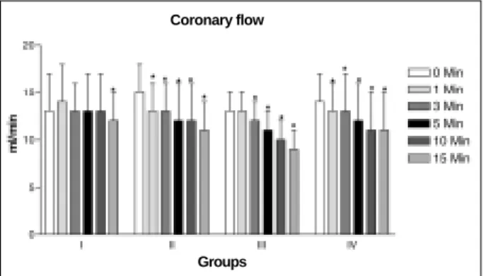
Figure 1 shows the percentage of change in contraction force, heart rate and coronary flow in isolated hearts perfused with normal Krebs-Ringer solution or normal Krebs- Ringer

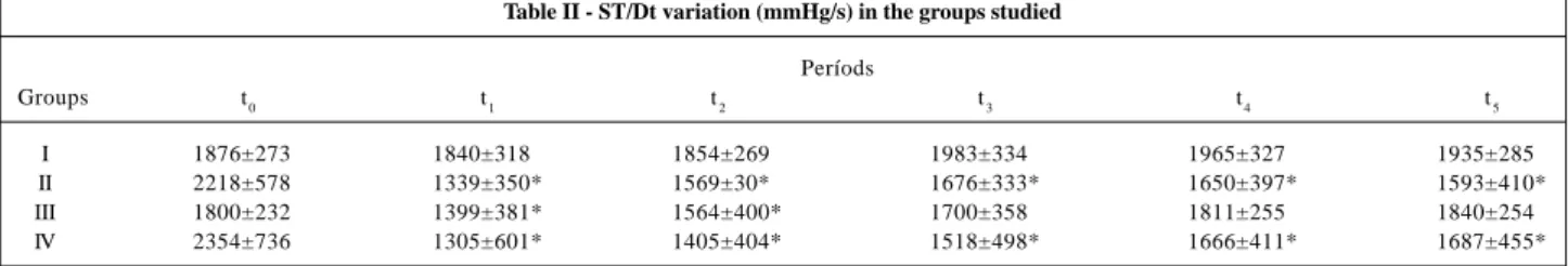
In addition, during reperfusion no further decrease was observed in +dT/dt or -dT/dt in isolated rat hearts perfused with Ang-(1-7), indicating a ben- eficial effect

The objective of the present study was to analyze the performance of widely used con- tractility indexes in the isovolumetrically beating isolated heart, in two experimental models

Atrial fibrillation and flutter are arrhythmias frequently observed in the first days following coronary artery bypass graft surgery.. Its incidence ranges from 17 to 33% 1-8

Regarding the multivariate analysis of the association between spontaneous echo contrast in the aorta and the presence of other risk factors for ischemic stroke, among the

Our study assessed the influen- ce of 2 single sessions of resistive (circuit weight training) and aerobic exercise training on changes in blood pressure and heart rate in a group

Comparison of methods of measure- ments of oxygen consumption in mechanically ventilated patients with multiple trauma: the Fick method versus indirect calorimetry. Flancbaun L,

Balloon valvuloplasty successfully used in severe mitral stenosis during pregnancy and in aortic stenoses in nonpregnant, but at high risk for aortic valve replacement 11,12 ,