Purpose: Pituitary adenomas (PA) correspond to 10-15% of intracranial tumors, and are generally benign slow-growing lesions. Our aims were: 1) to characterize its ophthalmic findings; 2) to correlate the findings of optical coherence tomography (OCT) with the presence of visual field defects; and 3) to establish a predictive model for imagiological tumor progression.
Methods: retrospective case-series of 100 patients with PA. The ophthalmic exam was recorded annually until 5-years of follow-up, including: visual acuity (VA), visual fields (VF), pupillary response (PR), ocular motility, retinal nerve fibre layer RNFL thickness (RNFLT) and ganglion cell layer thickness (GCLT).
Results: Mean age at diagnosis was 52-years [11-85]. Hormonal activity was present in 42.7% of cases (mostly prolactin production), and 48.3% were associated with ≥1 hormonal deficiency. Most common presenting symptoms were VF/VA defects (n=30), headache (n=29) and fatigue (n=13). VF defects respecting the vertical meridian occurred in 49 patients, of which 20 (40.8%) initially reported visual disturbances. The presence of baseline VF defects was associated with lower RNFLT (79.3µm vs 99.6µm, p<0.01), and higher bilateral asymmetry in RNFLT (12.8µ vs. 3.4µm, p<0.01) and MGCLT (4.8µm vs. 1.6µm, p<0.01). Imagiological tumor progression was accompanied by deterioration of the ophthalmic exam (visual function and/or OCT) in 50% of cases. The annual decrease in >15% RNFLT was a reliable predictor for imagiological tumor progression (p<0.01).
Conclusion: Though ophthalmic symptoms are frequent in the setting of PA, oftentimes the patient is unaware of visual disturbances. Collaboration with the ophthalmologist adds value to the management of these patients.
Keywords: pituitary neoplasms; pituitary adenoma; visual fields; optic chiasm; pituitary apoplexy
RESUMO
Objetivo: Os adenomas da hipófise (AH) correspondem a 10-15% de todos os tumores intracranianos, sendo geralmente tumores benignos e indolentes. Os nossos objetivos foram: 1) caracterizar os seus achados oftalmológicos; 2) correlacionar a presença de defeitos de campo visual com os achados da tomografia de coerência ótica; e 3) estabelecer um modelo preditivo para a progressão imagiológica tumoral.
Métodos: Série retrospetiva de 100 casos de AH. O exame oftalmológico multimodal foi registado anualmente até 5 anos de seguimento, incluindo: acuidade visual (AV), campos visuais (CV), resposta pupilar (RP), motilidade ocular, e espessura da camada de fibras nervosas (CFN) e da camada de células ganglionares (CCG).
Resultados: A idade média à altura do diagnóstico foi de 52 anos [11-85]. Atividade hormonal estava presente em 42.7% dos casos (sobretudo produção de prolactina), e 48.3% estavam associados com ≥1 défice hormonal. Os sintomas de apresentação mais frequentes foram defeitos AV/CV (n=30), cefaleia (n=29) e fadiga (n=13). Defeitos de CV respeitantes do meridiano vertical ocorreram em 49 doentes, dos quais 20 (40.8%) reportaram inicialmente perturbações visuais. A presença inicial de defeitos campimétricos associou-se a uma menor espessura média da CFN (79.3µm vs. 99.6µm, p<0.01), bem como a maior assimetria bilateral da CFN (12.8µm vs. 3.4µm, p<0.01) e da CCG (4.8µm vs. 1.6µm, p<0.01). Progressão imagiológica tumoral foi acompanhada por deterioração do exame oftalmológico (função visual e/ou OCT) em 50% dos casos. A diminuição anual da espessura da CFN em >15% foi estabelecida como preditor para a progressão imagiológica tumoral (p<0.01).
Conclusão: As manifestações oftalmológicas são frequentes no contexto de AH, mas frequentemente o doente não tem perceção das perturbações visuais. Colaboração com o oftalmologista acrescenta valor à gestão destes doentes.
Palavras-chave: Adenoma da hipófise; adenoma pituitário; campos visuais; quiasma ótico; apoplexia da hipófise
INTRODUCTION
Pituitary adenomas (PA) are the most common cause of optic chiasm compression, corresponding to 10-15% of all primary intracranial tumors.1-3 Prevalence rates are
probably still underestimated, as post-mortem and radiography studies have described PA in 14.4% and 22.5% of the overall population, respectively.4 These
studies reinforce PA are generally benign slow-growing tumors, often unnoticed or incidentally diagnosed. However, when clinically relevant, PA may be associated with a wide range of clinical manifestations, according to
their hormonal activity and compressive/invasive relation with adjacent structures.
Influence on endocrine axis may be exerted by two distinct mechanisms:1 hormonal hypersecretion by the
tumour itself (functioning adenomas), and2
hypopituitarism, caused by compression and/or destruction by the normal glandular tissue by the growing adenoma; compression of portal circulation; or inhibition of certain hormonal axes by hypersecreted secreted hormones (e.g.: hypogonadism secondary to hyperprolactinemia).5 Both
Subject of great interest to this paper is the mass effect produced by PA. These histologically benign tumors are generally intrasellar, possibly extending to para- and suprasellar adjacent regions. Its location in the middle cranial fossa ensures proximity to many vital structures, as the cavernous sinus, ventricular system and parenchymal brain, posing important clinical threats. The optochiasmatic tract closely located above is frequently among the first affected structures, for what ophthalmic manifestations are common and relatively early in the course of progressing disease.6,7
While optical coherence tomography (OCT) has been ascertained as a predictor for visual field recovery following PA surgery8-11, recent publications have also
discussed its role in the diagnosis of early chiasmal compression.12 Chronic optic neuropathies and
chiasmopaties associate with the subjective fundoscopic assessment of optic nerve pallor; as so, the measurement of ganglion and nerve fibre layers thickness may be more sensible in documenting optic nerve damage in this context, such as in correlating with visual field defects.12-14
Our paper intends to characterize the ophthalmic findings in the setting of PA, to study the correlation between the presence of visual field defects and OCT findings, and to establish a predictive model for imagiological tumor progression.
MATERIALS AND METHODS
We conducted a retrospective case-series of 100 patients with PA, followed in the Neurophthalmology department of a tertiary care centre (Hospital de Santa Maria, Lisbon, Portugal) between 2008 and 2018. Included subjects were pooled from the patient’s list of one doctor
(FC), and consecutively enrolled. All data were de-identified for patient’s privacy protection. The conduct
of this study was approved by the Institutional Review Board of the Lisbon Academic Medical Centre.
Clinical records were reviewed for the following data: demographics, hormonal activity and/or hypopituitarism, presenting symptoms, and efficacy and safety of chosen treatment modality (medical, surgical and/or radiation). We also collected data regarding the department from referral.
Ophthalmic exam was recorded annually until 5-years of follow-up, including: visual acuity (VA; Snellen
decimal), visual fields (VF; Humphrey Field Analyser II®,
Carl Zeiss Meditec, Jena, Germany; Octopus 900®,
Haag-Streit Diagnostics, Köniz, Switzerland), intraocular pressure (IOP), pupillary response, ocular motility, and retinal nerve fibre layer thickness (RNFLT), presence of nasal/temporal peripapillary atrophy, and macular ganglion cell layer thickness (MGCLT) assessed through spectral-domain optical coherence tomography (SD-OCT; Spectralis®, Heidelberg Engineering, Heidelberg,
Germany).The aforementioned VF and OCT devices were the same throughout the study timeline. Only threshold static automated techniques were used in patients’ visual field assessments. The presence of nasal/temporal peripapillary atrophy was determined from the Spectralis®
peripapillary RNFLT printout, whenever a nasal/temporal peripapillary sector was coded in red (outside normal limits); per definition, this color code implies the sector was thinner than the 201-subject reference database range at the level of 1%.15,16 Measurements of the ganglion cell
layer thickness were performed through automatical layer-by-layer segmentation, using the Spectralis® software.17
Automatic segmentation errors were manually corrected as needed.None of the included tomography exams presented incorrigible segmentation errors (i.e., abrupt deviations, coursing through one or several layers).
Considering the retrospective nature of this study, and that VF database might be incomplete for limited storage, VF defects were characterized from clinical records whenever necessary. Characterization was performed according to the respect for the vertical meridian, and number of affected quadrants. Always comparing to records of the previous year, clinical tumor progression was defined as one of the following:1 VA deterioration of
≥0.2, non-attributed to other causes (i.e. opacity of ocular media);2 de novo VF defect, or extension of previous
defects to ≥1 more quadrant;3 de novo pupillary or
oculomotor defect, or worsening of previously existing defect;4 decrease in RNFLT of ≥15%, or papilledema.
Imaging records as magnetic resonance imaging (MRI) and brain computed tomography (CT) were consulted for the anatomic characterization of the tumors, i.e. dimensions and extension to adjacent structures. Tumors were classified according to the Hardy-Wilson classification6 (Table 1), annually, for the first 5-years of
ophthalmic follow-up. Imagiological tumor progression was considered whenever there was a one grade/stage increase in the Hardy-Wilson classification, or a ≥20%
increase in the dimensions of the long tumor axis (average of 3 measurements, mm). Whenever imaging was missing (as when performed in external clinics), tumor classification was performed according to the imaging reports transcribed to patients’ clinical records.
Stage Grade
0: no suprasellar extension I: normal sella; tumor <10mm A: extension to suprasellar cistern II: enlarged sella; tumor ≥10mm B: recesses of third ventricle
obliterated III: local perforation of sellar floor C: third ventricle grossly displaced IV: diffuse sellar floor destruction D: intracranial V: spread via cerebrospinal fluid or
blood borne E: into/beneath the cavernous sinus
Table 1 - Hardy-Wilson classification
Chi-square tests were employed to compare binary variables, while 2-sided t-tests were used for continuous variables. Patients with past medical history of glaucoma, retinopathy or cerebral vascular accident with VF sequelae were excluded from any statistical tests involving perimetric defects, RNFLT and MCGLT.
A multivariate logistic regression analysis was used to find ophthalmological predictors for imagiological tumor progression. Tested variables were the following: initial presence of VF defects, progression of VF defects, nasal / temporal peripapillary atrophy, mean RNFLT, bilateral RNFLT asymmetry, mean MGCLT, bilateral MGCLT asymmetry, decrease in >15% RNFLT, and composed criteria of clinical progression. We controlled for the following potential confounders: age, sex, hormonal activity, and previous surgical or radiation therapy. A forward stepwise selection approach was used, adding variables one at a time whenever p-value was below 0.25.
Missing data were approached with a last observation carried forward (LOCF) method. Statistical analysis was performed with STATA® v15 software (StataCorp LLC,
College Station, TX, USA), at the significance level α of 0.05. All continuous outcomes results were presented as mean ± standard deviation (SD).
RESULTS
Baseline demographics
Mean age at diagnosis was 51.6±19.5 years-old [range: 11 to 85]. Forty-seven patients were female. Mean ophthalmological follow-up was 46.8±38.1 months, ranging from 0 (single appointment) to 121 months. Seventeen patients ultimately abandoned the Ophthalmology clinic and were lost to follow-up. During the first five years of ophthalmic follow-up, patients had an average of 5.3±3.3 Ophthalmology appointments [range: 1 to 15], rising further to 5.6±3.4 if excluding the group ultimately lost to follow-up.
From the 100 cases with hospital follow-up, ninety-one had clear documentation of department from referral: neurosurgery n=66; endocrinology n=16; internal medicine n=2; emergency department n=1; radiotherapy n=1. Five patients were diagnosed at the Ophthalmology department.
One patient had a family mutation associated with hereditary paraganglioma-pheochromocytoma (SDHB exon 3 deletion).
Hormonal activity
From the included cases, 41 were functioning adenomas (42.7%). Of these, most were prolactinomas (n=29, 70.7%); thirteen produced growth hormone (GH, 31.7%); one produced adrenocorticotropic hormone (ACTH, 2.4%); one produced the alpha-subunit of thyroid--stimulating hormone (α-TSH, 2.4%); and one produced antidiuretic hormone (ADH, 2.4%) with associated syndrome of inappropriate antidiuretic hormone secretion (SIADH).
Nearly half of PA were associated with ≥1 hormonal deficiency (n=42, 48.3%), the most common being secondary hypothyroidism (n=38, 44.2%) and hypogonadism (n=28, 32.9%). Some degree of adrenal failure was present in 21 patients (24.7%). Thirteen patients had no records regarding endocrinal insufficiency status.
Table 2 depicts the baseline demographics and main clinical characteristics of the included patients.
Female, n 47
Age at diagnosis, mean±SD 51.6±19.5
Age categories, n <20 years: 4 20-29 years: 13 30-39 years: 13 40-49 years: 15 50-59 years: 16 60-69 years: 9 70-79 years: 20 ≥80 years: 6 Unreported/Unknown: 4 Histological type, n Functioning 41 Non-functioning 55 Unreported/Unknown: 4 Pituitary apoplexy, n 11 Previous treatments, n Single surgery 21 Multiple surgeries 10 Radiotherapy 8
Table 2 - Clinical characteristics of the 100 included patients
Presenting symptoms
Most common presenting symptoms were VF/VA defects (n=30), of which only five patients had a normal ophthalmological exam (16.7%; VA, VF, PR and ocular motility), and only three had no imaging evidence of pituitary apoplexy or optic pathway compression (10%).
Twenty-nine patients complained of headache, 13 of fatigue, and 12 of galactorrhoea and/or menstrual irregularities (of which one mostly complained of breast pain). Acromegaly was detected in 9 patients, and 4 had decreased libido.
Overall, 11 patients were diagnosed with pituitary apoplexy, of which 8 had diplopia (out of 10 patients with this initial complaint); 4 had headache; 2 had altered level of consciousness; and 1 had nystagmus. From the 11 patients with apoplexy, one was pregnant (37 gestational weeks) and one had had recent adenoma surgery (2 weeks post-op).
Incidental diagnosis of PA occurred in 14 patients, through brain-CT performed in the context of trauma (n=11), vascular cerebral accidents (n=1), psychiatric disorder (n=1) and dementia (n=1).
Previous treatments
At first ophthalmic observation, 31 patients already had been submitted to at least one surgery (mostly transsphenoidal approach, TSA; one had gamma knife radiosurgery, GKRS); 10 to multiple surgeries; and 8 to cranial radiotherapy. Of these, 12 patients had no signs of tumor recurrence (38.7%).
Six of the patients previously submitted to TSA had significant surgical complications: diabetes insipidus n=2; nasal obstruction n=1; cerebrospinal fluid (CSF) fistula n=1; ischemic stroke n=1; and trigeminal neuralgia n=1, which surgery was complicated by facial palsy.
Baseline ophthalmic exam
Overall mean VA was 0.8±0.3, ranging from amaurosis to 1. Mean IOP was 14.6±2.5 mmHg. Abnormal pupillary responses (relative afferent pupillary defect, or bilaterally sluggish / absent reflexes) were found in 25 patients, and were associated with the presence of VF defects in 81% of cases (p<0.01).
Six patients had oculomotor defects, all with pituitary apoplexy or PA extending to the cavernous sinus. On the contrary, only 12.2% (6/49) of patients with cavernous sinus involvement or pituitary apoplexy presented with III cranial nerve paresis (Pearson’s Chi-squared p=0.01).
VF defects respecting the vertical meridian, or generalized reduction of the sensitivity thresholds non-attributed to ocular causes, were present in 49 patients - of which only 20 (40.8%) initially reported visual disturbances. Mean number of affected quadrants was 3.7±1.5 [range: 1 to 8], with 9 cases presenting defects compatible with involvement of the retrochiasmal optic pathway (all Hardy- Wilson grade E). Most common pattern of VF defect was bitemporal hemianopia. On the other side, among patients with imaging documentation of optic pathway compression, only 16.4% (10/61) did not present any VF defect. Fourty-five patients had records of normal VF, or VF non-compatible with optic pathway compression. Table 3 details the encountered VF defects.
Visual field defect n (% among total defects)
Bitemporal hemianopia 27 (55.1)
Complete 20 (41.8)
Incomplete (3 affected quadrants) 4 (8.3) Associated with unilateral amaurosis 3 (6.1) Bitemporal quadrantanopia 7 (14.3) Isolated temporal hemianopia 2 (4.1) Isolated temporal quadrantanopia 2 (4.1) Generalized reduction of sensitivity
thresholds 2 (4.1)
Defect associated with retrochiasmal
compression 9 (18.4)
Homonymous hemianopia 6 (12.2) Homonymous quadrantanopia 3 (6.1)
Total n: 49
RNFLT was reduced in 36.9% of patients at baseline, of which 91.7% had temporal peripapillary atrophy; 41.7% had nasal peripapillary atrophy; and 33.3% had peripapillary atrophy of both sectors. Table 4 illustrates baseline RNFLT reductions per sector, such as RFNLT progression during follow-up. Peripapillary atrophy was statistically significantly more frequent among patients with baseline VF defects (nasal peripapillary atrophy: 32% vs. 3.7%, chi-square p=0.010; temporal peripapillary atrophy: 64% vs. 11.1%, chi-square p<0.01). Accordingly, mean RNFLT in these patients was also significantly decreased (79.3µm vs 99.6µm, p<0.01). Bilateral RNFLT asymmetry was also superior in this set of patients (12.8µ vs. 3.4µm, p<0.01).
Decreased RNFLT at baseline, per sector
N (% among patients with decreased RNFLT)
Nasal 10 (41.7)
Temporal 22 (91.7)
Nasal and temporal 8 (33.3)
Total n: 24
Annual progression of RNFLT N (of which with imaging progression, n; %)
Reduction >15% 10 (7; 70%)
Stable 57 (12; 21.1%)
Insufficient information 33 (3; 9.1%)
Table 4 - Decreased RNFLT per sector
Mean MGCLT at baseline was 14.6±4.1 µm [range: 8 to 37] (n=31 subjects). Mean MGCLT was not statistically significantly different in patients with baseline VF defects (12.6µm vs. 15.0µm, p=0.14). However, bilateral asymmetry in MGCLT was significantly superior in this group (n=23, 4.8µm vs. 1.6µm, p<0.01). Sixty patients had an abnormal ophthalmic exam at baseline (VA, VF, pupillary response, ocular motility, and/or RNFLT), with 37% presenting 2 or more abnormalities among the aforementioned.
Clinical progression
Visual acuity. Eight patients had decreased VA non-attributed to other causes (median 0.3), all with optochiasmal compression. One patient decreased from 1.0 to counting fingers near the globe in <1 year. Of these 8 patients, 3 did not show other signs of clinical deterioration. Only one patient had imaging records with clear documentation of tumor progression.
Visual fields. From the 45 patients with normal baseline VF (or non-compatible with optic pathway compression), 32 maintained perimetric stability throughout the 5-year follow-up (71.1%) and 10 had insufficient information to
document progression (22.2%; single study, unreliable exams). From the remaining 3 who developed VF defects (6.7%), all had chiasmal compression - one had previously been submitted to surgery and was again operated with a TSA, and another is currently awaiting surgical treatment.
From the 49 patients with baseline VF defects, only 20 initially reported visual disturbances (40.8%). The defect further progressed in 8 cases (16.3%). Of these, 3 were recurrent tumors previously submitted to surgery and in one case to radiotherapy as well, and all had chiasmal compression. According to the detected clinical deterioration, 4 patients were then operated and one had also radiotherapy. Six patients had an unreliable first study, of which two were later documented with a VF defect.
Overall, 54 patients were documented with VF defects at some point during their 5-year ophthalmic follow-up. Eleven patients had documented VF deterioration (4 without other clinical signs of tumor progression), with decision to proceed to surgery in 5 of these cases. VF deterioration was associated with imagiology evidence of tumor progression (pairwise correlation coefficient 0.25, p=0.01).
Pupillary response. Baseline abnormal pupillary response was resolved in 4 patients (16%) – 2 had pituitary apoplexy with III cranial nerve involvement, and 3 had surgery soon after diagnosis.
Ocular motility. Of the 6 patients with baseline III cranial nerve palsy, 3 had complete recovery. Of these, two had had apoplexy with no tumor invasion of the cavernous sinus, and two patients (1 apoplexy, 1 tumor invasion of the cavernous sinus) had surgery soon after the beginning of diplopia.
RNFLT. Ten patients had an annual reduction of the RNFLT >15%, of which half had no other ophthalmological signs of PA progression. In 70% of cases, the reduction of the RNFLT was associated with imaging documentation of tumor progression (p<0.01). One third (n=33) did not have sufficient information to document progression (single study, fixation losses). RNFLT was maintained throughout the 5-year follow-up in 57 patients. Table 4 details RFNLT progression during follow-up and its relation with imaging tumor progression.
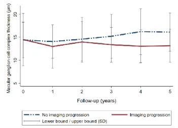
MGCLT. During the first year of follow-up, 27 patients had sufficient information to assess changes in the macular ganglion cell layer (21 with imaging tumor stability, and 6 with documented imaging progression during follow-up). While in the first group the MGCLT was maintained (baseline: 13.8±3.6 µm; year 1: 13.8±3.8 µm; p=0.88), a
small decrease was verified in the latter (baseline: 14.5±5.7 µm; year 1: 12.3±5.4 µm; p=0.23). This -2.2µm difference corresponded to a decrease in thickness of 15.2%. Image 1 depicts the changes in MGCLT across follow-up, according to imaging status.
Overall. The ophthalmological exam raised suspicion of PA progression in 21 cases, of which 11 (52.4%) were confirmed by brain imaging. As for the remaining 10 cases of clinical deterioration without imaging progression, clinical detail follows:1 giant adenoma with extension to cavernous
sinus, temporal lobe, lenticular nucleus and brainstem, and with no further clinical progression during follow-up (submitted to GKRS with significant tumor regression, and partial VF recovery);2 proposed to surgery, but ultimately lost
to follow-up;3 prolactinoma, decision to intensify medical
therapy with positive response;4 elderly patient with dementia
and reasonable clinical stability, currently under surveillance;5,6 postoperative early relapse, under
surveillance;7-10 uncompliant patients missing imaging exams
and/or appointments.
On the other hand, of the 22 cases of imaging deterioration, half were associated with altered ophthalmological exam (n=11, 50%, p<0.01): VA deterioration n=3 (13.6%, p=0.27); VF deterioration n=6 (27.3%, p=0.01); decreased RNFLT n=7 (31.8%, p<0.01).
The absence of ophthalmologic signs was negatively associated with visual pathway compression (one-sample test of proportion = 0.31, p=0.028), though it could not exclude it.
Predictive model for imagiological tumor
progression
In the initial single-variable tests, and controlling for age, gender, hormonal activity and previous surgical or radiation therapy, the following variables were associated with p<0.25: progression of VF defect, nasal peripapillary atrophy, temporal peripapillary atrophy, decrease in >15% RNFLT, and composed criteria of clinical progression. These proceeded to the forward stepwise approach.
In the combined and final models, only one variable was associated with a statistically significant predictive value for imagiological tumor progression, which was the decrease in >15% RNFLT (p<0.01). The remaining had their predictive value reduced when added to RNFLT decrease, proving their counfounding status (i.e., their effect on the outcome is itself caused by the decrease in RNFLT).
Surgical treatments during 5-year follow-up
During the ophthalmic 5-year follow-up, 33 patients were submitted to PA surgical approaches: TSA n=29; GKRS n=3; external ventricular drain n=1; transcranial approach, TCA n=1; CSF fistula closing n=1.
Excluding iatrogenic panhypopituitarism, surgical complications were detected in 9 patients. Table 5 depicts the surgical complications, according to surgical approach.
Of the 33 patients submitted to surgery during follow-up, 14 had improvements of pre-existing VF defect (42.4%). All of these were submitted to TSA, and 85.7% had a diagnosis-to-surgery interval <5 years. Two TSA patients had VF deterioration, both corresponding to Hardy-Wilson grade E. There was a postoperative improvement of VA in 10 patients, of which 7 achieved VA of 1.
Surgical approach Complications (n)
TSA
CSF nasal fistula (1) Diabetes Insipidus (3) Feeling of frontonasal pressure (1) Headache in early post-op period (3)
Nasal obstruction (2) Nosmic dysfunction (1) Voluminous epistaxis (1)
Total: 7 patients
TCA (transcallosal)
Headache in early post-op period (1) Ischemic stroke of the anterior cerebral artery
territory (1) Total: 1 patient
GKRS
Palsy of cranial nerves III and IV – 22gy, resolved in <6 months under oral
dexamethasone (1)
Medical therapy Complications (n)
Dopaminergic agonists Bromocriptine Headache (2) Nausea (2) Dizziness (1) Constipation (1) Flatulence (1) Total: 5 patients Cabergoline Headache (1) Nausea (2) Total: 2 patients Radiotherapy (n) Complications (n) Headache (2) Memory loss / forgetfulness (1)
Alopecia (1)
Transient worsening of diplopia (1) Decreased libido (1) Retro-orbital pain (1)
Total: 2
Medical treatment during 5-year follow-up
Fifty-eight patients were under any medical therapy. Replacement therapy for iatrogenic (post-op) hormonal insufficiencies was not accounted. The most frequently prescribed medical treatment was thyroid hormone (n=35), followed by dopaminergic agonists (n=32), oral corticosteroids (n=25), testosterone (n=13) and somatostatin analogue octreotide (n=10).
Intolerance to medical therapy was documented only for dopaminergic agonists. Sixteen patients were treated with bromocriptine, of which 5 manifested some degree of intolerance (31.3%), and 4 ultimately stopped/changed treatment (25%). Overall, patients were more tolerant to cabergoline: 2 patients (out of 25) manifested intolerance and changed treatment. Two patients stopped cabergoline for economic insufficiency. Table 5 details the reported adverse events, according to therapeutic approach.
Of the 15 patients with VF defects who were treated exclusively with medical therapy, only 2 had documented VF improvements (13.3%).
Radiotherapy during 5-year follow-up
Eleven patients were treated with radiotherapy during the 5-year ophthalmic follow-up, of which two reported side effects: headache (n=2); memory loss / forgetfulness (n=1), alopecia (n=1), transient worsening of diplopia (n=1, patient with cavernous sinus involvement), decreased libido (n=1), and retro-orbital pain (n=1).
DISCUSSION
Ophthalmological complaints, as VA/VF disturbances, are the most frequent presenting symptoms of PA followed in tertiary care centres, and correlate with optic pathway compression in 90% of cases. On the other hand, baseline VF defects were present in 84% of patients with imaging documentation of optic pathway compression. However, only 40% of patients with compromised VF actually reported visual symptoms, highlighting the importance of VF testing.
Peripapillary atrophy (mostly temporal) was more frequent among patients with VF defects related to chiasmal compression. These patients also showed a decreased average RNFLT, such as higher bilateral asymmetry in RNFLT and MGCLT, reinforcing the
uprising role of OCT in the initial management of PA patients.
Beyond its utility in PA diagnosis, our series indicate the Ophthalmology clinical exam identifies or raises suspicion of tumor progression in half the cases. Our results indicate OCT as being among the most useful clinical tests, as a >15% decrease in RNFLT was a significant predictor for tumor progression. While its value as been well established in predicting visual function recovery following surgery,8-11 no cut-off had been
previously defined to help monitoring for PA progression. On the other hand, VF deterioration was associated with the decision to proceed to surgery in nearly half the cases. The importance of the ophthalmological follow-up is thus not to be underestimated, as clinical information may be of useful value to patient management and decision making.
As for the macular ganglion cell layer analysis, it appears to be a useful exam in this setting. While the lack of normative databases precludes its use in PA diagnosis, its relevance may be clearer in the follow-up and clinical suspicion of progression in these patients. Though no statistical differences in average MCGLT were found in our tests, it is ought to remember they were significantly unpowered (only six patients with imaging progression). Nonetheless, differences were suggested regarding the mean difference in thickness since previous year. This was also depicted in the initial slope of our time-series graphic; the latter upward inflexions are the probable effect of therapeutic management (medical and surgical), applying for both groups.
Figure 1 - Changes in macular ganglion cell complex thickness across follow-up,
Ophthalmology appointments were also important in post-operative management, documenting VA/VF recovery mainly in patients with short diagnosis-to-surgery intervals (<5 years).
Our results highlight the importance of the Ophthalmologist in the follow-up of PA, such as in its associated therapeutic decisions.
REFERENCES
1. Ogra S, Nichols AD, Stylli S, et al. Visual acuity and pattern of visual field loss at presentation in pituitary adenoma. J Clin Neurosci 2014; 21:735-40.
https://www.jocn-journal.com/article/S0967-5868(14)00068-X/fulltext
2. Asa SL, Ezzat S. The pathogenesis of pituitary tumours. Nat Rev Cancer 2002; 2(11):836–49. https://www.annualreviews.org/doi/full/10.1146/annur
ev.pathol.4.110807.092259?url_ver=Z39.88-2003&rfr_id=ori%3Arid%3Acrossref.org&rfr_dat=cr_ pub%3Dpubmed
3. Blamires TL, Reeves BC: Vision defects in patients with peri-chiasmal lesions. Optom Vis Sci. 1996; 73:572-8.
https://insights.ovid.com/crossref?an=00006324-199609000-00002
4. Ezzat S, Asa SL, Couldwell WT, et al. The prevalence of pituitary adenomas: a systematic review. Cancer
2004: 101(3):613-9.
https://onlinelibrary.wiley.com/doi/full/10.1002/cncr.2 0412
5. Arahaf BM. Reversible hypopituitarism in patients with large nonfunctioning pituitary adenomas. J Clin Endocrinol Metab 1986; 62(6):1173-9.
https://academic.oup.com/jcem/article-abstract/62/6/1173/2674443?redirectedFrom=fulltext
6. Fredes F, Undurraga G, Rojas P, et al. Visual outcomes after endoscopic pituitary surgery in patients presenting with preoperative visual deficits. J Neurol Surg B Skull Base 2017; 78(6):461-5. https://www.ncbi.nlm.nih.gov/pmc/articles/PMC56800 29/
7. Sun M, Zhang ZQ, Ma CY, Xen SH, Chen XJ. Predictive factors of visual function recovery after pituitary adenoma resection: a literature review and meta-analysis. Int J Ophthalmol 2017; 10(11):1742-50.
https://www.ncbi.nlm.nih.gov/pmc/articles/PMC56863 75/
8. Newman SA, Turbin RE, Bodach ME et al. Congress of neurological surgeons systematic review and evidence-based guideline on pretreatment ophthalmology evaluation in patients with suspected nonfunctioning pituitary adenomas. Neurosurgery
2016; 79:E530–E532.
https://academic.oup.com/neurosurgery/article/79/4/E5 30/2837282
9. Danesh-Meyer HV, Papchenko T, Savino PJ, Law A, Evans J, Gamble GD. In vivo retinal nerve fiber layer thickness measured by optical coherence tomography predicts visual recovery after surgery for parachiasmal tumors. Invest Ophthalmol Vis Sci 2008; 49:1879– 1885.
https://iovs.arvojournals.org/article.aspx?articleid=212 5612
10. Danesh-Meyer HV, Wong A, Papchenko T et al. Optical coherence tomography predicts visual outcome for pituitary tumors. J Clin Neurosci 2015; 22:1098– 1104.
https://linkinghub.elsevier.com/retrieve/pii/S0967-5868(15)00077-6
11. Jacob M, Raverot G, Jouanneau E et al. Predicting visual outcome after treatment of pituitary adenomas with optical coherence tomography. Am J Ophthalmol
2009; 147:64–70 e62.
https://linkinghub.elsevier.com/retrieve/pii/S0002-9394(08)00543-6
12. Blanch RJ, Micieli JA, Oyesiku NM, Newman NJ, Biousse V. Optical coherence tomography retinal ganglion cell complex analysis for the detection of early chiasmal compression. Pituitary 2018; 21(5):515-23. https://link.springer.com/article/10.1007/s11102-018-0906-2
13. Monteiro ML, Hokazono K, Fernandes DB et al. Evaluation of inner retinal layers in eyes with temporal hemianopic visual loss from chiasmal compression using optical coherence tomography. Invest Ophthalmol Vis Sci 2014; 55:3328–3336. https://iovs.arvojournals.org/article.aspx?articleid=212 8964
14. Yum HR, Park SH, Park HY, Shin SY. Macular ganglion cell analysis determined by cirrus HD optical coherence tomography for early detecting chiasmal compression. PLoS ONE 2016; 11:e0153064.
https://journals.plos.org/plosone/article?id=10.1371/jo urnal.pone.0153064
15. Silverman AL, Hammel N, Khachatryan N, et al. Diagnostic accuracy of the Spectralis and Cirrus reference database in differentiating between healthy and early glaucoma eyes. Ophthalmology 2016; 123(2):408-14.
https://linkinghub.elsevier.com/retrieve/pii/S0161-6420(15)01144-6
16. Lumbroso B, Rispoli M. Practical handbook of OCT. New Delhi: Jaypee Brothers Medical Publishers; 2012. Chapter 9, Glaucoma; p.189-190.
17. Pazos M, Dyrda AA, Biarnés M, et al. Diagnostic accuracy of Spectralis SD OCT automated macular layer segmentation to discriminate normal from early glaucomatous eyes. Ophthalmology 2017; 124(8):1218-28.
https://linkinghub.elsevier.com/retrieve/pii/S0161-6420(17)30084-2
CONTACT
Raquel Esteves Marques Hospital de Santa Maria Departamento de Oftalmologia Av. Prof. Egas Moniz
1649-035 Lisboa, Portugal
email: raquelestevesmarques@gmail.com
Conflict of interest: No funding was received for the conduct of this work. The authors have no conflict of interests to declare.