UNIVERSIDADE DE LISBOA
Faculdade de Medicina Veterinária
CUTANEOUS AND RENAL GLOMERULAR VASCULOPATHY: A REVIEW OF CASES SEEN AT AN EMERGENCY VETERINARY PRACTICES IN UK
MARIANA DA SILVA SEQUEIRA MARQUES ABREU
ORIENTADOR
Doctor Sarah Ann Ambler
CO-ORIENTADOR
Doutor Rodolfo Assis Oliveira Leal
2019
LISBOA CONSTITUIÇÃO DO JÚRI
Doutora Maria Teresa da Costa Mendes Vitor Villa de Brito
Doutora Ana Mafalda Gonçalves Xavier Félix Lourenço
UNIVERSIDADE DE LISBOA
Faculdade de Medicina Veterinária
CUTANEOUS AND RENAL GLOMERULAR VASCULOPATHY: A REVIEW OF CASES SEEN AT AN EMERGENCY VETERINARY PRACTICES IN UK
MARIANA DA SILVA SEQUEIRA MARQUES ABREU
DISSERTAÇÃO DE MESTRADO INTEGRADO EM MEDICINA VETERINÁRIA
ORIENTADOR
Doctor Sarah Ann Ambler
CO-ORIENTADOR
Doutor Rodolfo Assis Oliveira Leal
2019
LISBOA CONSTITUIÇÃO DO JÚRI
Doutora Maria Teresa da Costa Mendes Vitor Villa de Brito
Doutora Ana Mafalda Gonçalves Xavier Félix Lourenço
Acknowledgments
First, I would like to thank my mentors, Sarah Ambler, and Rodolfo Oliveira Leal. Your support and knowledge through my journey towards being the best veterinary surgeon I can possibly be was incredible, and for that, I am forever thankful.
To my VetsNow team, you are the best! Rafal, Charlie, Nicky, Lauren, Charlotte Arthur, Joanna, Magda, Ada, Sophie, Anita, Janet, Gerry, locums, Jacqui, Sara Sampson, and Dave Leicester. Your help during my thesis, my two internships in VetsNow and two conferences, increased my passion for emergency and critical care. I would like to give a special thanks to Aoife Reid. Without you, my path would not have happened.
Ao Hospital Veterinário SOSVet, onde dei os meus primeiros passos no mundo da veterinária. Obrigada por me aceitarem como parte da equipa. Um grande obrigado à Dra. Ana Clotilde, em especial.
Aos meus professores. Pelos ensinamentos, aulas, e por reforçarem o meu amor pela veterinária.
Queria agradecer aos meus amigos da faculdade, ao grupo das meninas United Colours of FMVton, aos Amigos no UK e o Emplastro do Reis. Por todas as horas de estudo em conjunto, amizade e companheirismo. Sem vocês a faculdade não seria a mesma.
Aos meus afilhados, em especial ao Diogo. Estarás sempre comigo.
As minhas amigas do secundário. Amigas há mais de dez anos. Para a Alina, Daniela, Catarina Freire, Carolina Martins, Sara Ali, Ana Rita e Ana Sofia. O amor que sinto por vocês é enorme.
Um agradecimento especial a minha Joana Isabel. Chamar te irmã é um privilégio. Desde explicadora de físico-química a família. Adoro-te para sempre.
To my best friend Phil. Your friendship will last forever. Thank you for the best 6 months in England as my roommate. Many years are yet to come.
To Josh. Without you, I would not even be able to send an email in English. Your patience, love, and care are unremarkable. A simple thank you will never be enough.
À minha Dona Di, a minha primeira paciente. Adoro-te.
E por fim, a minha família. Aos meus avós Barata, por sempre me apoiarem no estudo e na carreira. Sem vocês as terças feiras não eram as mesmas.
Aos meus avós Sequeira pelo amor, ensinamentos ao longo da vida. Obrigada por serem os meus ídolos. Amo-vos.
Ao meu anjo da guarda, Nã. Sem ti a nossa família não era a mesma.
Às minhas primas, pela amizade incondicional.
Ao meu pai por me ter ensinado o espírito de aventura, perseverança e trabalho.
Aos meus irmãos Manel e Gonçalo. Serão para sempre os meus bebés. Amo-vos para sempre.
À minha irmã Nono. O meu maior orgulho e ídolo. A minha admiração por ti não tem limites.
Ao meu padrasto/pai Xuninho. Obrigada por tudo. Ter-te na minha vida e um privilégio, e serei sempre tua filha.
E para minha mãe. O maior exemplo de força, mulher, profissional que tenho na minha vida. Sem ti nunca teria chegado aqui. Amo-te muito.
E por fim, a todos os meus pacientes que já vi e verei. A minha razão para ser veterinária, e o que sou hoje.
Resumo
VASCULOPATIA CUTÂNEA E GLOMERULAR RENAL: UMA REVISÃO DE CASOS EM CLÍNICAS DE EMERGÊNCIA VETERINÁRIA NO REINO UNIDO A vasculopatia cutânea e glomerular renal (CRGV), mais conhecida por Alabama Rot, é uma doença que foi descrita pela primeira vez em galgos de corrida em 1988, mas reconhecida desde 1995 nos Estados Unidos da América. No Reino Unido, a sua ocorrência tem aumentado desde 2012. Esta doença manifesta-se sobretudo por eritema e edema das extremidades, progredindo rapidamente para úlceras cutâneas, trombocitopénia e insuficiência renal aguda (IRA). Quando o quadro de insuficiência renal aguda se instala, geralmente é fatal. A causa desta doença ainda não é conhecida. A principal alteração histopatológica renal que confirma CRGV é a Microangiopatia trombótica (TMA), descrita nos humanos e cães. Atualmente, é desconhecido se a CRGV é uma nova doença da espécie canina ou se é uma modificação da síndrome hemolítica urémica, síndrome atípica hemolítica urémica, púrpura trombótica trombocitopénica e coagulação intravascular disseminada, que são as microangiopatias descritas nos humanos. Este estudo tem como objetivo sistematizar casos de cães com suspeita de CRGV avaliando se as lesões cutâneas se correlacionam com o aparecimento de IRA e se esta está associada a um pior prognóstico da doença. Foi realizado um estudo retrospetivo que consistiu na análise de 40 casos consultados em 26 clínicas com uma unidade de cuidados intensivos analisando a anamnese, sinais clínicos, exames complementares de diagnóstico, tratamento e prognóstico. Vinte e sete cães exibiam apenas lesões de pele e 13 lesões de pele e IRA. As lesões mais comuns encontradas foram abrasões superficiais e úlceras cutâneas, sendo estas mais prevalentes no grupo com IRA, presença de inflamação e dermatite, variando no tamanho. Foram observados alopécia, eritema e edema, em especial quando localizados nos membros e dígitos. Lesões com dimensão igual ou superior a 5 cm estão significativamente correlacionadas com o aparecimento de IRA (p=0.029). A mediana do tempo decorrido entre o aparecimento das lesões e o diagnóstico de IRA foi de 3 dias ± 5 dias. Além da azotémia, os cães com IRA apresentaram anemia, proteinúria, hematúria, hipostenúria, hipocalcémia, trombocitopénia, neutrofilia, enzimas hepáticas elevadas e hiperbilirrubinémia. Cinco cães foram submetidos a eutanásia (38,5%), devido a azotémia, não-resposta á fluidoterapia e oligoanúria, sendo que os restantes sobreviveram. Este estudo revela que os cães com azotémia (p=0.001), oligoanúria (p<0.001), hipocalcémia (p=0.003) e hipofosfatémia (p<0.001) estão associados a um prognóstico reservado. Contudo, o tratamento médico intensivo é indicado nestes casos, uma vez que existem resultados positivos com recuperação de IRA completa, como analisado em sete cães (53.8%) neste estudo. Palavras-chave: CRGV, Alabama rot, insuficiência renal aguda, lesão cutânea, microangiopatia trombótica
Abstract
CUTANEOUS AND RENAL GLOMERULAR VASCULOPATHY: A REVIEW OF CASES SEEN AT AN EMERGENCY VETERINARY PRACTICES IN UK
Cutaneous and renal glomerular vasculopathy (CRGV), more commonly known as Alabama
rot is a disease first reported in racing greyhounds in 1988 but recognised since 1995 in the
USA, and with increasing occurrence, since 2012, in the UK. This disease is characterised with acute erythema and oedema progressing rapidly to cutaneous ulcers of the extremities, thrombocytopenia and clinically relevant acute renal failure (AKI). When acute renal failure develops it is usually fatal. The cause of this cutaneous and renal glomerular vasculopathy is not yet known. Thrombotic microangiopathy (TMA) is the main renal histopathological change that confirms CRGV and has been described in humans and dogs. It is currently undefined if CRGV is a new canine disease or if it is a variation of the haemolytic uremic syndrome, atypical haemolytic uremic syndrome, thrombotic thrombocytopenic purpura or disseminated intravascular coagulation which are the TMA’s reported in humans. The objectives of this present study are to review cases of dogs suspected with CRGV evaluating if the cutaneous lesions correlate with the developing of AKI and if this is associated with a worse outcome of CRGV. 40 cases from 26 first opinion emergency providers were analysed and their history, clinical signs, clinicopathological findings, diagnostics, treatment plan, and outcome observed. 27 dogs presented with only skin lesions and 13 with skin lesions and AKI. The most common macroscopic aspects of the skin lesions were superficial abrasions and cutaneous ulcers, particularly in the group of dogs with AKI, presence of inflammation and dermatitis, characterized by different sizes. Alopecia, erythema, and oedema were also observed, mainly when located in the limbs and digits. Lesions wider than five centimetres were significantly correlated with development of AKI (p=0.029). The median time between the presence of skin lesions and the diagnosis of AKI was 3 ± 5 days. Besides the azotaemia, dogs with AKI presented with anaemia, proteinuria, haematuria, hyposthenuria, hypocalcaemia, thrombocytopenia and neutrophilia, high serum liver enzyme activity, and hyperbilirubinaemia. Five animals were submitted to euthanasia (38.5%), due to azotaemia, no response to intravenous fluid therapy and oligoanuria. The remain survived. This study reveals that having azotaemia (0.001), oligoanuria (p<0.001), hypocalcaemia (p=0.003) and hypophosphatemia (p<0.001) was significantly correlated with a worse outcome. Nevertheless, intensive medical therapy is designated in these patients because successful outcomes with full recovery from AKI have been achieved as observed in seven dogs (53.8%) in this study.
Table of contents
Acknowledgments ... ii
Resumo ... iv
Abstract ... vi
Table of contents ... vii
List of Figures ... ix List of Tables ... x List of Graphics ... x List of Abbreviations ... xi Traineeship report ... 1 I. Introduction ... 3
1. Cutaneous and renal glomerular vasculopathy... 3
2. Aetiology and Differential Diagnosis ... 5
3. Pathogenesis ... 6
3.1 Thrombotic microangiopathies in humans ... 7
3.1.1 Haemolytic uraemic syndrome ... 8
3.1.2 Thrombotic thrombocytopenic purpura ... 10
3.1.3 Disseminated Intravascular Coagulation ... 11
4. Epidemiology of CRGV ... 12
5. Signalment ... 13
6. History and clinical signs ... 14
7. Clinicopathological findings ... 17 8.1 Haematological findings ... 17 8.2 Biochemical findings ... 18 8.3 Urinalysis ... 18 8.4 Imaging ... 18 8. Definitive diagnostic ... 19 9. Histopathology ... 19 10. Electron microscopy ... 23 11. Medical management ... 25 12.1 Management of cases... 25
12.1.1 Without apparent clinically relevant AKI ... 25
12.1.2 With apparent clinically relevant AKI ... 26
12.1.2.1 Fluid therapy ... 26
12.1.2.2 Urine Output ... 27
12.1.2.3 Medication ... 28
12.1.3 Wound management ... 29
12.1.4 Monitoring ... 30
12.1.5 Therapeutic Plasma Exchange ... 30
12. Prognosis ... 31
1. Introduction and objectives ... 33
2. Materials and Methods ... 33
2.1 Case selection criteria ... 33
2.2 Medical records review ... 33
2.3 Laboratory diagnostics ... 34 2.4 Imaging ... 35 2.5 Medical management ... 35 2.6 Statistical analysis ... 35 3. Results... 36 3.1 Signalment ... 36 3.2 Clinical History ... 37
3.3 Physical examination findings ... 38
3.4 Skin lesions ... 39 3.5 Diagnostics ... 40 3.6 Medical management ... 42 3.7 Outcome ... 43 4. Discussion ... 45 5. Conclusion ... 51 References ... 53
List of Figures
Figure 1- Map showing the distribution of confirmed cases of cutaneous and renal glomerular
vasculopathy (to the end of April 2019) (Adapted from https://www.vets4pets.com/stop-alabama-rot/)...13
Figure 2 - Ulcerated lesion situated in the digital and metatarsal pads in a dog confirmed with CRGV.
(Gently authorised by David Walker, Anderson Moores Veterinary Specialists)……….15
Figure 3 - Erosion to the carpal pad in a dog confirmed with CRGV (Gently authorised by David
Walker, Anderson Moores Veterinary Specialists)………15
Figure 4 - Ulcerated lesion situated in the craniolateral thigh in a dog confirmed with CRGV (Gently
authorised by David Walker, Anderson Moores Veterinary Specialists)…...………16
Figure 5 - Superficial ulcer located in the medial thigh in a dog confirmed with CRGV (Gently
authorised by David Walker, Anderson Moores Veterinary Specialists).…...………..…16
Figure 6 - Ulcerated lesions found in the prepuce, caudal thighs and scrotum of a dog confirmed with
CRGV (Gently authorised by David Walker, Anderson Moores Veterinary Specialists).………16
Figure 7 – Cheek superficial lesions in a dog confirmed with CRGV (Gently authorised by David
Walker, Anderson Moores Veterinary Specialists)………....17
Figure 8 – Tongue lesions in a dog confirmed with CRGV (Gently authorised by David Walker,
Anderson Moores Veterinary Specialists)………17
Figure 9 - Histopathology of a skin biopsy from a patient with CRGV, showing a dermal artery with
fibrinoid necrosis (arrow). Haematoxylin and eosin, x100. (Gently authorised by David Walker, Anderson Moores Veterinary Specialists)………..…20
Figure 10 - Necrotic hair follicle along with neutrophilic infiltrates. (Gently authorised by David
Walker, Anderson Moores Veterinary Specialists)……….…...21
Figure 11- Photomicrograph of small dermal artery with fibrinoid necrosis of the vessel wall (Gently
authorised by David Walker, Anderson Moores Veterinary Specialists)………...21
Figure 12 - Histopathological appearance of a skin biopsy from a patient with CRGV. Haematoxylin
and eosin, x40. (Gently authorised by David Walker, Anderson Moores Veterinary Specialists).…....22
Figure 13 (A, B, C and D)- Photomicrographs of a glomerulus and an intralobular artery from a dog
Figure 14 - Electron micrograph of a glomerular capillary loop from a dog with cutaneous and renal
glomerular vasculopathy (Gently authorised by David Walker, Anderson Moores Veterinary Specialists)………..……25
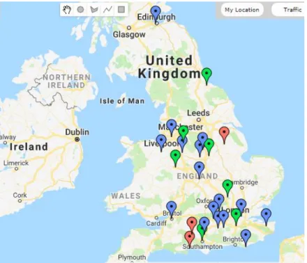
Figure 15 – Map of cases across the United Kingdom (original)………..……..…...37 Figure 16, A and B – Skin lesions in a dog confirmed with CRGV, from Macclesfield. Ulcerative
wound in the ventral aspect of the right hind, affecting the pad and skin (arrow) (photos taken by Lesley Moore, RVN)………..40
List of Tables
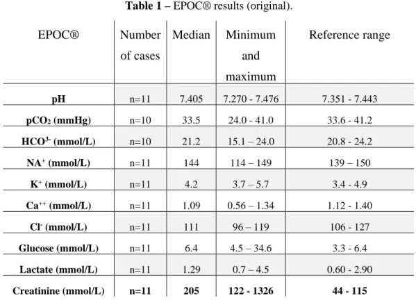
Table 1 - EPOC® results (original)……….…41 Table 2 – Biochemistry results in three dogs with AKI (original)……….42
List of Graphics
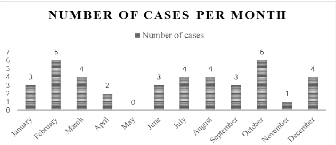
Graphic 1 – Number of cases between 29th April 2015 and 29th April 2019 (n=40) divided per month
(original)……….36
Graphic 2- Boxplot comparing the median BUN between two groups of dogs with CRGV. Group ‘Yes’
List of Abbreviations
ACEi – Angiotensin-converting-enzyme inhibitor
ADAMTS13 - a disintegrin and metalloproteinase with a thrombospondin type 1 motif, member 13
AFAST – Abdominal Focused Assessment with Sonography for Trauma aHUS – Atypical haemolytic uraemic syndrome
AKI – Acute renal injury ALKP – Alkaline phosphatase ALT – Alanine transaminase
aPTT - Activated partial thromboplastin time ARBs – Angiotensin II receptor blockers
AST – Aspartate transaminase or aspartate aminotransferase C3 - Complement component 3
CCB – Calcium channel blockers CFB - Complement factor B CFH - Complement factor H CFI - Complement factor I CK – Creatine kinase
CKD – Chronic kidney disease
CRGV – Cutaneous and renal glomerular vasculopathy D+HUS – Diarrheal HUS
D-HUS – Non-Diarrheal HUS
DIC - Disseminated intravascular coagulation
EPOC® – epoc Veterinary Blood Gas, Electrolyte and Critical Care Analyser FISH – Fluorescence in situ hybridization
GB – Great Britain
HUS - Haemolytic-uremic syndrome (HUS) i.m. – Intramuscular
i.v. – Intravenous
INR – International normalized ratio IQR – Interquartile range
IRIS – International Renal Interest Society KC – Kennel Club
MAT - Microscopic agglutination test MDB – Minimum Data Base
NSAIDs - Nonsteroidal anti-inflammatory drug OR – Odds Ratio
PCR – Polymerase chain reaction PEX– Plasma Exchange
PO – Per os
PT – Prothrombin time
RAAS – Renin–angiotensin–aldosterone system s.d. – standard deviation
STEC-HUS – E. coli serotype O157:H7 Shiga toxin-producing associated with haemolytic uremic syndrome
Stx-HUS – Shigella dysenteriae shiga toxin-producing associated with haemolytic uremic syndrome
TFAST – Thoracic Focused Assessment with Sonography for Trauma TMA – Thrombotic microangiopathy
TTP - Thrombotic thrombocytopenic purpura (TTP) UK – United Kingdom
UOP – Urine output
UPC – Urine Protein Creatinine Ratio US – Ultrasonography
Traineeship report
As part of the Integrated Master’s Degree in Veterinary Medicine from the Faculty of Veterinary Medicine of the University of Lisbon, I completed a six month internship in a ‘VetsNow’ clinic based in Stoke-on-Trent, Staffordshire, United Kingdom (UK). I fulfilled an estimate of 1700 hours in a high-quality provider of pet emergency care, working out-of-hours only, nights during the week, weekend and bank holidays.
My shifts started at six pm and finished at nine am, lasting 15 hours. During this time, we had a large case load, receiving patients from the more than 20 general practices around Staffordshire.
As I worked in emergency and critical care, I participated in different services throughout the night. These services were internal medicine where I often performed treatment plans for the patients solving clinical cases, treatment and monitoring the inpatients overnight and improved my skills in practical procedures such as placement of catheters, drug administration, blood sampling, blood transfusion, urinary catheterization, clinical examination, and consultation skills. I frequently triaged the patients independently or over the phone, deciding if the animals needed emergency care immediately.
I did also work in the surgery department where I regularly assisted all the emergency surgeries, being most commonly foreign bodies, caesareans, gastric dilation-volvulus, pyometras, amongst others. In this department, I was able to improve my knowledge in anaesthesia as I practiced procedures such as choosing and performing the most suitable pre-medication, induction, intubation and general anaesthesia for each patient.
In the diagnostics department I learned how to perform and to interpret emergency diagnostics such as EPOC, minimum data base (glucose, packed cell volume, total solids, lactate and urea), AFAST and TFAST (Focused Assessment with Sonography for Trauma, for abdomen and thorax, respectively), X-ray (positioning patients, using sedation when necessary and read the results), endoscopy and analyse blood and urine smears.
Every month, I had to prepare a Power Point presentation for my mentor with cases that I had assisted, including history, clinical exam, differential diagnostics, diagnostics, procedures performed, treatment and outcome. At the end of the presentation, I then discussed, in details, the cases that I presented.
During this period, I had the opportunity to integrate into the team that worked during the day at Lime Trees Veterinary hospital. I worked in general practice and a referral centre in small animal surgery and internal medicine, where I had the opportunity to assist surgeries such as pyometras, dog and cat castrations and ovariohysterectomies. I also performed independently with the supervision of a senior vet, a cat ovariohysterectomy using the flank technique. I then assisted the internal medicine referral consults with Doctor Hywel Parry (BVM&S CertSAM MRCVS RCVS).
On my first week of the traineeship, I prepared a case that I assisted during my traineeship about Addison’s disease and was selected to present it to a large audience and a panel of judges along with other six students and trainees, in the VetsNow Annual congress on the 8th of November 2018 in Harrogate, UK. During the congress, I had the opportunity to meet other qualified vets and assist the lectures about ECC.
I. Introduction
1. Cutaneous and renal glomerular vasculopathy
Cutaneous and renal glomerular vasculopathy (CRGV) is a disease firstly reported in racing greyhounds in 1988. It was recognised since 1995 in the USA (Carpenter, Andelman, Moore, & King, 1988), and with increasing occurrence, since 2012, in the UK (Holm, Hawkins, Jepson, Robin, Newton, Stanzani, McMahon, Cianciolo, Pesavento, Carr, Cogan, Couto, & Walker, 2015). This disease usually manifests itself with acute erythema and oedema progressing rapidly to cutaneous ulcers of the extremities, thrombocytopenia and clinically relevant acute renal failure (AKI). When acute renal failure develops it is usually fatal (Hertzke, Cowan, Schoning, & Fenwick, 1995). The cause of this cutaneous and renal glomerular vasculopathy is not yet known (Carpenter, et. al, 1988; Skulberg, Cortellini, Jepson, Chan, & Stanzani, 2018) however, the chronology of presentation, diagnostic testing and small sample size are limiting factors (Jepson, Cardwell, Cortellini, Holm, Stevens & Walker, 2019).
Terrestrial Animal Health Code of the World Organisation for Animal Health (OIE) defined
emerging disease as a
new occurrence in an animal of a disease, infection or infestation, causing a significant impact on animal or public health resulting from: a) a change of a known pathogenic agent or its spread to a new geographic area or species; or b) a previously unrecognised pathogenic agent or disease diagnosed for the first time (Moutou & Pastoret, 2015, p.41).
The outbreak pattern of CRGV in the UK accords with the definition of a newly emerging disease as no cases were reported prior to 2012. However, although this indicates the disease was not utterly known, it may not have been the case. It may simply not have been recognised owing to a very low incidence in the population prior to 2012 (Stevens, Jepson, Holm, Walker, & Cardwell, 2018). Up to this date, there are more than 180 confirmed cases since 2012, when CRGV was firstly reported in the UK (Anderson Veterinary Specialists), with only ten cases confirmed in 2019 (January until March). This contrasts with the year 2018 whereby the end of March 34 CRGV cases was confirmed. The leader investigators on CRGV in the UK (David Walker from Anderson Moors Veterinary Specialists), attributes this event to the different climate changes from January to March in 2018, compared to the year 2019, but currently, this has been the object of on-going research (Walker, D. personal communication, 2019). In 1988, Carpenter firstly characterized this disease syndrome which he observed only in racing training greyhounds since 1985 (n=168). These dogs manifested multifocal cutaneous
erythema, ulceration, and distal limb oedema. This syndrome was named ‘Alabama Rot’ or ‘Greenetrack disease’, as many cases were reported and affected many dogs in Greenetrack Racing Park in Alabama. Although this disease is commonly known by ‘Alabama Rot’ (Carpenter, et. al, 1988) or ‘New Forest disease’ (Hertzke, et. al, 1995) both diseases should more reasonably be entitled cutaneous and renal glomerular vasculopathy (CRGV) based on their histopathological features, rather than their geographic distribution (Cortellini & Humm, 2015).
It was thought this disease only affected greyhounds as this breed hold their own exclusive physiologic adaptations, owed to the fact they were racing sighthounds. As an example, these dogs have more muscular mass, higher haematocrit, elongated carpal, tarsal, metacarpal, and metatarsal bones and an improved sense of sight. All these features probably contributed to the unique hematologic and biochemical characteristics of Greyhounds compared with non-Greyhound breeds (Zaldívar-López et al., 2011). When the outbreak, however, occurred in the UK, CRGV was reported in a variety of breeds, including, the English springer spaniel, flat-coated retriever, whippet, border collie, Jack Russell terrier, Doberman, Labrador retriever, cocker spaniel, Staffordshire bull terrier, Hungarian vizsla, Weimaraner, Dalmatian, Tibetan terrier and crossbreds (Holm et al., 2015), with only one greyhound reported in the UK (Hendricks, 2000) and a Great Dane in Germany (Rotermund, Peters, Hewicker-Trautwein, & Nolte, 2002). Only two greyhounds have been affected with CRGV (with AKI) in the UK, therefore, it is still uncertain whether CRGV and ‘Alabama rot’ are identical or separate disease entities (Holm & Walker, 2018).
This disease presents itself in four different distinct manifestations (Carpenter, et. al, 1988): dogs can only manifest skin lesions without showing any signs of systemic illness. To this date, there are no studies that reveal how many dogs in the UK have CRGV with skin lesions only (Holm et al., 2015). The second course was seen in dogs with typical skin lesions, systemic illness signs and a quick onset of azotaemia; third manifestation is described as a cutaneous ulceration clinically normal with renal failure within ten days of onset of skin lesions; the fourth manifestation was identified in seven dogs that developed azotaemia before cutaneous ulceration (Carpenter, et. al, 1988). It is important to notice that the presence of skin lesions along with the presence of AKI due to other causes, is rarely reported in dogs (Jepson et al., 2019).
2. Aetiology and Differential Diagnosis
After CRGV been recognized in the UK, investigations were carried out to recognise whether the disease was attributable to any previously known causes of canine AKI, or whether the aetiology was analogous to human haemolytic uremic syndrome (HUS), atypical haemolytic uremic syndrome (aHUS) or thrombotic thrombocytopenic syndrome (TTP). No single unifying cause was able to be identified from these results (Holm et al., 2015).
When AKI is confirmed through laboratory analyses, the first method to approach is to rule out any known causes, before assuming CRGV is present. Chronic kidney disease (CKD) and post-renal causes should be excluded first. To assess post-post-renal causes abdominal imaging is helpful. This can be useful to evaluate renal architecture, which combined with clinical history can exclude CKD. Once these two illnesses been excluded, other potential causes of AKI and pre-renal causes must be considered (Holm et al., 2015)
Pre-renal causes are excluded through urine specific gravity, lack of response to intravenous fluids and exclusion of hypoadrenocorticism (basal cortisol blood test and ACTH stimulation test). A detailed clinical history regarding potential nephrotoxic substances ingestion should be obtained, full urinalysis including culture should be performed, leptospirosis testing (PCR on blood/urine before antibiotic therapy and microscopic agglutination test), abdominal imaging to assess renal architecture, analysis of serum electrolyte concentrations should all be considered (Holm & Walker, 2018).
When these patients present themselves to their primary veterinary practice with skin lesions, it is a common procedure to administrate nonsteroidal anti-inflammatory drugs (NSAIDs) in the management of these wounds (Holm et al., 2015) which are known to have indirect negative effects in the kidneys (Lomas & Grauer, 2015). Holm et al. (2015), stated that eleven of the dogs (36.7 per cent) received NSAIDs previously to the diagnosis of AKI. It is possible that their use aggravated AKI, but the histopathologic lesions were not consistent with NSAIDs being the particular cause of the AKI in these dogs (Holm et al., 2015).
In the largest case series of dogs with CRGV to date (Holm et al., 2015), many possible pathogens that can be the potential aetiology of CRGV were considered. One possible condition investigated was Leptospirosis. The predominant clinical signs of acute leptospirosis relate to the presence of acute kidney injury and liver damage (Schuller et al., 2015), which are similar to CRGV findings. Renal histopathology in dogs with leptospirosis (no typical hepatic lesions were identified) is not compatible with CRGV. Five dogs had positive titers, although at a low concentration (1:100–1:800). Nevertheless, all these dogs had been vaccinated less than one year before testing. Moreover, although it is discussable, only single titers greater than 1:1600 are considered relevant for indicating an infection in vaccinated dogs (Miotto et al., 2018;
Schuller et al., 2015).
In this same study, faecal culture was executed, and Escherichia coli was identified. Nevertheless, multiplex polymerase chain reactions (PCRs) for E. coli virulence genes were negative. Shiga toxin has not been identified in dogs with HUS before (Holloway, Senior, Roth, & Tisher, 1993).
A viral ethiopathogeny was hypothesized as well in this study and viral metagenomics and canine circovirus PCR were performed, although all results were negative, and histopathologically there was no evidence of viral cytopathic effect (cytoplasmic inclusion bodies) in any of the tissues examined. (Holm et al., 2015). Nevertheless, negative results for viral metagenomics do not eliminate a viral cause as these results could indicate that virus was present in low copy numbers, or that the virus was too weakly related to known viruses used for sequence alignment, or that the sample used was too autolyzed to preserve the virus. (Jepson et al., 2019)
Numerous other causes were deliberated in this UK case series such as Borrelia (PCR and serology were negative), and renal heavy metal concentrations (lead, arsenic, and cadmium; concentration below reported reference intervals) ( Holm et al., 2015).
Macdonald (2015), is conducting a research that hypotheses that the bacteria Aeromonas
hydrophila can be the pathogen of CRGV. These bacteria can live in areas were some dogs
normally walk and were confirmed with the disease. These areas feature substantial amounts of water (as result of an unusually high rainfall) and both the running water and the standing water were at 4°C for some weeks around the time of the cases. Only dogs appeared to be affected, with no registered mortalities in wild ponies, foxes, cattle or deer (Macdonald, 2015). Correspondingly, A. hydrophila in dogs can mimic Leptospirosis infections (Andre-Fontaine, Monfort, Buggin-Daubie, Filloneau, & Ganiere, 1995). Moreover, A. hydrophila is identified to cause ulcerative skin lesions in both ornamental and farmed fish, with consequent kidney failure. It is a very toxigenic organism and so the kidney failure will be a toxin response, making bacterial isolation from target tissues nearly impossible. Currently, studies are being conducted in order to confirm or not if this is the potential cause of CRGV (Macdonald, 2015).
3. Pathogenesis
Thrombotic microangiopathy (TMA) is the main renal histopathological change that confirms CRGV (Holm et al., 2015), and has been described in humans (Shatzel & Taylor, 2017) and dogs (Holloway et al., 1993). TMAs are characterised by inflammation and damage to the arteriolar endothelium, leading to platelet activation and aggregation and therefore to a widespread formation of microthrombi which constricts affected vessels. Erythrocytes suffer
shear injury and the resultant schistocytes are removed by the reticuloendothelial system, resulting in extravascular haemolysis. Intravascular haemolysis can similarly occur when the erythrocyte lesions are severe (Jepson et al., 2019). Fragmented red blood cells are probably cut as blood flows through turbulent extents of the microcirculation that are partially narrowed by microthrombi. This process leads to microangiopathic haemolytic anaemia (Moake, 2002). This acute haemolysis progresses to anaemia and the global formation of microthrombi outcomes in a consumptive thrombocytopenia. The microthrombi finally occlude the blood vessel lumen entirely, leading to reduced organ perfusion and death of the affected tissue (Holm & Walker, 2018) leading to an eventual multiorgan dysfunction (Hertzke et al., 1995).
Haemorrhage can occur when the platelet count is less than roughly 30-50 x 109/L. Classic laboratory reference ranges for platelet counts vary between 200–400 x 109/L (Hohenhaus & White, 2012).
Therefore, clinical manifestations of TMAs are thrombocytopenia, anaemia, fragmentation of erythrocytes (schistocytes), and particularly elevated serum levels of lactate dehydrogenase (LDH) (Moake, 2002). Haemolysis can be established over laboratory markers results, such as lactate dehydrogenase (LDH), haptoglobin, and bilirubin, which are all increased (Williams & Marques, 2016). Serum LDH level is increased due to the release of red blood cell LDH as a result of intravascular haemolysis (Cohen, Brecher, Bandarenko, Hill, & Carolina, 1998). Glomerular TMA in CRGV is characterised microscopically by hyaline thrombi within capillaries and sometimes the afferent arterioles, segmental to global congestion and necrosis, and thickening of glomerular capillary walls, which is consistent with findings in humans histopathology in TMAs (Hertzke et al., 1995).
Two major differential diagnosis for TMAs reported to occur in domestic animals are, more commonly CRGV and haemolytic uraemic syndrome (HUS) (Carpenter, et.al 1988; L. Holm & Walker, 2018; Skulberg et al., 2018).
3.1 Thrombotic microangiopathies in humans
TMA, in humans, include principally two syndromes, thrombotic thrombocytopenic purpura (TTP) and haemolytic uremic syndrome (HUS). Each of these syndromes appears to be caused by numerous distinct pathophysiologic mechanisms (Zheng & Sadler, 2008).
It is currently indefinite if CRGV is a new canine disease or a variation of the haemolytic uremic syndrome (HUS), atypical haemolytic uremic syndrome (aHUS), thrombotic thrombocytopenic purpura (TTP) and disseminated intravascular coagulation (DIC) which are the TMA’s reported in humans. The renal glomerular histopathologic findings resembling glomerular thrombosis, necrosis and haemorrhage are similar to the lesions found in CRGV (Carpenter, et. al, 1988).
3.1.1 Haemolytic uraemic syndrome
Haemolytic uraemic syndrome (HUS) is characterised by the presence of acute renal failure along with low platelet count and microangiopathic haemolytic anaemia (Zheng & Sadler, 2008).
Renal microcirculation becomes impaired due to the obstruction of small vessels from the fibrin thrombi that form in this particular area (Caprioli, Remuzzi, & Noris, 2011).
The term HUS was firstly reported by Gasser and colleagues in 1955. It was described as an acute haemolytic anaemia associated with kidney injuries in young children after an enteric or respiratory infection. HUS is categorized into three categories: HUS due to infections, often connected with diarrhoea (D+HUS), with the rare exception of HUS due to a severe disseminated infection caused by streptococcal organisms; HUS related to complement abnormalities or to factor ADAMTS13 deficiency, also recognised as ‘atypical HUS’ and is not diarrhoea associated (D-HUS); and HUS of unidentified aetiology that generally occurs in the course of systemic diseases or physiopathologic conditions. Examples of this are pregnancy, after transplantation or after drug assumption (Salvadori & Bertoni, 2013). Typical HUS (D+HUS) is associated with a prodromal diarrheal illness regularly caused by infection with E.
coli O157:H7 (STEC-HUS) or a different Shiga-toxin-producing strain of bacteria (e.g. Shigella dysenteriae) (Stx- HUS). This type of HUS constitutes about 95% of the HUS cases in children,
with the disease being rare to occur in adults (Zheng & Sadler, 2008). This Shiga-like verotoxins injure the endothelium, widely assumed to be the primary cause of renal dysfunction (Furlan & Lammle, 2001). Atypical HUS (aHUS) is quite sporadic and very heterogeneous as to its aetiology, age of onset and clinical presentations (Zheng & Sadler, 2008).
Infectious HUS, is related with prodromal diarrhoea followed by acute renal failure (with anuria), and considered a disease with a good outcome (Caprioli et al., 2011). This illness is confirmed by combining a positive stool test for Shiga-toxin-producing E. coli, a prodromal illness characterized by diarrhoea (often haemorrhagic) and the presence of TMA (Nalluru, Sridharan, Go, Said, & Marshall, 2018).
This type of infections requires prompt consideration of antibiotic treatment, before or after culture results are known. Nevertheless, such treatment may increase the risk of developing HUS (Freedman et al., 2016). Supportive treatment, such as isotonic volume replacement/ expansion, red blood cell, and platelet transfusion and, for severe AKI, haemo or peritoneal dialysis (Bitzan, 2009), yet plasma infusion or exchange are normally the chosen therapy to these patients (Schwartz et al., 2016).
Furlan’s findings confirmed that a deficit of ADAMTS13 activity is not a feature of HUS (Furlan & Lammle, 2001). These conclusions are consistent with previous clinical studies that
have demonstrated that plasma exchange (PEX) is not indicated in this disorder, however conservative therapy with fast volume repletion and dialysis when needed provides the best survival advantage and first-line treatment for Diarrheal HUS (D+HUS) (Michael, Elliott, Craig, Ridley, & Hodson, 2009; Bitzan, 2009). Nonetheless, a small number of cases may actually have aHUS or TTP⁄HUS and necessitate plasma therapy urgently (Clark, 2012). The microscopic and ultrastructural glomerular changes in patients affected with CRGV are extremely similar to lesions described in children with the classic form of HUS (Hertzke et al., 1995). Thickening of the glomerular capillary walls, inconstant in degree, caused by the deposition of an eosinophilic, faintly PAS-positive hyaline or granular material between the basement membrane and the endothelium, and hyaline or granular thrombi within glomerular capillaries and arterioles were the major histologic features (Vitsky, Suzuki, Strauss, & Churg, 1969). Even though HUS and CRGV have similar features, they contrast in some aspects. Cutaneous ulcerations are classically present in CRGV but are not related to HUS. Likewise, watery diarrhoea, followed by bloody diarrhoea, abdominal cramps along with vomiting and nausea marks the onset of HUS in children (Salvadori & Bertoni, 2013), but it is not seen in CRGV. Another feature that differs from HUS and CRGV is age. Whereas the CRGV appears in young adults, HUS is perceived in young children. (Hertzke et al., 1995; Holm & Walker, 2018). In domestic animals, HUS has been reported in calves (Valli & McSherry 1973), in a cow (Roby et al., 1987), four horses (Morris et al., 1987; Dickinson et al., 2008), three cats (Aronson & Gregory, 1999), three rabbits (García et al., 2002) and five dogs (Holloway et al., 1993; Chantrey, Chapman, & Patterson-Kane, 2002; Orco, Bertazzolo, Pagliaro, Roccabianca, & Comazzi, 2005).
Five to ten percent of all cases of HUS in humans are unrelated to infections by Shiga toxin- producing E. coli and are classified as atypical. aHUS is an ultra-rare disease regularly associated with progressive renal function deteriorating, microangiopathy disease with anaemia, thrombocytopenia and is characterized by a high mortality rate (Caprioli et al., 2011). It results of uninhibited activation of the alternative complement system due to genetic mutations impacting complement regulatory proteins such as complement factor H (CFH) and membrane cofactor protein (MCP), factor I and thrombomodulin (THBD) (Salvadori & Bertoni, 2013; Stevenson, Leung, & Winters, 2016). Immunosuppressive therapy should also be considered in patients with aHUS due to factor-H auto-antibodies (Fakhouri, Frémeaux-Bacchi, & Loirat, 2013). Cyclophosphamide pulses along with steroids administration and PEXs led to a prolonged decrease in CFH antibody titters and a favourable outcome in patients with aHUS (Boyer et al., 2010).
has not been described with STEC-HUS (Ardissino et al., 2014). Atypical HUS has not been reported in dogs, although, CRGV may have some similarity to this illness, particularly given the concurrent findings of skin lesions and AKI identified in both diseases. An infectious or environmental trigger for CRGV may be suspected, given the number of contact between dogs that developed skin lesions with or without AKI (Holm et al., 2015).
3.1.2 Thrombotic thrombocytopenic purpura
Thrombotic thrombocytopenic purpura (TTP) is an unusual hematologic disorder, characterized by microangiopathic haemolytic anaemia (MAHA) with schistocytes in the blood smear, thrombocytopenia, and variable stages of organ damage, including renal function impairment, neurological symptoms including stroke, and fever (Shatzel & Taylor, 2017). The aggregation of platelets in the microvascular system leads ischaemia in the brain, causing neurological signs, and other organs (Ruggenenti, Noris, & Remuzzi, 2001). Despite the fact that AKI is common on HUS, it is not a severe condition in TTP (Phillips, Westwood, Brocklebank, Marchbank, & Gale, 2016).
It has been described in both acquired and inherited deficiencies forms, in the activity of von Willebrand factor (vWF) cleaving protease (ADAMTS13) (Lewis & Meyers, 1996; Caprioli et al., 2011; Jepson et al., 2019). The acquired form is the most common and ensues when IgG autoantibodies bind and remove ADAMTS13 (Stevenson et al., 2016). Furlan, et. al (1996) and Tsai (1996), independently isolated and characterized this new metalloprotease from human plasma that cleavages vWF at the Y1605- M1606 bond of the subunit. This metalloprotease was identified in 1996 as the 13th member of the ADAMTS family (A Disintegrin And Metalloprotease with Thrombospondin type 1 repeats) (Fujikawa, Suzuki, Mcmullen, & Chung, 2001). ADAMTS13 is the cleaving protein of von Willebrand factor (vWF). vWF is crucial for primary haemostasis. It allows adhesion of circulating platelets and thrombi formation where the endothelium suffered an injury (Zheng & Sadler, 2008). Physiologically, vWF is degraded into smaller circulating forms by ADAMTS13 with the influence of shear stress (Stokol, 2008). In patients diagnosed with TTP, ADAMTS13 level is less than 5% (normal range: 60–123%) (Park, Waldrum, & Marques, 2010). The activity of this protease is deficient, increasing the accumulation of ultra large vWF multimers that are highly reactive with platelets. As a result, large, potentially occlusive platelet thrombi are formed (Moake, 2002).
According to the Guidelines on the Use of Therapeutic Apheresis in Clinical Practice plasma exchange (PEX) is the first-line treatment either as a primary standalone treatment or in conjunction with other modes of treatment (Category I) (Schwartz et al., 2016). PEX removes ADAMTS13 autoantibodies and unusually large multimers of von Willebrand factor besides
replenishment ADAMTS13 (Brocklebank, Wood, & Kavanagh, 2018). Using PEX as a therapy, 91 percent of these patients survive an episode of TTP (Bell, Braine, Ness, & Kickler, 1991); Schwartz et al., 2016).
Acquired TTP, commonly requires immunosuppression as the treatment as 80 percent of these patients enters remission with plasma therapy, however, one-third of them have a relapse. The relapse rate is greater amongst survivors with ADAMTS13 activity <10 percent (estimated risk for relapse at 7.5 years, 41 percent) than survivors with ADAMTS13 activity of 10 percent or more (Hovinga, Vesely, Terrell, Lammle, & George, 2010).
Rituximab, a monoclonal anti-CD20 antibody has been used efficaciously in patients with thrombotic thrombocytopenic purpura (Bresin et al., 2009) and anti–ADAMTS-13 autoantibodies refractory to standard therapies (Fakhouri et al., 2005).
Although TMA is a characteristic feature of both aHUS and TTP, they differ in terms of clinical manifestations. In aHUS, the lesions and clinical symptoms are mostly restricted to the kidneys, whereas the pathologic changes of TTP are more extensively distributed, more often with central nervous system signs (Salvadori & Bertoni, 2013).
3.1.3 Disseminated Intravascular Coagulation
DIC is an acquired syndrome defined by the intravascular systemic activation of coagulation pathways and excessive microthrombi formation locally and at areas distant from the site of the original injury in small and medium-sized vessels, and eventually organ dysfunction. Consumption and exhaustion of platelets and coagulation factors lead to bleeding and haemorrhage into tissues (Taylor, Toh, Hoots, Wada, & Levi, 2001).
DIC occurs as a complication of infections, solid cancers, hematologic malignancies, obstetric diseases, trauma, aneurysm, and liver diseases, amongst others, and each nature of DIC presents distinguishing features associated to the primary disorder (Wada et al., 2013).
TMA is similar to DIC (Skulberg et al., 2018), however clinical pathologic findings in dogs with CRGV do not suggest this condition. Coagulation profiles were within normal limits, and fibrin degradation products were negative (Hertzke et al.,1995) whereas in DIC prothrombin time, in 50 to 75 percent of cases, is prolonged and activated partial thromboplastin time (aPTT) is prolonged in 50–60 percent of patients with DIC, but a normal or shortened aPTT may also be seen (Bick, 1996). Fibrin-related markers, such as fibrinogen and fibrin degradation products are elevated (Prisco et al., 1989).
4. Epidemiology of CRGV
There are case reports from throughout the UK, but 36.7 percent of the cases come from The New Forest, Hampshire. This percentage can be explained by the locality of the investigators that conducted this study and the increased interest and awareness amid local veterinarians in that area (Holm et al., 2015). The North-east of England and the New Forest region of south England have the highest five-year density of CRGV cases (Stevens, et al., 2018).
A possible winter/spring seasonality is proposed, as the case incidence has been the highest these months (92 percent of cases are reported between November and May), yet there are cases all the rest of the months, but less in number (Stevens et al., 2018). During the colder months in the UK maximum temperatures are increasing and may have provided a favourable habitat for an evolving organism or a new ecological niche for a pathogen that had always been existing in the environment but was beforehand unable to flourish in the comparatively cooler conditions of previous decades (Stevens et al., 2018).
There has been a distribution of cases all over the UK, more specifically in 39 counties. Only a single case has been identified in Ireland (Figure 1) (Holm & Walker, 2018).
D+HUS is often sporadic, but reports of large outbreaks have been reported (Salvadori & Bertoni, 2013) which is similar to the epidemiological findings of CRGV (Stevens et al., 2018). Outbreaks of E. coli O157 have been reported in beef cattle and their products (Hussein, 2007) with cattle and sheep being the main reservoirs. The most important route of transmission is thought to be food contaminated with animal faeces (Ferguson et al., 2005), although in CRGV, pastures were the habitat least linked with CRGV occurrence. Combined with the decreasing domestic livestock densities, it suggests that it is unlikely CRGV is the result of a livestock-related pathogen to which dogs are exposed while walking across pastures, either from contact with the livestock themselves or their excretions (Stevens et al., 2018). This is, however, contrary to the findings in human HUS (Ferguson et al., 2005).
The incidence rate of D+HUS varies according to countries and climate and is higher in colder countries. For example, the incidence rate in Scotland (3.4 × 10⁵ children under age 5) is higher than the overall incidence rate in Great Britain (1.54 × 10⁵ children under age 5) (Lynn et al., 2005), which differs from CRGV, with only two cases reported in Scotland (Figure 1). Habitat, woodlands, and lowland dry heath communities were the variable identified to have the highest relative contribution to CRGV occurrence. The woodlands provide a rich habitat for a wide range of wildlife, plants, and fungi and this diversity makes it very difficult to isolate a single pathogen that might be the cause of CRGV (Stevens et al., 2018).
In conclusion, the uppermost relative probability of CRGV occurrence is associated with a range of agroecological factors more specifically, woodland and heath habitats, decreasing cattle and sheep densities, increasing maximum temperatures in winter and, to a lesser scale, spring, and autumn, and higher mean rainfall in winter and spring (Stevens et al., 2018).
5. Signalment
A previous case series conducted by Stevens et. al (2018), investigated the signalment risk factors for CRGV in UK dogs. It was the first and only study including 101 dogs diagnosed with CRGV showing that breed, kennel club (KC) breed group, and neuter status are significantly associated with a confirmed diagnosis of the disease, and that age group is not an important risk factor. Specific breeds have more probability of being a CRGV case compared with crossbreeds (80% of dogs in this study were purebreds and only 19 percent were crossbreds). Between the breeds more likely to be affected, there are specific ones with
Figure 1- Map showing the distribution of confirmed cases of cutaneous and renal glomerular
vasculopathy (to the end of April 2019) (Adapted from https://www.vets4pets.com/stop-alabama-rot/).
Caption: The area from which cases have been reported covers much of the south and west of the UK, while the east appears to have a lower incidence of cases
increased odds ratio such as the flat-coated retriever (Odd Ratio (OR) = 84.48), Hungarian vizsla (OR = 40.98), Manchester terrier (OR = 41.41), Saluki (OR = 27.46), whippet (OR = 22.43), English springer spaniel (OR = 11.41) and bearded collie (OR= 10.85). Breeds with decreased odds included German shepherd dogs (OR = 0.45), Jack Russell terriers (OR= 0.37) and Staffordshire bull terriers (OR= 0.50). It remains mysterious whether this is due to a truly increased breed-associated risk, or due to the increased popularity of these breeds in areas experiencing a higher case incidence of CRGV (Stevens et al., 2018).
Gundogs1 and hounds2, two KC breed groups, are nine and ten times more likely to diagnosed
with CRGV than terriers3, according to this same study. Toy dogs4 were not among the breeds
affected by CRGV, therefore this group was excluded from the study (Stevens et al., 2018). Female and neutered dogs are more likely to be diagnosed with CRGV, but the reasons behind these results are unclear. Nevertheless, previous reports have shown being female is a risk factor for HUS in human beings (George & Nester, 2014), although this does not appear to be the same in the other TMAs. Being neutered is also a risk factor with neutered dogs being 3.36 times more likely to be diagnosed with CRGV (Stevens et al., 2018).
The age range for dogs with CRGV is between 1.73 and 4.11 years old (Stevens et al., 2018), although in previous studies can range between 1.00 and 11.75 years (median 4.90 years) (Holm et al., 2015) and between10 months and 8 years, median 4 years old (Skulberg et al., 2018).
6. History and clinical signs
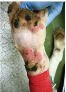
Clinical signs have acute onset before presentation to the first-opinion practice and the patients show a rapid clinical deterioration (Skulberg et al., 2018). Typically, skin lesions develop before AKI occurs. After 11 to 21 days of the presence of these skin lesions, AKI rarely occurs (Holm & Walker, 2018). Systemic signs develop a median of four days later (Holm et al., 2015), however, a proportion of dogs do not develop AKI, and only have lesions restricted to the skin, recovering from them uneventfully (Carpenter, et. al, 1988; Holm & Walker, 2018). These injuries involve the distal extremities (Figure 2 and 3) (Carpenter, et. al, 1988; Holm &
1 Gundogs – “Dogs that were originally trained to find live game and/or to retrieve game that had been shot and wounded. This group is divided
into four categories - Retrievers, Spaniels, Hunt/Point/Retrieve, Pointers and Setters - although many of the breeds are capable of doing the same work as the other sub-groups. They make good companions, their temperament making them ideal all-round family dogs.” https://www.thekennelclub.org.uk/activities/dog-showing/breed-standards/
2 Hounds – “Breeds originally used for hunting either by scent or by sight. The scent hounds include the Beagle and Bloodhound and the sight
hounds such breeds as the Whippet and Greyhound. Many of them enjoy a significant amount of exercise and can be described as dignified, aloof but trustworthy companion” https://www.thekennelclub.org.uk/activities/dog-showing/breed-standards/
3 Terriers – “Dogs originally bred and used for hunting vermin. 'Terrier' comes from the Latin word Terra, meaning earth. This hardy collection
of dogs was selectively bred to be extremely brave and tough, and to pursue fox, badger, rat and otter (to name but a few) above and below ground. Dogs of terrier type have been known here since ancient times, and as early as the Middle Ages, these game breeds were portrayed by writers and painters” https://www.thekennelclub.org.uk/activities/dog-showing/breed-standards/
4 Toy – “The Toy breeds are small companion or lap dogs. Many of the Toy breeds were bred for this capacity although some have been placed
Walker, 2018). These locations feature an increased number of smaller calibre vessels and an increased predisposition to infarction (Holm et al., 2015). Injuries start with an erythematous and mild cutaneous swelling, situated around the tarsus, stifle, or inner thigh most commonly (Figure 4, 5 and 6) (Carpenter et. al.,1988). Less frequently, lesions can be found in the head with both cutaneous and oral lesions (Figure 7 and 8) (Skulberg et al., 2018). First opinion veterinarians oftenconsider these lesions as a typical pyoderma, pododermatitis, bite/sting, or wound (Holm et al., 2015). These wounds were typically treated with systemic antimicrobial agents which did not appear to have any improvement on the skin lesion. The healing is indeed very slow, usually taking often up to two months (Carpenter et. al.,1988).
Figure 3 - Erosion to the carpal pad in a dog
confirmed with CRGV (Gently authorised by David Walker, Anderson Moores Veterinary Specialists).
Figure 2 - Ulcerated lesion situated in the digital
and metatarsal pads in a dog confirmed with CRGV. (Gently authorised by David Walker, Anderson Moores Veterinary Specialists).
According to the study conducted by Holm (2018) in 30 dogs that develop systemic involvement, including AKI, presented with anorexia (n=20), vomiting (n=20), lethargy (n=19), hypothermia (n=19), lameness (n=10), icterus (n=6), tachycardia, pyrexia (n=6), diarrhoea (n=4), petechiae (n=4), seizures (n=3), haematochezia (n=2), haematemesis (n=1), epistaxis (n=1), polyuria/polydipsia (n=1), ataxia (n=1) and behavioural changes (n=1) (Holm, 2015). Moreover, dogs can develop signs of volume overload (weight gain in comparison to the weight reported in the history when healthy, the presence of cutaneous oedema or cavitary effusion and/or peritoneal effusion) and some patients can be hypertensive (Skulberg et al., 2018).
Figure 5 - Superficial ulcer located in the medial
thigh in a dog confirmed with CRGV (Gently authorised by David Walker, Anderson Moores Veterinary Specialists).
Figure 4 - Ulcerated lesion situated in the
craniolateral thigh in a dog confirmed with CRGV. (Gently authorised by David Walker, Anderson Moores Veterinary Specialists).
Figure 6 - Ulcerated lesions found in the prepuce, caudal thighs and scrotum
of a dog confirmed with CRGV (Gently authorised by David Walker, Anderson Moores Veterinary Specialists).
7. Clinicopathological findings
8.1 Haematological findings
A recent study, conducted by Holm & Walker (2018), described 52 percent of dogs with neutrophilia, 77.5 percent with thrombocytopenia and 22.2 percent with anaemia which is consistent to previous reports (Carpenter, et. al, 1988; Hertzke, et. al, 1995).
Dogs that have skin lesions, but do not consequently develop AKI are likely to have results within the normal range in haematology at the time of presentation, however mild neutrophilia can be found in some cases. A percentage of these patients will present with thrombocytopenia and/or anaemia. Contrary to these findings, dogs that develop AKI have at least one abnormality of packed cell volume/haematocrit, neutrophil or platelet count at the time of initial assessment (Holm & Walker, 2018). Typically, thrombocyte counts appear to decrease sharply immediately prior to the rise in serum urea nitrogen and creatinine (Hertzke et al., 1995; Holm & Walker, 2018).
Dogs affected with CRGV usually present with anaemia (Carpenter et. al.,1988) or became anaemic after presentation (Holm et al., 2015). The anaemia is pre- or non-regenerative. The possible aetiologies are gastrointestinal haemorrhage secondary to uraemia or microangiopathic red cell injury (Hertzke et al., 1995). Hypoalbuminaemia also occurs, and supports gastrointestinal haemorrhage, without excluding other possible causes (Holm et al., 2015). Blood smears show evidence for burr cells, schistocytes or acanthocytes ( Holm et al., 2015).
Figure 8 – Tongue lesions in a dog confirmed
with CRGV (Gently authorised by David Walker, Anderson Moores Veterinary Specialists)
Figure 7 – Cheek superficial lesions in a dog
confirmed with CRGV (Gently authorised by David Walker, Anderson Moores Veterinary Specialists)
8.2 Biochemical findings
In general, dogs that do not develop clinically significant AKI have no biochemical abnormalities on initial presentation, however, a slight increase in the serum liver enzymes can be present: elevated alanine aminotransferase (ALT) and alkaline phosphatase (ALKP). The dogs developing azotaemia at any point during their illness have raised serum urea concentration and serum creatinine concentrations (Holm et al., 2015). In the study conducted by Holm & Walker (2018), serum urea concentration and serum creatinine were elevated in 95.7 percent and 93.5 percent of the dogs, respectively, at the time of initial presentation. Hyperphosphatemia was observed in 78.3 percent of dogs, as well as hyperbilirubinemia, mildly elevated serum liver enzyme activities (ALT and ALKP), mildly elevated serum muscle enzyme activity, creatine kinase (CK) and aspartate aminotransferase (AST). Also, an abnormal specific canine pancreatic lipase may also be identified in 79 percent of the cases, but the significance of this finding is still unclear (Holm & Walker, 2018).
Dogs presenting with AKI should be evaluated according to the International Renal Interest Society (IRIS) AKI guidelines system. Most of these patients belong to Grade III (creatinine 221-439 µmol/l) and Grade IV (440-880 µmol/l) during the first 24 to 48 hours of hospitalization (Skulberg et al., 2018).
Standard coagulation profile including activated partial thromboplastin time (aPTT) and prothrombin time (PT) are within normal reference intervals (Skulberg et al., 2018).
8.3 Urinalysis
The results of the urinalysis of dogs with CRGV with AKI are consistent with results found in patients with AKI of any cause (Piech & Wycislo, 2019). The abnormalities commonly detected at initial presentation include proteinuria, median urine protein: creatinine ratio (UPC) 3.42 (range 1.81 to 7.64; reference range <0.5), haematuria/haemoglobinuria, glucosuria, and urinary casts, mostly granular or hyaline. Urine specific gravity can vary but its median is 1.015 which according to IRIS corresponds to a minimally concentrated urine specific gravity. Patients that do not develop clinically AKI have generally unremarkable urinalysis, except for some cases which can present mild proteinuria (Holm & Walker, 2018).
8.4 Imaging
When haematology, biochemistry and/or urinalysis detect any abnormalities, it is always useful to pursue further diagnostics. In azotaemic cases, ultrasonography (US) is a useful tool for
excluding other causes of AKI. In general, ultra-sonographic findings in animals affected by CRGV are unremarkable. Some animals present hyperechoic renal cortices. Free abdominal fluid due to volume overload or haemorrhage can be found (Holm & Walker, 2018). Abdominal US findings more commonly reported are peritoneal effusion as well as, enlarged and oedematous pancreas with mixed echogenicity. Thoracic radiographs report no specific abnormalities on intrathoracic structures (Skulberg et al., 2018).
8. Definitive diagnostic
The definitive and accurate diagnosis of CRGV is only via renal histopathology, and there is no single test available which can predict which dogs will develop clinically significant AKI. Veterinarians, however, must be aware of this illness if multiple skin lesions are present, with extensive oedema and/or bruising, systemic signs like lethargy, pyrexia, anorexia, and presence of laboratory abnormalities such as anaemia, neutrophilia, thrombocytopenia, hyperbilirubinemia, and proteinuria, should complement to build a strong suspicion regarding the development of AKI. Nevertheless, some dogs have innocuous lesions and no other laboratory abnormalities and still develop AKI. This inconsistency leads to a challenging antemortem diagnosis. If there is a high suspicion of CRGV, based on time of the year of the lesion display, then further laboratory tests such as haematology, biochemistry, and a urinalysis may provide more useful information that supports the diagnostic, and a baseline for ongoing monitoring. If all these results are unremarkable, then just monitoring is the appropriate procedure in most cases (Holm & Walker, 2018). The dogs suspected with CRGV were all diagnosed at post-mortem analysis, as there are concerns about the invasiveness of renal biopsy in patients with AKI (Ross, 2011). One of the most common complication of renal biopsy is haemorrhage (DiBartola, 2010). In addition, patients with severe azotaemia, which occurs in CRGV, are more likely to have major complications such as severe haemorrhage and hydronephrosis. Abnormal bleeding in patients with uraemia is described by increased bleeding time and platelet-function abnormalities (Vaden et al., 2010).
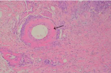
9. Histopathology
CRGV is a disease reported to cause ulceration of the distal extremities in dogs. It is variably associated with clinically acute kidney injury, so the most remarkable and characteristic lesions are allocated to the skin and kidneys (Holm et al., 2015). Previous studies, however, demonstrate that macroscopic lesions can occur in other organs as well, and can include: ascites and/or pleural effusion, haemorrhages on serosa surfaces, mural oedema of the stomach, small
intestine, and/or colon, ulcerative glossitis, red-black flecked gastric and small intestinal contents and tarry colon contents (Hertzke, et. al, 1995). The microscopic changes of these structures reported in this study consisted of rare thrombi in submucosal arterioles of the stomach, small intestine, and colon; small intestine crypts can be occasionally dilated and filled with necrotic debris and neutrophils; heart had widely scattered fibrinoid vascular necrosis, with rare foci of myocardial necrosis; hemosiderin inclusions in macrophages were found commonly within lymph nodes, liver, and spleen. This diffuse finding can be a nonspecific indicator of increased erythrocyte destruction that may result from the elimination of damaged erythrocytes, as occurs in microangiopathic processes; central nervous system lesions were limited to the mild expansion of peri-vascular spaces (Hertzke et al., 1995). Yet, in the study conducted by Holm et al. (2015), other tissues were evaluated and analysed, and changes appeared unremarkable, with mild and non-specific changes.
Histopathology of the skin samples is not a reliable diagnostic tool for CRGV. Most commonly the findings are non-specific, even though skin biopsies may be diagnostically useful as a means to reject other differential diagnostics, such as immune-mediated and neoplastic diseases. (L. Holm & Walker, 2018). Multifocal subcutaneous haemorrhages, oedema, and multifocal fibrinoid necrosis of small to medium-sized arterioles, with abrupt, full-thickness necrosis of the epidermis were found, and this suggests that cutaneous vasculitis with ischemic necrosis is a possible pathogenic mechanism (Hertzke et al., 1995).
The subcutaneous and deep dermal arterial lesions varied from mild to severe and ranged from increased eosinophilia of the tunica media to pyknosis, karyorrhexis and occasionally fibrinoid necrosis (Figure 9) (Carpenter, et. al, 1988), with, rare fibrinocellular thrombi. The subjacent dermis was often undergoing coagulative necrosis (Holm et al., 2015).
Figure 9 - Histopathology of a skin biopsy from a patient with CRGV, showing a dermal artery with
fibrinoid necrosis (arrow). Haematoxylin and eosin, x100. (Gently authorised by David Walker, Anderson Moores Veterinary Specialists).
At the level of the adnexa, the affected hair follicles are necrotic in the entire pilosebaceous units, leading to reduced or absent sebaceous glands, reduce cellularity and separate by an increase fibrous tissue and an attenuate follicular epithelium (Figure 10 and 11). The affected follicles were often bordered by variable numbers of neutrophils, foamy macrophages, and karyorrhectic debris; this often obscured the follicular epithelium interface and sebaceous gland units. (Hertzke et al., 1995; Holm et al., 2015).
In samples from the oral cavity lesions, similar ulceration of the mucosa was observed with associated necrosis, inflammation and fibrovascular change of the submucosa (Holm et al., 2015).
In conclusion, skin biopsies are not a reliable method to confirm the definitive diagnostic of CRGV but are a very helpful tool towards the final diagnosis and choice of treatment. The biopsies reveal necroulcerative dermatitis with vasculitis and thrombosis. The principal findings include extensive dermal ulceration with necrotizing folliculitis and perifolliculitis and adnexal necrosis with occasional vasculitis. The lesions were mostly centred on the hair follicles and adnexa (Figure 12) (Skulberg et al., 2018).
Figure 11 - Photomicrograph of small dermal
artery with fibrinoid necrosis of the vessel wall (Gently authorised by David Walker, Anderson Moores Veterinary Specialists)
Figure 10- Necrotic hair follicle along with
neutrophilic infiltrates. (Gently authorised by David Walker, Anderson Moores Veterinary Specialists)
Figure 12 - Histopathological appearance of a skin biopsy from a patient with CRGV. Haematoxylin
and eosin, x40. (Gently authorised by David Walker, Anderson Moores Veterinary Specialists).
Macroscopically, kidneys are uniformly swollen, unremarkable or slightly pale, congested, with numerous, irregularly distributed, barely visible, cortical and capsular petechiae (Carpenter, et. al, 1988; Hertzke et al., 1995).
The most consistent and severe microscopic lesions were found in the kidneys (Hertzke et al., 1995), with the most striking changes involving the glomeruli (Holm et al., 2015).
Carpenter et al. (1988) found in all kidneys of the greyhound interstitial congestion, oedema, and small haemorrhages originating at or near glomerular vascular poles. This specific area and adjacent to interlobular and intralobular veins a mild, multifocal, lymphocytic, and plasmocytic interstitial infiltrate were present. Most of the glomeruli are affected and for individual glomeruli, these changes range from mild and segmental to global and severe (Holm et al., 2015). These changes consist of hyaline thrombi within capillaries and afferent arterioles, segmental to global congestion and ischemic necrosis (Hertzke et. al, 1995). Fibrinoid necrosis occurs frequently and is defined by a distortion of vessel walls with eosinophilic, hyalinised, smudgy material, intermingled with low numbers of degenerate and viable neutrophils, fragmented red blood cells and mild amounts of karyorrhectic debris (Holm et al., 2015). Glomerular tufts are enlarged and congested, partially occluded by haemorrhage, obliterating the urinary space, and are mildly hypercellular. Glomerular capillary walls are thickened by finely fibrillar material. A minority of glomeruli are small, shrunk, and hypocellular. The interstitium has scatter periglomerular haemorrhages and mild oedema. Medullary congestion is also present. (Hertzke et al., 1995; Holm et al., 2015). Tubular changes are multifocal and can alter in extent (Hertzke et al., 1995) which include, hypoxic nephrosis, striking hyaline droplet change, and a variety of casts (hyaline, granular, bile-stained, and red blood cell) (Figure 13) (Carpenter et. al, 1988).
Caption - This is haired skin demonstrating extensive ulceration (black arrows) and necrosis of the adnexa, with secondary pyogranulomatous inflammation near disrupted hair follicles (white arrow).