a DNA Base Lesion through Base Excision Repair
Yanhao Lai1,2., Meng Xu1., Zunzhen Zhang2
, Yuan Liu1*
1Department of Chemistry and Biochemistry, Florida International University, Miami, Florida, United States of America,2Department of Environmental and Occupational Health, West China School of Public Health, Sichuan University, Chengdu, People’s Republic of China
Abstract
Trinucleotide repeat (TNR) expansions and deletions are associated with human neurodegeneration and cancer. However, their underlying mechanisms remain to be elucidated. Recent studies have demonstrated that CAG repeat expansions can be initiated by oxidative DNA base damage and fulfilled by base excision repair (BER), suggesting active roles for oxidative DNA damage and BER in TNR instability. Here, we provide the first evidence that oxidative DNA damage can induce CTG repeat deletions along with limited expansions in human cells. Biochemical characterization of BER in the context of (CTG)20 repeats further revealed that repeat instability correlated with the position of a base lesion in the repeat tract. A lesion located at the 59-end of CTG repeats resulted in expansion, whereas a lesion located either in the middle or the 39-end of the repeats led to deletions only. The positioning effects appeared to be determined by the formation of hairpins at various locations on the template and the damaged strands that were bypassed by DNA polymeraseb and processed by flap endonuclease 1 with different efficiency. Our study indicates that the position of a DNA base lesion governs whether TNR is expanded or deleted through BER.
Citation:Lai Y, Xu M, Zhang Z, Liu Y (2013) Instability of CTG Repeats Is Governed by the Position of a DNA Base Lesion through Base Excision Repair. PLoS ONE 8(2): e56960. doi:10.1371/journal.pone.0056960
Editor:Giovanni Maga, Institute of Molecular Genetics IMG-CNR, Italy
ReceivedOctober 17, 2012;AcceptedJanuary 16, 2013;PublishedFebruary 26, 2013
Copyright:ß2013 Lai et al. This is an open-access article distributed under the terms of the Creative Commons Attribution License, which permits unrestricted use, distribution, and reproduction in any medium, provided the original author and source are credited.
Funding:This work was supported by NIH grant ES017476 to Y.L.(http://www.nih.gov/) and partially was supported by the grant No. 81172632 from the National Natural Science Foundation of China to Z.Z.(http://www.nsfc.gov.cn/Portal0/default166.htm). The funders had no role in study design, data collection and analysis, decision to publish, or preparation of the manuscript.
Competing Interests:The authors have declared that no competing interests exist. * E-mail: [email protected]
.These authors contributed equally to this work.
Introduction
Trinucleotide repeat (TNR) expansions are identified as the cause of more than 40 neurodegenerative diseases [1], and their deletions are implicated in cancer development [2]. TNRs associated with human diseases include (CAG)n/(CTG)n,
(CTG)n/(CAG)n, (CGG)n/(CCG)n, and (GAA)n/(TTC)n.
Expan-sions of these repeats are responsible for Huntington’s disease (HD), spinocerebellar ataxia, myotonic dystrophy type 1 (DM1), fragile X syndrome, and Friedreich’s ataxia [1]. Epidemiological studies also suggest a correlation between CAG repeat deletions in the androgen receptor and prostate and ovarian cancers [2,3], implying that TNR deletions are equally as important as TNR expansions in causing human diseases.
Over the past 20 years, substantial progress has been made in understanding the mechanisms underlying TNR expansions and deletions using model systems such as bacteria [4,5], yeast [6], mammalian cells [7], and mouse models of TNR-related human diseases [8]. TNR instability is considered to be mediated by the formation of a series of non-B form DNA secondary structures and their metabolism by DNA replication [9], repair [10], and recombination [11]. Typical non-B form DNA structures include hairpins and tetraplexes that are usually generated by CAG, CTG, and CGG repeats due to their propensity of self-base pairing [12]. Hairpin structures generated on a strand with newly synthesized DNA usually cause expansions, whereas hairpins formed on a template strand usually cause repeat deletions [13]. Therefore,
factors that can facilitate the formation and stability of TNR hairpins could lead to TNR instability. For example, the length of a TNR tract appears to be critical for TNR expansion. It has been found that expansions can occur when the repeat length is greater than 35242 units. This is called the threshold of TNR expansions [14] that presumably allows the formation of stable secondary structures, and further evades cellular repair mechanisms for removing the structures [15]. However, the outcomes for TNR expansions or deletions are ultimately determined by DNA replication [1,14,16,17], repair, and double-stranded DNA repair-mediated recombination [18], during which TNR second-ary structures are processed for their genome integration [19,20]. Thus, the stability of TNRs may be modulated by the interactions between dynamic DNA structures and replication, repair, and recombination machinery.
One of the most important features of TNRs is that they all are composed of a stretch of guanines, which allow them to become the hotspots of oxidative DNA damage. A link between oxidative DNA damage and TNR instability has been established in bacteria [5,21], mammalian cells, tissues [22,23], and mouse models [24]. Exposure of bacteria to hydrogen peroxide (H2O2)
increased the deletions of TNRs [5]. H2O2significantly increased
CAG repeat expansions in the striatum of HD transgenic mouse models [22,25]. In addition, potassium bromate, an environmental oxidative DNA damaging agent, increased the level of 8-oxoG and CGG repeat expansions in the germ cells of fragile X syndrome pre-mutation mice [24]. Thus, oxidative DNA damage is actively involved in causing TNR instability, and its repair appears to play crucial roles in modulating TNR instability. This hypothesis is supported by a recent finding that 8-oxoG DNA glycosylase (OGG1), an enzyme that specifically removes 8-oxoG, is required for the age-dependent somatic CAG repeat expansions in the striatal neurons of a HD mouse model [22]. Moreover, an essential enzyme of base excision repair (BER), DNA polymeraseb(polb) binds to CAG repeats in vivo in the striatum of HD mice [26], suggesting an important role of pol b-mediated BER in modulating CAG repeat instability. Our previous study demon-strated that removal of an 8-oxoG in the context of CAG repeats by OGG1 induced single-stranded DNA (ssDNA) breaks leading to DNA strand slippage and the formation of a 59-hairpin [27]. This disrupts efficient long-patch BER that is mediated by the "hit-and-run" mechanism through pol b and flap endonuclease 1 (FEN1) [27,28], thereby resulting in an inefficient long-patch BER that involves polbmulti-nucleotide gap-filling synthesis and FEN1 alternate flap cleavage [27]. In support of this possibility, low levels of FEN1 along with normal levels of polbin the striatum of HD mice were associated with CAG repeat expansions [26]. Thus, it appears that inefficient BER is associated with TNR expansions.
Oxidative DNA damage may preferentially occur at specific locations of TNR tracts. This could modulate DNA repair efficiency and affect the outcomes of TNR instability. Oxidized DNA base lesions are preferentially induced at the loop region of a hairpin by a DNA damaging agent directly [29] and by repositioning of the lesions located in the stem of a repeat hairpin [30]. However, the lesion at this specific location was found to be resistant to OGG1 activity [31], allowing its escape from BER, leading to multiple rounds of "toxic oxidation cycles" for TNR expansion [1,31]. An abasic lesion located at the 59-end of CAG repeats was removed by BER with a much lower efficiency than the abasic lesion located either at the 39-end or in the middle of the repeats [32]. These results suggest that the positions of an oxidized base lesion in TNR tracts alter its repair efficiency that modulates accumulation of ssDNA breaks and hairpin structures at specific locations. Consequently, this would direct the damage repair path towards repeat expansions or deletions.
Here, we asked several important questions with regard to the positions of DNA base lesions and TNR instability. Can a specific location of a base lesion determine whether TNR repeat tracts are expanded or deleted through BER? If so, how are BER enzymes involved in mediating the positioning effect of base lesions, and how can TNR instability be regulated when multiple base lesions occur in TNR tracts simultaneously? In this study, we show for the first time that oxidative DNA damaging agents induce various sizes of CTG repeat deletions and limited sizes of expansions in human cells. We demonstrate that the position of an oxidative base lesion governs the instability of CTG repeats through the imbalanced activities of polbDNA synthesis and FEN1 alternate flap cleavage. Our study provides new insights into the molecular mechanisms underlying TNR expansion and deletion induced by oxidative DNA damage.
Materials and Methods
Materials
Potassium chromate (K2CrO4, purity$98.0%) and potassium
bromate (KBrO3, purity$98.0%) were obtained from Alfa Aesar
(Ward Hill, MA). Thirty percent (w/w) H2O2 was from BDH
(London, England). Dulbecco’s modified eagle medium (DMEM), fetal bovine serum (FBS), L(+)-glutamine, and 0.25% trypsin-EDTA were purchased from Life Technologies (Grand Island, NY). DNA oligonucleotides were synthesized by Integrated DNA Technologies Inc. (Coralville, IA). The radionucleotide [c232
P] ATP (6000 mCi/mmol) and cordycepin 59-triphosphate 39 -[a232P] (5000 mCi/mmol) were purchased from PerkinElmer Inc. (Boston, MA). Micro Bio-Spin 6 chromatography columns were from Bio-Rad (Hercules, CA). Deoxynucleoside 59 -triphos-phates (dNTPs) were from Roche Diagnostics (Indianapolis, IN). T4 polynucleotide kinase and terminal nucleotidyltransferase were from Fermentas (Glen Burnie, MD). Mung Bean Nuclease was from Epicenter (Madison, WI). All other reagents were purchased from Sigma-Aldrich (St. Louis, MO) and Fisher Scientific (Pittsburgh, PA). Purified recombinant human apurinic/apyrimi-dinic endonuclease 1 (APE1), polb, FEN1, and DNA ligase I (LIG I) were generous gifts from Dr. Samuel Wilson at the National Institute of Environmental Health Sciences, National Institutes of Health (Research Triangle Park, NC) or were expressed and purified as described previously [28].
Oligonucleotide Substrates
DNA oligonucleotide substrates containing a tetrahydrofuran (THF), an abasic site analog in the context of (CTG)20 repeats
were used to mimic an oxidized abasic site. The guanines in the first, tenth, twentieth, or both the first and eleventh CTG unit were substituted with a THF residue. Substrates were constructed by annealing an oligonucleotide strand with one or two base lesions to its template strand at a molar ratio of 1:2. A DNA fragment that contained (CTG)20without any DNA base lesions
was used as a size marker for DNA fragment analysis. The sequences and descriptions of the oligonucleotides are shown in Supplemental Table S1.
Cell Culture and Transfection of (CTG)35/(CAG)35 and (CTG)20/(CAG)20-containing Plasmids
Human embryonic kidney (HEK) 293-H cells (Life Technolo-gies, Grand Island, NY) were cultured in DMEM supplemented with 10% FBS and 4 mM L(+)-glutamine at 37uC under 5% CO2.
A plasmid containing (CTG)35/(CAG)35 or (CTG)20/(CAG)20
repeats was constructed by inserting a fragment containing a (CTG)35/(CAG)35 or (CTG)20/(CAG)20 tract flanked by the 59
-and the 39-side random DNA sequences into pcDNA3.1/CT-GFP-TOPO vector (Life Technologies), respectively. A DNA fragment containing a random sequence with the same length as the (CTG)35/(CAG)35 repeat-containing fragment (225 nt) or
(CTG)20/(CAG)20repeat-containing fragment (100 nt) was cloned
into pcDNA3.1/CT-GFP-TOPO for constructing the plasmids used as the random sequence control. Plasmids (12mg) were
pre-incubated with 36ml lipofectamine 2000 (Life Technologies), for 20 min at room temperature. The mixture of plasmids and lipofectamine was subsequently transferred to the medium supplied for culturing 46105HEK293-H cells. Cellular
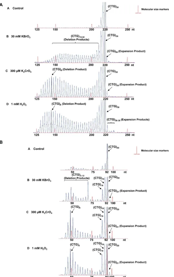
transfec-(CTG)20repeats (12mg). PanelAillustrates the result from untreated cells. PanelsB,C,andDrepresent the results from the cells treated with KBrO3, K2CrO4,and H2O2, respectively. The repaired products are illustrated as peaks. The height of a peak indicates the abundance of a specific repair product. The sizes of repair products are illustrated in nucleotides. Size standards are indicated.
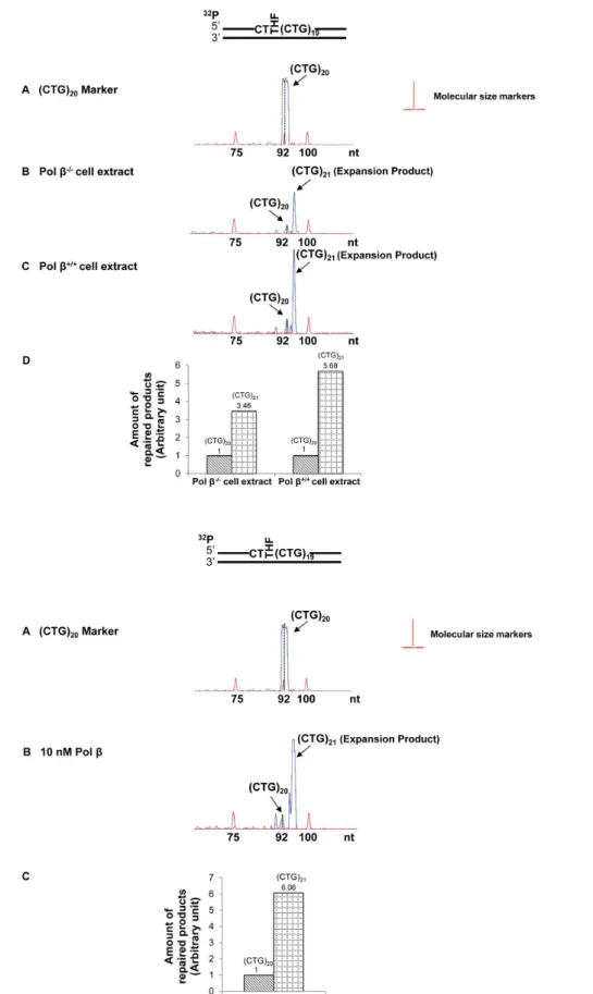
Figure 2. CTG repeat expansion resulting from an abasic lesion located at the 59-end of (CTG)20repeats.(A) A substrate containing
(CTG)20repeats with a THF inserted for substituting the guanine of the first CTG was incubated with cell extracts of polb2/2or polb+/+MEFs under the conditions described in the Materials and Methods. PanelArepresents the result of PCR amplification of a DNA marker containing (CTG)20repeats without any damage. PanelsBandCillustrate the results from BER mediated by polb2/2and polb+/+
tion efficiency was determined using a fluorescent microscope (Leica, Wetzlar, Germany). For all the experiments, the transfec-tion efficiency was greater than 95%.
Measurement of Instability of (CTG)35/(CAG)35 and (CTG)20/(CAG)20Induced by Oxidative DNA Damage in HEK293-H cells
Instability of (CTG)35/(CAG)35 or (CTG)20/(CAG)20 repeats
induced by oxidative DNA damage was examined by treating 46105HEK293-H cells, transfected with the (CTG)35/(CAG)35or
(CTG)20/(CAG)20-containing plasmids, using three well known
environmental and endogenous oxidative DNA-damaging agents, KBrO3, K2CrO4, and H2O2 at concentrations of 30 mM,
300mM, and 1 mM, respectively for 2 hr. Cells were washed
twice with phosphate-buffered saline (PBS), supplied with fresh medium, and grown for 2 days to allow recovery from DNA damage. The treatment was repeated three times before cells were harvested. In the control experiment, HEK293-H cells transfected with plasmids that contain random DNA sequences were treated by three DNA damaging agents under the same conditions used for treatment of cells bearing repeat-containing plasmids. At the end of the experiments, cells were trypsinized using 0.25% trypsin-EDTA and harvested by centrifugation at 3000 rpm for 15 min. Plasmids were isolated from cells using Qiagen Miniprep Kits (Qiagen, Valencia, CA), dissolved in Tris-EDTA (TE) buffer (10 mM Tris-HCl, pH 7.5, and 1 mM EDTA), and stored at 220uC for subsequent size analysis. Untreated cells served as a negative control. The experiments were repeated at least 3 times.
In VitroBER in Mouse Embryonic Fibroblast Cell Extracts
Polbnull (polb2/2) and wild type (polb+/+
) mouse embryonic fibroblasts (MEFs) were grown to near confluence. Cells were washed twice with PBS, harvested by cell scrapers, and centrifuged at 3000 rpm for 15 min. Cell extracts were made as described previously [33] and were dialyzed into BER reaction buffer containing 50 mM Tris-HCl, pH 7.5, 50 mM KCl, 0.1 mM EDTA, 0.1 mg/ml bovine serum albumin, and 0.01% Nonidet P-40. Substrates were pre-incubated with 50 nM purified APE1 at 37uC for 30 min, and completely converted into ssDNA break intermediates for subsequent BER reactions. In vitro BER of a THF in polb2/2 and pol b+/+cell extracts was performed by incubating APE1 precut (CTG)20repeat-containing substrate with
60mg cell extracts under the conditions described previously [27].
Reaction mixtures were assembled on ice and incubated at 37uC for 30 min. BER reactions were terminated by transferring to 95uC for 5 min. Reaction mixtures were subsequently digested with protease K at 55uC for 30 min. Repair intermediates and products were precipitated and dissolved in stopping buffer containing 95% formamide and 2 mM EDTA, and were separated by 15% urea-denaturing polyacrylamide gel electro-phoresis (PAGE). Repair products were further isolated from the gel and eluted with TE buffer through rotation at room temperature overnight. The products were precipitated with ethanol, dissolved in TE buffer, and stored at 220uC for subsequent size analysis.
In vitroBER Reconstituted with Purified Enzymes
BER of ssDNA break intermediates was reconstituted by incubating 50 nM purified APE1, 10 nM pol b, 10 nM FEN1, and 5 nM LIG I with 25 nM (CTG)20 repeat-containing
substrates with one or two THF residues. The 20ml reaction
mixture contained BER buffer with 50mM dNTPs, 5 mM Mg2+,
2 mM ATP, and indicated concentrations of BER enzymes and substrates. Reaction mixtures were assembled on ice, and incubated at 37uC for 15 min. Reactions were terminated by transferring to 95uC for 5 min in stopping buffer. Repair products and intermediates were separated by 15% urea-denaturing PAGE. Repair products were isolated from the gel and eluted with TE buffer through rotation at room temperature overnight. The products were precipitated with ethanol, dissolved in TE buffer, and stored at220uC for subsequent sizing analysis.
Probing of Hairpin Structures by Mung Bean Nuclease Digestion
Hairpin formation on the damaged and template strands of (CTG)20-containing substrates were probed by Mung Bean
Nuclease digestion. Substrates (200 nM) containing one or two THF residues at different locations of (CTG)20repeats were
pre-cut by 10 nM APE1 and were subjected to digestion with 1 U Mung Bean Nuclease at 37uC for 1, 2, 3, 5, and 8 min. The 10-ml
reaction was conducted in buffer containing 30 mM sodium acetate (pH 4.6), 50 mM NaCl, 1 mM zinc acetate, and 0.01% Triton X-100. Enzymatic reactions were terminated by 2mg proteinase K digestion at 55uC for 30 min. Reaction mixtures were subjected to 95uC for 10 min to denature DNA. Substrates and digestion products were separated by 15% urea-denaturing PAGE and detected by a Pharos FX Plus PhosphorImager from Bio-Rad. Synthesized DNA size markers were used to indicate the size of nuclease cleavage products.
Enzymatic Activity Assay
PolbDNA synthesis during BER of ssDNA break intermediates was measured by using 25 nM oligonucleotide substrates contain-ing (CTG)20 with one or two THF residues as illustrated in
Supplemental Table S1. Polbactivity was examined at 37uC in a 20ml reaction mixture that contained BER reaction buffer with 50mM dNTPs and 5 mM Mg2+. FEN1 cleavage activity in the absence or presence of pol b was examined under the same conditions used for determining polbactivity. Repair intermedi-ates and products were separated by 15% urea-denaturing PAGE and detected by a PhosphorImager. Synthesized size markers for illustrating the size of pol b DNA synthesis products or FEN1 cleavage products were run in parallel with repair products.
Sizing Analysis of CTG Repeats by DNA Fragment Analysis and PeakScanner Software
The size of repaired products was determined by capillary electrophoresis using an ABI 3130XL Genetic Analyzer (Applied Biosystems, Foster City, CA) and DNA fragment analysis with PeakScanner version 1.0 software (Applied Biosystems, Foster City, CA) with assistance from the DNA Sequencing Core of Florida International University. A 225 nt- or 100 nt-DNA fragment in plasmids containing (CTG)35/(CAG)35 repeats or
quantitative analysis of the results of panelsBandC. (B) The THF at the 59-end of (CTG)20repeats was repaired by BER reconstituted with 10 nM purified polbas described in the Materials and Methods (panelB). PanelAillustrates the result of PCR amplification of a (CTG)20repeat-containing marker without any DNA damage, and panelCillustrates the quantitative analysis of the results from panelB. Substrates were32P-labeled at the 59 -end of the damaged strand as indicated. Size standards are indicated.
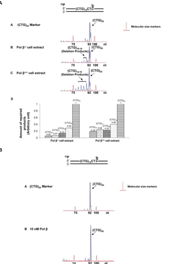
Figure 3. CTG repeat deletions induced by an abasic lesion in the middle of (CTG)20repeats.(A) A substrate containing (CTG)20repeats with a THF, inserted for substituting the guanine of the tenth CTG, was incubated with 60mg cell extracts of polb2/2or polb+/+
MEFs for 30 min. PanelArepresents the result of PCR amplification of a DNA marker with (CTG)20repeats. PanelsBandCillustrate the results from BER of the THF residue mediated by polb2/2and polb+/+
(CTG)20/(CAG)20repeats or random sequences was amplified by
PCR using a forward primer with a 59–6-carboxyfluorescein tag and a reverse primer (Supplementary Table S1). PCR amplifica-tion condiamplifica-tions were as follows: 95uC for 10 min, 1 cycle; 95uC for 30 s, 50uC (for repeats) or 55uC (for random sequences) for 30 s, and 72uC for 1.5 min, 35 cycles; final extension at 72uC for 1 hr. Size standards, MapMarker 1000 (Bioventures, Murfreesboro, TN) were run in parallel with PCR-amplified repair products.
Results
CTG Repeat Deletions and Expansions Induced by Oxidative DNA Damage in Human Cells
To determine how oxidative DNA damage may affect TNR instability in human somatic cells, we initially examined the effects of environmental and endogenous oxidative DNA damaging agents on CTG repeat instability in HEK293-H cells. We reasoned that these agents can result in a stretch of 8-oxoGs and ssDNA breaks in TNRs that lead to accumulation of ssDNA break intermediates, DNA slippage, and the formation of hairpin structures on both the damaged and template strands of TNR tracts. The repair of ssDNA break intermediates including hairpin structures could allow integration of the hairpins into the genome, thereby causing expansions and deletions.
To explore this possibility, we examined the instability of (CTG)35/(CAG)35 and (CTG)20/(CAG)20 repeats induced by
oxidative DNA damaging agents, KBrO3, K2CrO4,and H2O2
[34,35] in HEK293-H cells. The results showed that the length of (CTG)35/(CAG)35repeats in the untreated cells varied between 33
to 35 repeat units, although a small portion of plasmids containing 32 and 36 repeats were detected (Figure 1A, panel A). The length of (CTG)20/(CAG)20 repeats in the untreated cells ranged from
(CTG)18to (CTG)20repeats. Exposure of cells to 30 mM KBrO3
resulted in a series of deletion products with (CTG)10to (CTG)32
repeats for (CTG)35-containing plasmids (Figure 1A, panel B) and
deletion products with (CTG)3to (CTG)6or (CTG)12or (CTG)18
to (CTG)19 repeats for (CTG)20-containing plasmids (Figure 1B,
panel B). Thus, bromate-induced DNA damage led to deletion of (CTG)35repeats by 3 to 25 repeat units and deletion of (CTG)20
by 1–17 repeat units. Three hundred micromoles of K2CrO4and
1 mM H2O2led to deletion products with repeat length mainly
ranging from (CTG)4to (CTG)32for (CTG)35repeats (Figure 1A,
panels C and D) and (CTG)4 to (CTG)19 for (CTG)20 repeats
(Figure 1B, panels C and D). In addition, all of the DNA damaging agents led to small amounts of expansion products with (CTG)36to
(CTG)39 repeats for (CTG)35 repeat-containing plasmids
(Figure 1A, panels B, C, and D) and an expansion product with (CTG)21 repeats for (CTG)20 repeat-containing plasmids
(Figure 1B, panels B, C, and D).
Bromate, chromate and hydrogen peroxide failed to induce any length change in a 225 nt- and 100 nt-fragment that contained random DNA sequence (Supplementary Figure S1) indicating that oxidative DNA damage-induced CTG repeat instability was TNR sequence specific.
Interestingly, KBrO3-induced deletion products from (CTG)35
repeats exhibited a pattern with even size distribution, which suggests that a single 8-oxoG may be induced at different repeats in a randomized manner. KBrO3-induced deletion products from
(CTG)20 repeats contained (CTG)18–19, (CTG)12 and (CTG)3–6
repeats that correspond to small, middle and large size deletion, respectively, suggesting that the agent resulted in a similar deletion pattern in (CTG)20 repeats as the one in (CTG)35 repeats. In
contrast, K2CrO4and H2O2predominantly induced the deletions
with a peak size of (CTG)9 or (CTG)10 repeats. The size
distribution pattern of deletions and expansions suggests that a specific oxidative DNA damaging agent may induce a single or multiple base lesions/ssDNA breaks at specific positions in a CTG repeat tract, preferentially leading to either repeat deletions or expansions.
We designated the damage position-specific effect on the instability of CTG repeats as the ‘‘DNA damage positioning effect.’’ Because oxidative DNA damage is mainly repaired by BER, it is possible that the positioning effects of oxidative DNA damages are accomplished through BER of oxidative DNA base lesions in the context of CTG repeats. To test this possibility, we examined the effects of the position and the number of DNA base lesions on CTG repeat instability duringin vitrocell extract-based and reconstituted BER.
A Specific Location of a DNA Base Lesion on (CTG)20 Repeats Correlated with Repeat Expansion or Deletion
The position of a DNA base lesion or ssDNA break in CTG repeats may be classified as at the 59-end, in the middle, or at the 39-end of the repeat tract. To examine how a base lesion at these positions may modulate repeat instability, we used a series of (CTG)20 repeat-containing substrates with an abasic lesion
represented by a THF residue that substituted the guanine of the first, tenth, and twentieth CTG unit. These substrates mimic the scenarios wherein a single oxidized base lesion occurs at the 59 -end, in the middle, and at the 39-end of a (CTG)20repeat tract,
respectively. A substrate containing two THF residues embedded in the first (the 59-end) and the eleventh CTG (the middle) of (CTG)20repeats was used to mimic a situation in which more than
one DNA base lesion occurs simultaneously in the repeat tract. The effects of an abasic lesion at these locations on the instability of (CTG)20 repeats during BER were initially
deter-mined with cell extracts made from polb2/2 or polb+/+
MEFs (Figure 2A, 3A, 4A, 5A), and were verified by reconstituted BER (Figure 2B, 3B, 4B, 5B). The results revealed that a lesion located at the 59-end of (CTG)20repeats resulted in a (CTG)21expansion
product through BER mediated by pol b2/2 and pol b+/+
cell extracts (Figure 2A, panels B and C). The expansion product was also generated by BER reconstituted with10 nM purified polbin the presence of APE1, FEN1, and LIG I (Figure 2B, panel B). Quantitative analysis showed that the ratio between the amount of (CTG)21 expansion product and that of (CTG)20 unexpanded
products was increased from 3.5 to 6 by the presence of pol b
during BER (Figure 2A, panel D and Figure 2B, panel C). This indicates that polbpromoted repeat expansion during BER of an abasic lesion at the 59-end of the damaged strand. PCR amplification of a DNA marker without any damage gave no repeat expansions or deletions (Figure 2A and 2B, panel A). In addition, PCR amplification of a (CTG)20-containing substrate
with an intact or APE1-preincised abasic site, failed to produce any amplified products (Figure S5). The results demonstrate that the expansion product was from BER rather than from PCR
of a THF in the middle of (CTG)20repeats was performed by BER reconstituted with 10 nM purified polb(panelB). PanelAis the result of PCR amplification of a (CTG)20repeat-containing marker, and panelCillustrates the quantitative analysis of the results from panelB. BER reactions and repeat sizing analysis were performed under the conditions described in the Materials and Methods. Substrates were32P-labeled at the 59-end of the damaged strand as indicated. Size standards are indicated.
Figure 4. CTG repeat instability resulting from an abasic lesion at the 39-end of (CTG)20repeats.(A) A substrate containing (CTG)20 repeats with a THF that substituted the guanine of the twentieth CTG was incubated with 60mg cell extracts of polb2/2or polb+/+
MEFs (panelsB andC) for 30 min. PanelArepresents the result of PCR amplification of a DNA marker with (CTG)20repeats. PanelDrepresents the quantitative analysis of the results of panelsBandC. (B) The repair of a THF at the 39-end of (CTG)20repeats was performed by BER reconstituted with 10 nM purified polb(panelB). PanelAis the result of PCR amplification of a (CTG)20repeat-containing marker without any DNA damage. Substrates were 32
P-labeled at the 59-end of the damaged strand as indicated. Size standards are indicated. doi:10.1371/journal.pone.0056960.g004
artifacts. In conclusion, our results indicated that a base lesion located at the 59-end of (CTG)20led to repeat expansion through
BER.
Interestingly, we found that cell extract-based BER of an abasic site located in the middle resulted in deletion products with (CTG)4-(CTG)6repeats (Figure 3A, panels B and C). Quantitative
analysis demonstrated that pol b increased (CTG)4 and (CTG)5
deletion products by about 2- to 4-fold, but did not affect the production of (CTG)6 deletion product (Figure 3A, panel D).
Surprisingly, reconstituted BER of a lesion in the middle of the repeat tract only resulted in small deletion products containing (CTG)18and (CTG)19repeats (Figure 3B, panel B). These results
suggest that the large size deletions from cell extracts involve other repair enzymes/proteins in addition to the BER core enzymes, APE1, polb, FEN1, and LIG I.
Cell extract-based BER of a 39-end abasic lesion resulted in small deletion products containing (CTG)16 to (CTG)19 repeats
(Figure 4A, panels B and C). The amount of deletion products was increased by approximately 3-fold in pol b+/+
cell extracts (Figure 4A, panel D). However, reconstituted BER of the damaged products gave neither deletion nor expansion (Figure 4B). This indicated that the repair of a 39-end base lesion by the core BER enzymes was not sufficient to cause CTG repeat deletions. This further suggests that deletions may essentially be mediated by the cooperation between core BER enzymes and other repair proteins that can shorten CTG repeats from the 39-end of the damaged strand.
For the scenario in which two base lesions located at both the 59-end and in the middle of the repeat, large deletion products containing (CTG)5to (CTG)10repeats were detected during cell
extract-based and reconstituted BER (Figure 5A, panels B and C, and Figure 5B). Quantitative analysis indicated that deletions were increased by 5- to 7-fold in the presence of endogenous pol b
(Figure 5A, panel D). All these results indicate an active role of pol
bin promoting both expansions and deletions (Figure 2A, 3A, 4A, 5A). Interestingly, for all the positions, base lesions induced CTG repeat deletions and expansions in polb2/2cell extracts. Absence of pol b also facilitated the formation of (CTG)18 and (CTG)19
repeat deletion products (Figure 5A) suggesting a role of pol b -independent BER pathways in modulating both small and large size of TNR deletions and expansions.
It should be noted that the size of both the (CTG)20size marker
and the (CTG)20unexpanded repaired products was calculated by
DNA fragment analysis to be 92 nt which was 8 nt shorter than its actual length of 100 nt. This is because the (CTG)20-containing
size marker and repaired products contain stretches of CTG repeats, and the standards for calculating the sizes of DNA fragments contain random sequences. Such difference in DNA sequences resulted in a difference between the mobility of the CTG repeat-containing DNA fragments and that of random sequence DNA fragments during capillary electrophoresis. This led to the difference between the calculated size of a DNA fragment and its actual size.
Various sizes of Hairpins Formed on the Damaged and Template Strands of (CTG)20Repeats
Because the formation of hairpin structures has been proposed as the basis underlying TNR instability [13,27], the propensity of a base lesion at specific positions to lead to CTG repeat deletion or expansion could be due to the formation of hairpins at different locations in the repeat tract that favors deletion or expansion. To test this idea, we examined the formation of hairpins on both strands of the (CTG)20repeat-containing substrates after APE1 59
-incision of a THF residue located at different positions in the CTG repeat tract, using Mung Bean Nuclease, the enzyme that preferentially cleaves at a single-stranded hairpin loop as well as at the sites with mismatched base-pairs in the stem region of a hairpin.
For the substrate containing a THF at the 59-end, the cleavage by Mung Bean Nuclease on the template strand resulted in products of 22 nt, 29 nt, 32 nt, 34 nt, 37 nt, and 40 nt (Figure 6A, panel A). The cleavage pattern indicated the formation of a (CAG)7hairpin with a loop constituted by (CAG)3repeats and a
stem consisting of two pairs of CAG repeats (Figure 6A, panel D). The hairpin was located adjacent to the 39-end flanking region of the template strand. The nuclease cleavage on the damaged strand resulted in products with 20 nt, 22 nt, 25 nt, 28 nt, 31 nt, 34 nt, 37 nt, 40 nt, 43 nt, 46 nt, and 49 nt (Figure 6A, panel B), indicating the formation of a (CTG)10 repeat-containing hairpin
with a loop composed of two CTG repeats and a stem containing four pairs of CTG repeats (Figure 6A, panel D). The hairpin was adjacent to the 39-side random sequence of the damaged strand.
For a lesion located in the middle of (CTG)20 repeats, the
nuclease cleavage on the template strand led to products with 22 nt, 28 nt, 31 nt, 34 nt, 37 nt, 40 nt, 67nt, 68 nt, and 74 nt (Figure 6B, panel A). This cleavage pattern indicates the coexistence of two hairpins on the template strand with seven repeats in between them. One was a (CAG)7-repeat containing
hairpin adjacent to the 39-end flanking region and composed of a loop with one CAG and a stem consisting of three pairs of CAG repeats. The other was a (CAG)4-repeat-containing hairpin that
was two CAG repeats away from the 59-end flanking region, and contained a loop with two repeats and a short stem with only one pair of CAG repeats (Figure 6B, panel E). For the damaged strand, hairpins forming on both the upstream strand and the downstream strand were probed. The nuclease cleavage on the upstream strand resulted in products with 20 nt, 25 nt, 28 nt, 31 nt, 34 nt, 37 nt, 40 nt, and 43 nt (Figure 6B, panel B), indicating the formation of a (CTG)8repeat hairpin with a loop containing two CTG repeats
and a stem with three pairs of repeats (Figure 6B, panel E). The nuclease cleavage on the downstream CTG repeats resulted in products with 20 nt, 22 nt, 25 nt, 28 nt, 31 nt, 34 nt, 37 nt, 40 nt, and 43 nt (Figure 6B, panel C), indicating the formation of a downstream (CTG)8 hairpin adjacent to the 39-flanking region
(Figure 6B, panel E).
For a THF located at the 39-end of (CTG)20 repeats, Mung
Bean Nuclease cleavage on the template strand resulted in products with 22 nt, 28 nt, 31 nt, 34 nt, 37 nt, and 40 nt (Figure 6C, panel A), indicating the formation of a (CAG)7repeat
Figure 5. CTG repeat deletions from the abasic lesions located at the 59-end and in the middle of (CTG)20repeats.(A) A substrate
containing (CTG)20repeats with two THF residues that substituted the guanines of the first and the tenth CTG, was incubated with 60mg cell extracts of polb2/2or polb+/+
MEFs for 30 min (panelsBandC). PanelArepresents the result of PCR amplification of a DNA marker with (CTG)20repeats. PanelDrepresents the quantitative analysis of the results of panelsBandC. (B) The repair of two THF residues was performed by BER reconstituted with 10 nM purified polb(panelB). PanelAillustrates the result of PCR amplification of a (CTG)20repeat-containing marker without any DNA damage. BER reactions were performed under the conditions described in the Materials and Methods. Substrates were32P-labeled at the 59-end of the damaged strand as indicated. Size standards are indicated.
doi:10.1371/journal.pone.0056960.g005
Figure 6. The formation of hairpins resulting from an abasic lesion located at different positions of (CTG)20repeats.The hairpins
formed on both the damaged and template strands of the (CTG)20repeat-containing substrate with a base lesion located at different positions of repeat tracts, were probed by Mung Bean Nuclease digestion under the conditions described in the Materials and Methods. The results of hairpin probing from a damage located at the 59-end, in the middle, or at the 39-end of the repeat tract are illustrated in (A), (B), and (C), respectively. The results from the damages located at both the 59-end and in the middle of the repeats are illustrated in (D). The relative amount of hairpins was illustrated as the percentage of Mung Bean Nuclease cleavage products. The percentage of Mung Bean Nuclease cleavage products for the damaged strand were calculated by the amount of Mung Bean Nuclease cleavage products that accounted for the formation of hairpin over the total amount of APE1 cleavage products at 1-min interval of enzymatic digestion of hairpins. The percentage of Mung Bean Nuclease cleavage products that represent the formation of a template hairpin induced by the damage at the 59-end, in the middle, at the 39-end, and at both the 59-end and the middle of (CTG)20repeat, was calculated based on 55%, 50%, 56% and 75% of the total amount of the template strand, respectively. A deduced hairpin is illustrated schematically along with specific nuclease digestion sites as indicated. Lane1represents the undigested substrates. Lanes226 represent the digestion products generated at various time intervals. Lane7represents a series of synthesized size markers (M) for illustrating the size of the digestion products.
hairpin on the template strand adjacent to the 39-flanking region. The nuclease cleavage also resulted in products with larger size (.80 nt). This indicated that the nuclease cleavage in the random sequence flanking region of the template. This may be because of transient dissociation of the 20 nt random DNA sequence from its template after APE1 59-incision of the THF residue on the damaged strand. This resulted in a single strand region in the template strand that was subsequently captured and cleaved by the nuclease. The nuclease digestion on the damaged strand resulted in products with 20 nt, 25 nt, 31 nt, 34 nt, 37 nt, 40 nt, 43 nt, 46 nt, and 49 nt (Figure 6C, panel B), indicating the existence of a (CTG)10hairpin adjacent to the 59-side random sequence region
with a loop containing two CTG repeats and a stem containing (CTG)8(Figure 6C, panel D).
Finally, for the substrate that contained two base lesions at both the 59-end and in the middle of the damaged strand, the nuclease cleavage on the template strand resulted in two groups of products. One group contained products of 22 nt, 28 nt, 32 nt, 34 nt, 37 nt, and 40 nt, and the other contained products of 67 nt, 71 nt, and 73 nt (Figure 6D, panel A). This indicates the formation of both (CAG)9and a (CAG)4repeat-containing hairpin on the template
strand. As illustrated in panel D of Figure 6D, the (CAG)9hairpin
consisted of a loop with (CAG)3repeats and a stem with three pairs
of mismatched CAG repeats, and the (CAG)4hairpin contained a
loop with two CAG repeats and a stem with one pair of mismatched CAG repeats. The enzymatic cleavage on the damaged strand of the substrate generated products with 22 nt, 25 nt, 28 nt, 31 nt, 34 nt, 37 nt, and 40 nt (Figure 6D, panel B), indicating the formation of a (CTG)7 hairpin with a loop
containing only one CTG and a stem with three pairs of CTG repeats (Figure 6D, panel D).
Quantitative analysis of Mung Bean Nuclease cleavage products from all of the substrates showed that approximately 16.3962.65% to 79.2562.20% of cleavage products were generated from the template strand, the upstream and down-stream strands (panel C of Figure 6A, 6C, and 6D; panel D of Figure 6B), indicating that the formation of hairpins during BER is significant.
It also should be noted that a 17 nt product cleavage product was observed during probing of the hairpins induced by a base lesion in the middle or at both the 59-end and in the middle of (CTG)20repeats (Figure 6B, panels A and C; Figure 6D, panel B).
This indicated that Mung Bean Nuclease also made the cleavage in the random sequence regions that flanked the repeats. This could be due to transient dissociation of the random sequence strand from its template strand after Mung Bean Nuclease made the cleavage at the base of hairpins. The dissociated random sequence strand was then captured and cleaved by the enzyme.
To verify the specificity of Mung Bean Nuclease, we examined the enzyme cleavage on a substrate containing a template hairpin composed of a loop of six adenosines and a stem with 15 nt-matched base pairs. The results showed that the enzyme only specifically cleaved at the ssDNA loop region of the hairpin (Supplemental Figure S2). We failed to detect any cleavage products on both the damaged strand and the template strand of a random sequence substrate (Supplemental Figure S3). We also failed to detect Mung Bean Nuclease cleavage products on all of the (CTG)20 repeat substrates in the absence of APE1
(Supple-mental Figure S4). These results indicated that the formation of hairpins is CTG repeat-specific and exclusively dependent on ssDNA breaks.
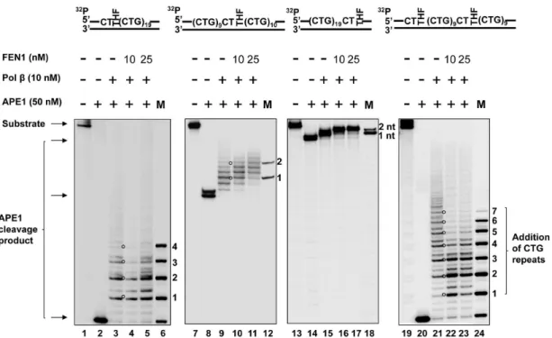
Figure 7. PolbDNA synthesis during BER of an abasic lesion located at different sites of (CTG)20repeats.PolbDNA synthesis with the
substrates containing one or two THF residues located at the 59-end, or/and in the middle, or at the 39-end of (CTG)20repeats was determined in the presence of 10 nM of polbalong with 10 nM and 25 nM FEN1. Lanes1, 7, 13,and19correspond to substrates only. Lanes2, 8, 14,and20 correspond to reaction mixtures with 50 nM APE1. Lanes325,9211,15217,and21223correspond to reaction mixtures with 10 nM polbin the absence or the presence of 10 nM and 25 nM FEN1. Lanes6,12,18, and24correspond to a series of synthesized size markers (M) for illustrating the size of polbDNA synthesis products. Substrates were32P-labeled at the 59-end of their damaged strands. Substrates are illustrated schematically above the gel.
doi:10.1371/journal.pone.0056960.g007
PolbMulti-nucleotide Gap-filling DNA Synthesis during BER of a THF Residue Located at Different Sites of (CTG)20 Repeats
Because our previous study suggested that disruption of polb
and FEN1 coordination during long-patch BER led to TNR expansion [27], and our current results also indicated that polb
promoted TNR deletions (Figures 3, 4, 5), we asked whether the positioning effects of a base lesion on CTG repeat instability could be due to different efficiencies of polbDNA synthesis and FEN1 flap cleavage activity in the context of various numbers, sizes, and positions of hairpin structures.
To address this question, we initially characterized polbDNA synthesis during the repair of a THF located at the 59-end, in the middle, and at the 39-end of (CTG)20repeats as well as during the
repair of the two damages located at the 59-end and in the middle. The results revealed that in the absence or the presence of 10 nM and 25 nM FEN1, 10 nM pol b mainly inserted three CTG repeats for repairing the 59-end THF group (Figure 7, lanes 3, 4, 5) and four repeats for repairing the base lesions located at both the 59-end and in the middle of the repeats (Figure 7, lanes 21, 22, 23). However, the same concentration of the enzyme only caused insertion of two repeats for repairing the damage in the middle of the repeat tract (Figure 7, lanes 9, 10, 11), and one or two nucleotides in repairing the 39-end base damage (Figure 7, lanes 15, 16, 17). The results indicated that pol b mainly performed limited multi-nucleotide DNA synthesis for repairing a base lesion in the middle or at the 39-end of CTG repeat tracts, but exhibited efficient DNA synthesis for repairing base lesions located at the 59 -end or at both the 59-end and in the middle of the repeats. It is possible that efficient DNA synthesis of polbleads to expansions, whereas inefficient DNA synthesis causes deletions through
coordination with FEN1 cleavage of CTG flaps. Interestingly, we observed that FEN1 cleavage slightly reduced pol b DNA synthesis by 1 to 3 repeat units during BER of an abasic site located at both the 59-end and in the middle of (CTG)20repeats
(Figure 7, lanes 22–23). This may be due to FEN1 binding to a repair intermediate with a 59-CTG flap attached to a downstream hairpin that dislodged pol b from the intermediate, thereby inhibiting DNA synthesis by the polymerase. This was also demonstrated by our recent finding showing that FEN1 inhibited polbDNA synthesis in the presence of a (CTG)7flap, but failed to
affect polbactivity in the absence of the flap [36].
FEN1 Cleavage on CTG Repeat Flaps during the Repair of a Base Lesion at Various Locations of (CTG)20 Repeats
Because FEN1 alternate cleavage for processing the 59-end of a hairpin is critical for TNR expansion [27], and repeat deletions during BER requires a cleavage of repeats for shortening repeat length, we reasoned that FEN1 flap cleavage activity plays a critical role in mediating both repeat expansions and deletions during the repair of an abasic lesion located at specific sites in (CTG)20repeats.
To test this idea, we determined FEN1 flap cleavage on the substrates with a THF at different locations of (CTG)20repeats in
the presence of 10 nM polb(Figure 8). The results revealed that FEN1 removed two CTG repeats for repairing a 59-end THF and six repeats for repairing a THF in the middle and for repairing two THF residues (Figure 8, lanes 324, lanes 829, and lanes 18219). However, FEN1 cleaved up to 12 nucleotides in the 39-side random sequence region in a stepwise manner for repairing a THF located at the 39-end of the repeat tract (Figure 8, lanes 13214). This indicated that FEN1 cleaved relatively larger sizes of Figure 8. FEN1 cleavage during BER of an abasic lesion located at various sites of (CTG)20repeats.Substrates containing one or two THF
residues located at the 59-end, or/and in the middle, or at the 39-end of (CTG)20repeats were pre-incubated with 50 nM APE1. FEN1 cleavage activity was determined in the presence of 10 nM and 25 nM FEN1 plus 10 nM polbunder the conditions described in the Materials and Methods. Lanes1, 6,11,and16correspond to substrates only. Lanes2,7,12,and17correspond to reaction mixtures with 50 nM APE1. Lanes324,829,13214, and18219correspond to reaction mixtures with 10 nM and 25 nM FEN1 with 10 nM polb. Lanes5,10,15, and20correspond to a series of synthesized size markers (M) for illustrating the size of FEN1 cleavage products. Substrates were32P-labeled at the 39-end of their damaged strands. Substrates are illustrated schematically above the gel.
repeats for repairing the base lesion located in the middle of the repeat tracts than for repairing the damage at the 59-end.
Discussion
In this study, we provide the first evidence that CTG repeat expansions and deletions can be induced by oxidative DNA damage (Figure 1) and mediated through BER (Figure 2–5). We demonstrate that the repeat instability is correlated with the positions and the numbers of DNA base lesions in repeat tracts. We further demonstrate that the positioning effect is mediated by the formation of multiple hairpins with varying sizes on both the template and damaged strands of (CTG)20repeats (Figure 6). This
is accomplished by different efficiencies of pol b DNA synthesis
and FEN1 flap cleavage (Figure 728). Pol b exhibited more efficient DNA synthesis (Figure 7, lanes 325) than FEN1 flap cleavage (Figure 8, lanes 324) to repair damage located at the 59 -end of the repeats, thereby leading to expansion. In contrast, FEN1 exhibited more efficient cleavage (Figure 8, lanes 829) than polbDNA synthesis (Figure 7, lanes 9, 10, 11) to repair a damage located in the middle of CTG repeats, leading to one or two repeat deletions. For the two damages located at both the 59-end and in the middle of CTG repeats, high efficiency of polbDNA synthesis displaces the repeat strand in between the two damaged sites from the template, and high efficiency of FEN1 cleavage removes more repeats. The synergistic effect of efficient polband FEN1 activity leads to large deletions. For a base lesion located at the 39-end of the repeats, the repair event is carried out in the context of a Figure 9. Hypothetical models illustrating CTG repeat instability governed by the positions of oxidative DNA base lesions through polband FEN1.Oxidative stress can result in an oxidized DNA base lesion, 8-oxoG that is located either at the 59-end, in the middle, or at the 39-end of CTG repeats. Removal of the damaged site by OGG1 leaves an abasic site that is subsequently 59-incised by APE1 generating ssDNA breakage, results in the formation of multiple hairpins on repeat tracts. A base lesion that occurs at the 59-end of the repeats allows the formation of a multi-nucleotide gap and subsequently a 59-hairpin along with a template hairpin. Polbpasses through the template hairpin and synthesizes more repeats than those removed by FEN1, thereby causing repeat expansions (sub-pathway1). A base lesion located in the middle of CTG repeats results in the formation of an upstream hairpin and a downstream hairpin in the damaged strand along with two template hairpins. The upstream hairpin leads to inefficient polbDNA synthesis that inserts one or two repeats, but allows FEN1 to remove more repeats than those synthesized by polb, leading to small repeat deletions (sub-pathway2). A base damage located at the 39-end of repeats immediately adjacent to the random sequence region, allows the formation of a large upstream hairpin and a template hairpin. Polbinserts one to three nucleotides to fill a single-nucleotide gap. In this scenario, repeats in the upstream strand cannot be processed by repair activities, thereby leading to maintenance of repeat length (sub-pathway3). When two base lesions simultaneously occur at the 59-end and in the middle of CTG repeat tracts, two hairpins in the damaged strand and two template hairpins would form. This leads to efficient polbDNA synthesis that can strand-displace the hairpin in between two damage sites, resulting in loss of large numbers of repeats. Efficient FEN1 cleavage of the 59-downstream CTG repeats leads to additional shortening of repeats. The cooperation between polbstrand-displacement synthesis and FEN1 cleavage leads to large repeat deletions (sub-pathway4).
doi:10.1371/journal.pone.0056960.g009
random DNA sequence. Polband FEN1 activity fail to affect the repeat instability.
Our results support the idea that differences in the efficiency of polbDNA synthesis and FEN1 cleavage underlies the positioning effect of a base lesion on TNR instability. Based on the results, we can suggest hypothetical models that illustrate the positioning effect of a DNA base lesion mediated by polband FEN1 during long-patch BER (Figure 9). The paths for inducing TNR instability by a base lesion or a ssDNA break located at the 59 -end, in the middle, and at the 39-end of CTG repeats are illustrated in sub-pathways 1, 2, and 3 (Figure 9). The path for TNR instability induced by the lesions located at both the 59-end and in the middle of the repeats is illustrated in sub-pathway 4 (Figure 9).
Substantial expansions of CTG repeats in the 39-untranslated region of the myotonic dystrophy protein kinase gene have been implicated as the cause of DM1 [37]. Interestingly, CTG repeat deletions have also been identified during replication and transcription in bacterial and human cells [5,7,38], and this is proposed as one avenue for DM1 treatment [39]. Therefore, understanding of the mechanisms underlying CTG repeat deletions will help identify new targets for disease treatment. Herein, we observed predominantly large deletions, but also small expansions in the tract of (CTG)35and (CTG)20repeats induced
by environmental and endogenous oxidative DNA damaging agents in human cells (Figure 1). This is consistent with the finding that H2O2increased the rate of CTG/CAG repeat deletions in
bacteria [5]. Our attempt to explore the molecular basis underlying CTG instability revealed that the positions and the numbers of DNA base lesions and/or ssDNA breaks governed the formation of hairpins with varying sizes at specific locations of TNRs. This subsequently modulated the efficiency of BER enzymes, causing either repeat expansions or deletions. Our results suggest that CTG repeat expansions or deletions can be regulated by the interactions between dynamic DNA structures in CTG repeats and BER proteins. We demonstrated that BER mediated by polb+/+cell extracts led to significant amounts of expansion and deletion products (Figures 2A, 3A, and 4A) when compared to pol b2/2 cell extracts, indicating that pol b can promote CTG repeat-expansion and deletion. This further suggests an involvement of pol b-mediated BER in modulating TNR instability.
Interestingly, we observed some expansion and deletion products resulting from the repair of an abasic lesion by polb2/ 2cell extracts with the same sizes as the ones from polb+/+
cell extracts (panel B of Figures 2A, 3A, and 4A). This suggests that pol
b-independent BER pathways also play an important role in modulating CTG repeat instability. Because pol b-independent BER usually involves replication and other repair DNA polymer-ases such as pold, pole[40], and poll[41], it appears that these DNA polymerases may substitute polbto perform effective DNA synthesis to repair base lesions located at various positions in TNRs. Therefore, it will be important to explore the roles of replicative DNA polymerases, pol l, and other X family polymerases in modulating TNR expansions and deletions during BER of oxidative DNA damage.
Larger sizes of repeats (15 repeats) were deleted through cell extract-based BER than through BER reconstituted with purified BER enzymes (one to ten repeats, Figures 3 and 5). This indicated that BER mediated by MEF cell extracts caused much more severe TNRs deletions than BER reconstituted by the core BER enzymes, suggesting that other DNA repair proteins in cell extracts may facilitate the large repeat deletions. This could result from the
activity of other nucleases that cooperate with FEN1 cleavage activity.
This possibility is supported by a recent study showing that a 59– 39 exonuclease, exonuclease 1 (EXO1) cooperated with FEN1 to remove flaps of CTG and CGG repeats during DNA replication [42]. This suggests that a synergistic effect from EXO1 and FEN1 may promote large TNR deletions. In addition, TNRs might also be processed by 39–59exonucleases, such as Mre 11 [43]. This can lead to shortening of the repeats by cleaving the 39-end of TNR, thereby contributing to large repeat deletions in coordination with EXO1 and FEN1 flap cleavages. Large deletions could also be promoted by stabilizing a template hairpin via hairpin binding proteins, MSH2/MSH3 [44]. This may stimulate polb hairpin bypass synthesis, resulting in loss of large numbers of repeat units. The roles of various nucleases and hairpin binding proteins in causing large TNR deletions during BER in a coordinated manner need to be elucidated in further studies.
In this study, we also provide the first evidence that a base lesion located at various positions on (CTG)20repeats can result in the
formation of multiple hairpins with varying sizes at specific positions on CTG/CAG repeat tracts (Figure 6). The coexistence of multiple hairpin structures on both the template and the damaged strand allowed the formation of a cluster of hairpins that may resemble the clustered slip-outs as described in a recent study [45]. Such clustered slip-outs have been found to act as roadblocks for their repair by hMutSb (MSH2/MSH3) complexes, and therefore are proposed to be responsible for CTG/CAG repeat expansion. It is conceivable that clustered hairpin structures generated during BER may also function as blockages to promote inefficient BER that ultimately leads to expansions and deletions. Consistent with this notion, we found inefficient activity of polb
DNA synthesis (Figure 7, lanes 9211), or limited FEN1 cleavage of 59-hairpins (Figure 8, lanes 324) in the presence of multiple hairpins on both DNA strands. The role of BER in governing TNR instability for coordination with MSH2/MSH3 through repairing clustered hairpins must be further elucidated.
In conclusion, we demonstrated for the first time that oxidative DNA damage can lead to both CTG deletions and expansions in human somatic cells andin vitro. We found that BER of a base lesion at a specific position on (CTG)20 repeats correlates with
either deletion or expansion, i.e., a damage at the 59-end of repeats preferentially leads to repeat expansion, whereas a damage in the middle results in deletions. A lesion located at the 39-end mainly leads to maintenance of repeat lengths. The positioning effect results from the formation of multiple hairpin structures with varying sizes at different locations on repeat tracts. Hairpins at specific locations lead to different efficiency of polbDNA synthesis and FEN1 cleavage activity that governs whether the repeats can be expanded or deleted. We propose that the positioning effect of oxidative DNA base damage on TNR instability is a consequence of the interaction between hairpin structures and BER enzymes and cofactors. Our study defines a mechanism underlying oxidative DNA base lesion-induced TNR instability.
Supporting Information
Figure S1 Oxidative DNA damage does not alter the length of random DNA sequences in human cells. (A) Plasmids containing a fragment with random DNA sequence that has the same length as (CTG)35/(CAG)35 repeat-containing
results from the cells treated with KBrO3, K2CrO4,and H2O2,
respectively. (B) Plasmids containing a fragment with random DNA sequence that has the same length as (CTG)20/(CAG)20
repeat-containing fragment (100 nt) were transfected into HEK293-H cells that were treated with oxidative DNA-damaging agents as described in the Materials and Methods. PanelAis the result from untreated cells. Panels B, C, and D represent the results from the cells treated with KBrO3, K2CrO4,and H2O2,
respectively. Size standards are illustrated. (TIF)
Figure S2 Mung Bean Nuclease specifically cleaves the loop region of a stable hairpin. A substrate containing a template hairpin composed of a loop of six adenines and a stem with 15 nt base pairs was radiolabeled at the 59-end of its template strand. The substrate was incubated with 0.01 U Mung Bean Nuclease at 0.5- and 1-min time intervals (Lanes2and3). Lane1
represents substrate only. Lane4represents synthesized markers (M) with 30, 45, 48, 51, 66 nucleotides, respectively.
(TIF)
Figure S3 No hairpin forms in the context of random sequences. The formation of a hairpin on both the template strand and the damaged strand of random sequence of a substrate with a THF residue at the 59-end of its damaged strand was probed by Mung Bean Nuclease digestion. The substrate was radiolabeled at either the 59-end of its template strand (panelA) or the 39-end of its damaged strand (panel B). The substrate was precut by 10 nM APE1 and was incubated with 1 U Mung Bean Nuclease at 1-, 2-, 3-, 5-, 8-min time intervals (lanes2–6of panelA
and lanes8–12of panelB)under the conditions described in the Materials and Methods. Lane 1 represents the undigested substrate.
(TIF)
Figure S4 No hairpin forms in the absence of ssDNA breakage. (A) Hairpin formation on both the template strand and the damaged strand of a (CTG)20-containing substrate with a
THF residue at the 59-end was probed by Mung Bean Nuclease in the absence of APE1 cleavage. The substrate was radiolabeled at the 39-end of its template strand (left panel) or its damaged strand (right panel). The substrate was incubated with 1 U Mung Bean Nuclease at 1-, 2-, 3-, 5-, 8-min time intervals. Lane 1 and 8
represent substrate without enzyme digestion. Lane 7represents synthesized markers (M) with 18, 29, 38, 56, 74 nucleotides. Lane
14 represents synthesized markers with 18, 29, 38, 47, 56, 74 nucleotides. (B) The formation of hairpins on the template strand and the damaged strand of a (CTG)20-containing substrate with a
THF residue in the middle of CTG repeats was probed by Mung Bean Nuclease in the absence of APE1 cleavage under the condition described in (A). The substrate was radiolabeled at the 39-end of its template strand (left panel) or the 59- or the 39-end of
its damaged strand (middle and right panels). Lanes1,8and15
represent substrate only. Lanes7, 14and21represent the same synthesized markers (M) described in (A). (C) Hairpin formation on the template strand and the damaged strand of a (CTG)20
-containing substrate with a THF residue at the 39-end of the repeat track was probed by Mung Bean Nuclease in the absence of APE1. The substrate was radiolabeled at the 39-end of its template (left panel) or the 59-end of its damaged strand (right panel). The substrate was incubated with 1 U Mung Bean Nuclease under the condition described in (A). Lane1and8represent substrate only. Lane 7 and 14 represent the same synthesized markers (M) described in (A). (D) Hairpins on the template strand and the damaged strand of (CTG)20-containing substrate with two THF
residues were probed by Mung Bean Nuclease in the absence of APE1 under the condition described in (A). The substrate was radiolabeled at the 39-end of either its template (left panel) or its damaged strand (right panel). Lane1 and 8 represent substrate only. Lane7and 14 represent the same synthesized markers (M) as used in (A).
(TIF)
Figure S5 PCR amplification of a (CTG)20 repeat-containing substrate with an AP site at the 59-end of the damaged strand with or without APE1 59-incision.
PCR reactions were performed with the substrate containing a THF residue at the 59-end with or without APE1 59-incision under the conditions described in the Materials and Methods. Lane 2 represents the result of PCR amplification of the damage-containing substrate without APE1 59-incision. Lane 3 represents the result of PCR amplification of the substrate preincised by 50 nM APE1. Lane 4 represents the result of PCR amplification of a (CTG)20-contaning marker without any damage. Lane 1 and 5
represent DNA size markers ranging from 100 bp to 1000 bp. (TIF)
Table S1 Oligonucleotides sequences. (DOCX)
Acknowledgments
We thank Samuel H. Wilson, Laboratory of Structural Biology, National Institute of Environmental Health Sciences (NIEHS), National Institutes of Health (NIH) for generously providing purified BER enzymes and vectors
for expressing BER enzymes, as well as polb2/2and polb+/+
MEF cell lines. We thank Justin Torner and Shyama Ramjagsingh for technical assistance.
Author Contributions
Construction of ideas: Y. Lai Y. Liu MX ZZ. Conceived and designed the experiments: Y. Lai Y. Liu MX ZZ. Performed the experiments: Y. Lai MX. Analyzed the data: Y. Lai MX Y. Liu ZZ. Wrote the paper: Y. Lai Y. Liu ZZ MX.
References
1. McMurray CT (2010) Mechanisms of trinucleotide repeat instability during human development. Nat Rev Genet 11: 786–799.
2. Nelson KA, Witte JS (2002) Androgen receptor CAG repeats and prostate cancer. Am J Epidemiol 155: 883–890.
3. Schildkraut JM, Murphy SK, Palmieri RT, Iversen E, Moorman PG, et al. (2007) Trinucleotide repeat polymorphisms in the androgen receptor gene and risk of ovarian cancer. Cancer Epidemiol Biomarkers Prev 16: 473–480. 4. Kang S, Jaworski A, Ohshima K, Wells RD (1995) Expansion and deletion of
CTG repeats from human disease genes are determined by the direction of replication in E. coli. Nat Genet 10: 213–218.
5. Hashem VI, Sinden RR (2002) Chemotherapeutically induced deletion of expanded triplet repeats. Mutation Research 508: 107–119.
6. Freudenreich CH, Kantrow SM, Zakian VA (1998) Expansion and length-dependent fragility of CTG repeats in yeast. Science 279: 853–856.
7. Pelletier R, Farrell BT, Miret JJ, Lahue RS (2005) Mechanistic features of CAG*CTG repeat contractions in cultured cells revealed by a novel genetic assay. Nucleic Acids Res 33: 5667–5676.
8. Savouret C, Brisson E, Essers J, Kanaar R, Pastink A, et al. (2003) CTG repeat instability and size variation timing in DNA repair-deficient mice. EMBO J 22: 2264–2273.
9. Kang S, Ohshima K, Shimizu M, Amirhaeri S, Wells RD (1995) Pausing of DNA synthesis in vitro at specific loci in CTG and CGG triplet repeats from human hereditary disease genes. J Biol Chem 270: 27014–27021.
10. Oussatcheva EA, Hashem V.I., Zou Y., Sinden R.R. and Potaman V.N. (2001) Involvement of the nucleotide excision repair protein UvrA in instability of CAG*CTG repeat sequences in Escherichia coli. J Mol Biol 279: 1101–1110. 11. Jakupciak JP, Wells R.D. (2000) Genetic instabilities of triplet repeat sequences
by recombination. IUBMB life 50: 355–359.
12. Wells RD, Dere R, Hebert ML, Napierala M, Son LS (2005) Advances in mechanisms of genetic instability related to hereditary neurological diseases. Nucleic Acids Res 33: 3785–3798.
13. Mirkin SM (2007) Expandable DNA repeats and human disease. Nature 447: 932–940.
14. Wells RD (1996) Molecular basis of genetic instability of triplet repeats. J Biol Chem 271: 2875–2878.
15. Lahue RS, Slater DL (2003) DNA repair and trinucleotide repeat instability. Front Biosci 8: S653–665.
16. Pearson CE, Nichol Edamura K, Cleary JD (2005) Repeat instability: mechanisms of dynamic mutations. Nat Rev Genet 6: 729–742.
17. McMurray CT (1999) DNA secondary structure: a common and causative factor for expansion in human disease. Proc Natl Acad Sci U S A 96: 1823–1825. 18. Pluciennik A, Iyer RR, Napierala M, Larson JE, Filutowicz M, et al. (2002) Long
CTG.CAG repeats from myotonic dystrophy are preferred sites for intermo-lecular recombination. J Biol Chem 277: 34074–34086.
19. Liu Y, Bambara R (2003) Analysis of human flap endonuclease 1 mutants reveals a mechanism to prevent triplet repeat expansion. J Biol Chem 278: 13728–13739.
20. Liu Y, Wilson SH (2012) DNA base excision repair: a mechanism of trinucleotide repeat expansion. Trends Biochem Sci 37: 162–172.
21. Jackson AL, Loeb LA (2000) Microsatellite instability induced by hydrogen peroxide in Escherichia coli. Mutat Res 447: 187–198.
22. Kovtun IV, Liu Y, Bjoras M, Klungland A, Wilson SH, et al. (2007) OGG1 initiates age-dependent CAG trinucleotide expansion in somatic cells. Nature 447: 447–452.
23. Gomes-Pereira M, Monckton DG (2004) Chemically induced increases and decreases in the rate of expansion of a CAG*CTG triplet repeat. Nucleic Acids Res 32: 2865–2872.
24. Entezam A, Lokanga AR, Le W, Hoffman G, Usdin K (2010) Potassium bromate, a potent DNA oxidizing agent, exacerbates germline repeat expansion in a fragile X premutation mouse model. Hum Mutat 31: 611–616. 25. Bogdanov MB, Andreassen OA, Dedeoglu A, Ferrante RJ, Beal MF (2001)
Increased oxidative damage to DNA in a transgenic mouse model of Huntington’s disease. J Neurochem 79: 1246–1249.
26. Goula AV, Berquist BR, Wilson DM, 3rd, Wheeler VC, Trottier Y, et al. (2009) Stoichiometry of base excision repair proteins correlates with increased somatic CAG instability in striatum over cerebellum in Huntington’s disease transgenic mice. PLoS Genet 5: e1000749.
27. Liu Y, Prasad R, Beard WA, Hou EW, Horton JK, et al. (2009) Coordination between polymerase beta and FEN1 can modulate CAG repeat expansion. J Biol Chem 284: 28352–28366.
28. Liu Y, Beard WA, Shock DD, Prasad R, Hou EW, et al. (2005) DNA polymerase beta and flap endonuclease 1 enzymatic specificities sustain DNA synthesis for long patch base excision repair. J Biol Chem 280: 3665–3674. 29. Jarem DA, Wilson NR, Delaney S (2009) Structure-dependent DNA damage
and repair in a trinucleotide repeat sequence. Biochemistry 48: 6655–6663.
30. Volle CB, Jarem DA, Delaney S (2012) Trinucleotide repeat DNA alters structure to minimize the thermodynamic impact of 8-oxo-7,8-dihydroguanine. Biochemistry 51: 52–62.
31. Jarem DA, Wilson NR, Schermerhorn KM, Delaney S (2011) Incidence and persistence of 8-oxo-7,8-dihydroguanine within a hairpin intermediate exacer-bates a toxic oxidation cycle associated with trinucleotide repeat expansion. DNA Repair (Amst) 10: 887–896.
32. Goula AV, Pearson CE, Della Maria J, Trottier Y, Tomkinson AE, et al. (2012) The nucleotide sequence, DNA damage location, and protein stoichiometry influence the base excision repair outcome at CAG/CTG repeats. Biochemistry 51: 3919–3932.
33. Biade S, Sobol RW, Wilson SH, Matsumoto Y (1998) Impairment of proliferating cell nuclear antigen-dependent apurinic/apyrimidinic site repair on linear DNA. J Biol Chem 273: 898–902.
34. Kawanishi S, Murata M (2006) Mechanism of DNA damage induced by bromate differs from general types of oxidative stress. Toxicology 221: 172–178. 35. Messer J, Reynolds M, Stoddard L, Zhitkovich A (2006) Causes of DNA single-strand breaks during reduction of chromate by glutathione in vitro and in cells. Free Radic Biol Med 40: 1981–1992.
36. Xu M, Gabison J, Liu Y (2012) Trinucleotide repeat deletion via a unique hairpin bypass by DNA polymerase beta and alternate flap cleavage by flap endonuclease 1. Nucleic Acids Res doi: 10.1093/nar/gks1306.
37. Pineiro E, Fernandez-Lopez L, Gamez J, Marcos R, Surralles J, et al. (2003) Mutagenic stress modulates the dynamics of CTG repeat instability associated with myotonic dystrophy type 1. Nucleic Acids Res 31: 6733–6740. 38. Nakamori M, Pearson CE, Thornton CA (2011) Bidirectional transcription
stimulates expansion and contraction of expanded (CTG)*(CAG) repeats. Hum Mol Genet 20: 580–588.
39. Lopez Castel A, Cleary JD, Pearson CE (2010) Repeat instability as the basis for human diseases and as a potential target for therapy. Nat Rev Mol Cell Biol 11: 165–170.
40. Wilson SH, Beard WA, Shock DD, Batra VK, Cavanaugh NA, et al. (2010) Base excision repair and design of small molecule inhibitors of human DNA polymerase beta. Cell Mol Life Sci 67: 3633–3647.
41. Braithwaite EK, Kedar PS, Stumpo DJ, Bertocci B, Freedman JH, et al. (2010) DNA polymerases beta and lambda mediate overlapping and independent roles in base excision repair in mouse embryonic fibroblasts. PLoS One 5: e12229. 42. Vallur AC, Maizels N (2010) Complementary roles for exonuclease 1 and Flap
endonuclease 1 in maintenance of triplet repeats. J Biol Chem 285: 28514– 28519.
43. Paull TT, Gellert M (1998) The 39to 59exonuclease activity of Mre 11 facilitates repair of DNA double-strand breaks. Mol Cell 1: 969–979.
44. Owen BA, Yang Z, Lai M, Gajec M, Badger JD, 2nd, et al. (2005) (CAG)(n)-hairpin DNA binds to Msh2-Msh3 and changes properties of mismatch recognition. Nat Struct Mol Biol 12: 663–670.