Objective:To validate the quantiication of T-cell receptor excision circles (TRECs) and kappa-deleting recombination excision circles (KRECs) by real-time polymerase chain reaction (qRT-PCR) for newborn screening of primary immunodeiciencies with defects in T and/or B cells in Brazil.
Methods: Blood samples from newborns and controls were collected on filter paper. DNA was extracted and TRECs, and KRECs were quantified by a duplex real-time PCR. The cutoff values were determined by receiver operating characteristic curve analysis using SPSS software (IBM®, Armonk, NY, USA).
Objetivo:Validar a quantiicação de T‑cell receptor excision circles (TRECs) e kappa‑deleting recombination circles (KRECs) por reação em cadeia de polimerase (polymerase chain reaction, PCR) em tempo real (qRT-PCR), para triagem neonatal de imunodeiciências primárias que cursam com defeitos nas células T e/ou B no Brasil. Métodos:Amostras de sangue de recém-nascidos (RN) e controles foram coletadas em papel-iltro. O DNA foi extraído e os TRECs e KRECs foram quantiicados por reação duplex de qRT-PCR. O valor de corte foi determinado pela análise de Receiver Operating Characteristics Curve, utilizando-se o programa Statistical Package for the Social Sciences (SSPS) (IBM®, Armonk, NY, EUA).
ABSTRACT
RESUMO
*Autor correspondente. E-mail: [email protected] (L.A. Barreiros).
aDepartment of Immunology, Universidade de São Paulo (USP), São Paulo, SP, Brazil. bHospital Municipal Dr. José de Carvalho Florence, São José dos Campos, SP, Brazil.
cDepartment of Pediatrics, Universidade Federal de São Paulo (UNIFESP), São Paulo, SP, Brazil.
dHospital da criança Santo Antonio, Porto Alegre, RS, Brazil. eAPAE-SP, São Paulo, SP, Brazil.
fAmparo Maternal, São Paulo, SP, Brazil.
gHospital Regional da Asa Sul, Brasilia, DF, Brazil.
hHospital Geral de Carapicuíba, Carapicuíba, SP, Brazil. iHospital Municipal São Luiz Gonzaga, SP, Brazil.
jClinic of Recurrent Infections, Faculdade de Medicina do ABC, Santo André, SP, Brazil. kHospital da criança Conceição, Porto Alegre, RS, Brazil.
lClínica de Medicina Preventiva do Pará (CLIMEP), Belém, PA, Brazil.
First authorship is shared between Marilia P. P. Kanegae and Lucila A. Barreiros.
Received on May 12, 2016; accepted on October 2, 2016; available online on March 08, 2017.
NEWBORN SCREENING FOR SEVERE COMBINED
IMMUNODEFICIENCIES USING TRECS AND KRECS:
SECOND PILOT STUDY IN BRAZIL
Triagem neonatal de imunodeficiências graves combinadas
por meio de TRECs e KRECs: segundo estudo piloto no Brasil
Marilia Pyles P. Kanegae
a*, Lucila Akune Barreiros
a*, Jusley Lira Sousa
a,
Marco Antônio S. Brito
a, Edgar Borges de Oliveira Junior
a, Lara Pereira Soares
b,
Juliana Themudo L. Mazzucchelli
c, Débora Quiorato Fernandes
d, Sonia Marchezi Hadachi
e,
Silvia Maia Holanda
f, Flavia Alice T. M. Guimarães
g, Maura Aparecida P. V. V. Boacnin
h,
Marley Aparecida L. Pereira
i, Joaquina Maria C. Bueno
b, Anete Sevciovic Grumach
j,
Regina Sumiko W. Di Gesu
k, Amélia Miyashiro N. dos Santos
c, Newton Bellesi
l,
26
INTRODUCTION
Severe combined immunodeiciency (SCID) is a heterogeneous group of diseases characterized by a low number or absence of T lymphocytes. A defect in the antibody production, which may be due to intrinsic defects of B lymphocytes or inappro-priate activity of T cells, is usually present.1 Patients with SCID
are healthy at birth; however, they develop bacterial, viral, or fungal infections in the irst months of life and, if not prop-erly treated, they will die before reaching one year of age.2,3
Curative therapy is the hematopoietic stem cell transplantation (HSCT) or, in some cases, gene therapy. he earlier HSCT is performed, the better the prognosis.4
Since 2008, newborn screening for SCID has been avail-able in the United States of America. he methodology con-sists in quantifying T-cell receptor excision circles(TRECs), which are small markers produced during the development of T lymphocytes in the thymus. During the recombination of T-cell receptor genes, DNA segments are excised, forming small circles named TRECs, which can be ampliied by poly-merase chain reaction (PCR). Its quantity in the peripheral blood directly relects thymic activity.3,5
Recently, Borte et al.6 described a multiplex assay
capa-ble of detecting TRECs and kappa-deleting excision circles (KRECs) in the same reaction for newborn screening of
primary immunodeiciencies. KRECs and TRECs are simi-larly formed during the development of B lymphocytes and their quantiication enables early diagnosis of defects in B cells, such as X-linked agammaglobulinemia (XLA) or auto-somal recessive. hese are diseases characterized by B-cell deiciency caused by mutations in genes that encode compo-nents of the B-cell receptor (BCR) or precursor thereof (pre-BCR) and lead to decreased serum immunoglobulin levels. he predominant defect is XLA, corresponding to 85% of cases. Carriers of this disease are susceptible to viral and bac-terial infections, and the most common causes of infection are the encapsulated bacteria Haemophilus inluenzae and
Streptococcus pneumonia. he onset of symptoms in agam-maglobulinemia occurs between three and six months of age, as maternal immunoglobulin levels fall. If there is no family history, the diagnosis is usually late.7,8 Early identiication
of carriers of agamaglobulinemias is very advantageous, as these children are prone to develop chronic and debilitating respiratory infections.3,6
A previous work carried out by our group, which was pio-neer in the country, has validated the TRECs quantiication methodology for neonatal screening for SCID.9 his study
aimed at validating the quantiication of TRECs and KRECs in the same reaction by quantitative real-time PCR (qRT-PCR) Results: Around 6,881 samples from newborns were collected
and TRECs and KRECs were quantiied. The TRECs values ranged between 1 and 1,006 TRECs/µL, with mean and median of 160 and 139 TRECs/µL, respectively. Three samples from patients with severe combined immunodeiciency (SCID) showed TRECs below 4/µL and a patient with DiGeorge syndrome showed undetectable TRECs. KRECs values ranged from 10 to 1,097 KRECs/µL, with mean and median of 130 and 108 KRECs/µL. Four patients with agammaglobulinemia had results below 4 KRECs/µL. The cutoff values were 15 TRECs/µL and 14 KRECs/µL and were established according to the receiver operating characteristic curve analysis, with 100% sensitivity for SCID and agammaglobulinemia detection, respectively.
Conclusions: Quantiication of TRECs and KRECs was able to diagnose children with T- and/or B-cell lymphopenia in our study, which validated the technique in Brazil and enabled us to implement the newborn screening program for SCID and agammaglobulinemia.
Keywords: Severe combined immunodeficiency (SCID); Agammaglobulinemia; Newborn screening; Immunologic deiciency syndromes; Child.
Resultados: 6.881 amostras de RN foram analisadas quanto à concentração de TRECs e KRECs. Os valores de TRECs variaram entre 1 e 1.006 TRECs/µL, com média e mediana de 160 e 139 TRECs/µL, respectivamente. Três amostras de pacientes diagnosticados com imunodeiciência grave combinada (severe combined immunodeiciency, SCID) apresentaram valores de TRECs abaixo de 4/µL e um paciente com Síndrome de DiGeorge apresentou TRECs indetectáveis. Os valores de KRECs encontraram-se entre 10 e 1.097 KRECs/µL, com média e mediana de 130 e 108 KRECs/µL, e quatro pacientes com diagnóstico de agamaglobulinemia tiveram resultados abaixo de 4 KRECs/µL. Os valores de corte encontrados foram 15 TRECs/µL e 14 KRECs/µL, e foram estabelecidos de acordo com a análise da Receiver Operating Characteristics Curve, com sensibilidade de 100% para detecção de SCID e agamaglobulinemia, respectivamente. Conclusões: A quantificação de TRECs e KRECs foi capaz de diagnosticar crianças com linfopenias T e/ou B em nosso estudo, validando a técnica e dando o primeiro passo para a implementação da triagem neonatal em grande escala no Brasil.
for neonatal screening of primary immunodeiciencies which occur with defects in T and/or B cells. his enabled to widen the range of diseases screened.
METHOD
This cross-sectional study included samples from new-borns (NB), whose parents/guardians agreed to partici-pate in the research project. Blood samples were collected between September 2014 and July 2015 on filter paper from heel puncture in the NB, in compliance with current ethical norms (CAAE: 36364214.8.0000.5467). Samples from three hospitals of the metropolitan region of São Paulo (Amparo Maternal, Hospital Geral de Carapicuíba, and Hospital Municipal São Luiz Gonzaga), from a clinic in Belém (PA – CLIMEP), a hospital in São José dos Campos (SP – Hospital Municipal Dr. José de Carvalho Florence), and a hospital in Porto Alegre (RS – Hospital Nossa Senhora da Conceição) were collected and analyzed. Twenty-three sam-ples from patients with suspected primary immunodefi-ciency (Table 1)10 were referred for investigation by
pediat-ric immunologists/allergists across the country. As positive controls for test validation, samples from three patients previously diagnosed with SCID and four patients with agammaglobulinemia were used.
For PCR, we performed DNA elution from discs of 3.2 mm in diameter from the samples collected on ilter paper.11
he DNA was then ampliied in a duplex qRT-PCR for TRECs and KRECs in inal volume of 20 uL, containing 10 uL of
TaqMan Gene Expression Master Mix, 8 uL of DNA solution, 0.8 uL of 10 mg/mL bovine serum albumin, and primer and probes as described by Sottiniet al.12 he inal concentrations of
primers for TRECs and KRECs were 300 and 600nM, respec-tively, and probes were used at concentration of 200 nM. he ampliication of beta-actin was performed on the same micro-plate, only for samples with TRECs and/or KRECs below the cutof value, with primers and probe described by Baker et al.,5
at inal concentrations of 250 and 150 nM, respectively. he concentrations of TREC, KREC, and beta-actin molecules (endogenous control) were calculated using one standard curve built by dilution of plasmids containing the speciic sequences described by Sottini et al.12
To determine whether a sample was within the normal parameters, we used an initial cutof value for TRECs and KRECs of 25 copies/uL, which was based on the study of Baker et al.5 If the sample showed values below this concentration in
the initial analysis, we repeated the whole process of extraction and ampliication, along with the quantiication of beta-actin. After this second step, if TRECs and/or KRECs remained below normal, with beta-actin above 8,000 copies/uL, patients were contacted and referred to pediatric immunologist/allergist for evaluation and conirmatory testing (immunophenotyping of lymphocytes and subpopulations).
he inal data analysis was performed using descriptive sta-tistics. We determined the non-Gaussian distribution of the sample by means of the Kolmogorov–Smirnov test, and data were presented according to median and interquartile val-ues. Concentrations of TRECs and KRECs in full-term and preterm NBs were analyzed using the Mann–Whitney test in the software GraphPad Prism 5.0 (San Diego, CA, USA). Finally, we analyzed the receiver operating characteristic curve (area under the curve) to determine a inal cutof value, with 100% sensitivity for SCID and agammaglobulinemia detec-tion, using the Statistical Package for Social Sciences(SPSS) (IBM®, Armonk, NY, USA).
RESULTS
Around 6,881 samples from NBs were collected and analyzed for the concentration of TRECs and KRECs. Of the total sample, 1,853 (26.90%) were from CLIMEP (Belém/PA), 146 (2.10%) from the Hospital Municipal Dr. José de Carvalho Florence (São José dos Campos/SP), and 40 (0.58%) from the
Hospital Nossa Senhora da Conceição (Porto Alegre/RS), and the remaining samples were from hospitals located in the metro-politan region of São Paulo.
he TRECs values ranged between 1 and 1,006 molecules/ µL, with mean and median of 160 and 139 molecules µL, Table 1 Ten signs of primary immunodeficiencies
in infants.
Two or more pneumonia in the last year Four or more new otitis in the last year
Recurrent stomatitis or moniliasis for more than two months
Recurrent abscesses or ecthyma
An episode of severe systemic infection (meningitis, osteoarthritis, septicemia)
Recurrent intestinal infections/chronic diarrhea/ giardiasis
Severe asthma, collagen disease, or autoimmune disease Adverse efect to BCG and/or mycobacterial infection Clinical phenotype suggestive of a syndrome associated with immunodeiciency
Family history of immunodeiciency
28
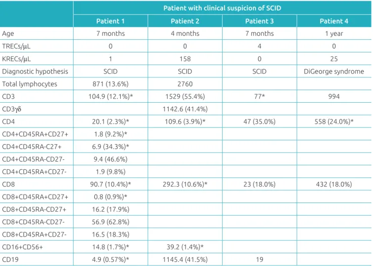
respectively. Three samples of patients diagnosed with SCID showed values of TRECs below 4/µL, and a patient with DiGeorge syndrome presented undetectable TRECs despite normal amplification of beta-actin (Table 1).13
KRECs values ranged between 10 and 1,097 molecules/µL, with mean and median of 130 and 108 molecules/µL. Four patients with agammaglobulinemia had results below 4 molecules/µL.
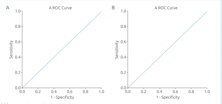
he cutof values were 15 TRECs/µL and 14 KRECs/µL, established according to the analysis of the receiver operating characteristic curve, with sensitivity of 100% for detection of SCID and agammaglobulinemia, respectively (Fig. 1). Samples with results below the cutof values described ear-lier were subjected to new DNA extraction and were re-ex-amined aggregating the beta-actin analysis for the extraction quality control. Only two samples from the newborn screen-ing remained with low values of TRECs and/or KRECs, as shown in Fig. 2. hese patients were contacted and referred
to a pediatric immunologist/allergist, who was a collaborator to this work. he irst patient, whose results were 1 TREC/ µL and 211 KRECs/µL, died on the sixth day after birth as a result of a pleural efusion occurred at six months of pregnancy. As a consequence, parents received genetic coun-seling and a blood sample followed for genetic sequencing. he second patient (157 TRECs/µL and 10 KRECs/µL) attended the irst medical appointment. No history related to primary immunodeiciencies or consanguinity was found on this occasion; however, this patient did not return to col-lect samples for speciic tests. Twenty-three samples from patients with suspected primary immunodeiciencies (black triangles in Fig. 2) were referred by pediatric immunolo-gists/allergists throughout Brazil. Of these, ive resulted in TRECs and/or KRECs values below the cutof. his result corroborated the clinical suspicion. One of these patients is carrier of trisomy 21 and presented values of 6 TRECs/µL and 14 KRECs/µL.
Table 1 Immunophenotyping of lymphocytes from three patients with severe combined immunodeiciency and
one with diagnosed DiGeorge syndrome who was referred as control in neonatal screening.
Patient with clinical suspicion of SCID
Patient 1 Patient 2 Patient 3 Patient 4
Age 7 months 4 months 7 months 1 year
TRECs/μL 0 0 4 0
KRECs/μL 1 158 0 25
Diagnostic hypothesis SCID SCID SCID DiGeorge syndrome
Total lymphocytes 871 (13.6%) 2760
CD3 104.9 (12.1%)* 1529 (55.4%) 77* 994
CD3γδ 1142.6 (41.4%)
CD4 20.1 (2.3%)* 109.6 (3.9%)* 47 (35.0%) 558 (24.0%)*
CD4+CD45RA+CD27+ 1.8 (9.2%)* CD4+CD45RA-C27+ 6.9 (34.3%)* CD4+CD45RA-CD27- 9.4 (46.6%) CD4+CD45RA+CD27- 1.9 (9.8%)
CD8 90.7 (10.4%)* 292.3 (10.6%)* 23 (18.0%) 432 (18.0%)
CD8+CD45RA+CD27+ 0.8 (0.9%)* CD8+CD45RA-CD27+ 16.2 (17.9%) CD8+CD45RA-CD27- 56.9 (62.8%) CD8+CD45RA+CD27- 16.5 (18.3%)
CD16+CD56+ 14.8 (1.7%)* 39.2 (1.4%)*
CD19 4.9 (0.57%)* 1145.4 (41.5%) 19
*Below p10 for Brazilian infants, according to Moraes-Pinto et al.14.
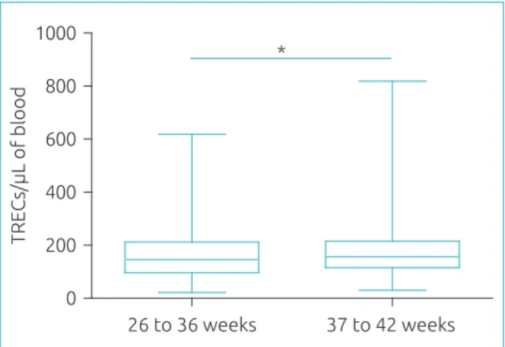
A total of 1,548 samples from Belém (PA) were analyzed for gestational age and TRECs and KRECs values, because there are reports in the literature of preterm infants who have
lower TRECs values,13,15 which would represent a
false-pos-itive risk. Fig. 3 shows that TRECs values were signiicantly lower in premature infants (p<0.05). hey showed median of
Figure 1 (A) Receiver operating characteristic curve for cutof value of T-cell receptor excision circles of 15/µL,
with area under the curve of 1.00. (B) Receiver operating characteristic curve for cutof value of kappa-deleting recombination circles of 14/µL, with area under the curve of 1.00.
0.0 0.2 0.4 0.6 0.8 1.0
A ROC Curve A
Sensitivity
1.0
0.8
0.6
0.4
0.2
0.0
1 - Speciicity
0.0 0.2 0.4 0.6 0.8 1.0
A ROC Curve B
Sensitivity
1.0
0.8
0.6
0.4
0.2
0.0
1 - Speciicity
ROC curve: receiver operating characteristic curve.
Figure 2 Concentration of T-cell receptor excision circles and kappa-deleting recombination circles of 6,881
samples of newborn screening and of 23 patients referred by pediatric immunologists/allergists for suspected primary immunodeiciency. T-cell receptor excision circles and kappa-deleting recombination excision circles were quantiied by quantitative real-time polymerase chain reaction. Samples from patients already diagnosed with severe combined immunodeiciency, agammaglobulinemia, hypogammaglobulinemia, and DiGeorge syndrome were analyzed as controls for validation of the method. Dashed lines represent the cutof values resulting from the receiver operating characteristic curve analysis.
Newborn screening
Referred samples
Agammaglobulinemia
SCID
Hypogammaglobulinemia
DiGeorge Syndrome
KRECs/µL
10000
1000
100
10
1
0,1
0,1 1 10 100 1000 10000
TRECs/µL
30
146 TRECs/µL, whereas those born at term had median of 156 TRECs/µL. KRECs values did not vary according to gestational age.
DISCUSSION
We analyzed 6,881 samples from diferent states of Brazil, along with positive controls of SCID and agammaglobulin-emia, which validated the assay to quantify the concentration of TRECs and KRECs.
For the first 1,000 samples analyzed, cutoff values of 25 TRECs/µL and 25 KRECs/µL were initially applied, which resulted in a high repetition rate – 2.5% of the sam-ples had concentrations below the cutof values in a irst anal-ysis – compared to a previous work of our group,9 but still
within the range reported by other authors (0.20–3.26%).4
One possibility for this high repetition rate was the use of a plasmid which was diferent from the previously employed – only contained the sequence of TRECs. herefore, it was decided to analyze a larger number of samples along with positive controls of the diseases under investigation in order to establish an appropriate cutof value. hus, after quantii-cation of almost 7,000 samples, receiver operating character-istic curve was analyzed,and cutof values of 15 TRECs/µL
Figure 3 Concentration of TRECs/μL in 180 preterm
newborns (26–36 weeks) and 1,368 full-term infants (37–42 weeks), quantiied by real-time polymerase chain reaction. Preterm infants presented lower concentrations of T-cell receptor excision circles than those born at term (146 versus 156 TRECs/ μL, respectively; *p<0.05, Mann–Whitney test). The
concentration of T-cell receptor excision circles is represented in box plot showing median, 25th and 75th percentiles, and minimum and maximum.
0 200 400 600 800 1000
*
TRECs/µL of blood
37 to 42 weeks 26 to 36 weeks
TRECs: T cell receptor excision circles.
and 14 KRECs/µL, with 100% sensitivity for detection of SCID and agammaglobulinemia, respectively, were found. hese values are very close to those reported by Borteet al.8
(15 TRECs/uL and 10 KRECs/uL) and resulted in a repe-tition rate of 0.49% (34 samples), which was very close to the value found in our previous work.9 After the reanalysis
of these 34 samples that were below the new cutof values, only two samples (0.03%) remained altered. Conirmation of diagnosis of these two individuals was not possible owing to the absence of follow-up.
he repetition rate of the applied methodology, that is, the percentage of samples to be reanalyzed, is directly related to the restriction imposed by the cutof value used. he lower the cutof value, the smaller the number of samples to be resubmit-ted to all the procedure. Moreover, another important factor to be considered in determining cutof values are the patholo-gies the newborn screening intends to detect. A recent review of newborn screening for SCID/T-cell lymphopenia showed that several other diseases that occur with T-cell lymphope-nia can be detected by quantifying TRECs; however, some of these cases do not present undetectable or very low TRECs, as in classic SCID.16
Another interesting study quantiied TRECs in carriers of 22q11 deletion and showed that cutof variation leads to diferent success rates in the neonatal diagnosis of patients with this syndrome, and the lower the cutoff value, the lower the number of identiied carriers.17 In our study, a
patient with tetralogy of Fallot, hypocalcemia, and lymph-openia (CD3+:994/mm3) was referred to the Laboratório
de Imunologia Humana (ICB – USP) owing to suspected DiGeorge syndrome, which was later confirmed by the detection of 22q11 deletion. TRECs were undetectable in this patient, being compatible with the number of T cells, which showed the usefulness of the assay in the detection of not only SCID, but also other diseases that progress with low number of T lymphocytes.
Similarly to TRECs, quantifying KRECs enables early detection of immunodeficiencies with defects in B-lymphocyte development. The methodology described in this study not only enables the identification of patients with agammaglobulinemia, but also assists in the classifi-cation of the type of SCID (T-B+; T-B-).6 With the advent
of newborn screening for T- and B-cell lymphopenia, these children can be diagnosed soon after birth, as confirmed by the results of our study.
previous immunophenotyping of lymphocytes. As expected, the patient with DiGeorge syndrome presented normal value of KRECs.
Physicians who were collaborators of BRAGID network (Brazilian Group of Primary Immunodeiciencies, www. bragid.org.br) were responsible for the referral of 23 sam-ples with suspected primary immunodeiciency. Of the total samples received, ive had TRECs and/or KRECs values below the cutof, which conirmed the clinical suspicion. One of these patients, who was carrier of trisomy 21, pre-sented TRECs values below the cutof, and consistent with previous American reports of newborn screening for SCID.16
Associated immunodeiciency was also found in this patient, warning physicians and caregivers about the additional pre-cautions to be taken.
Analysis of samples from preterm and full-term infants con-irmed data previously reported showing that preterm infants have lower TRECs values (146 versus 156 TRECs/µL).13,15
herefore, we established that preterm infants with altered TRECs values at birth should undergo a second newborn screening for T-cell lymphopenia at an adjusted gestational age of 37 weeks. With regard to KRECs, the values were not statistically diferent between preterm and at term infants, as previously reported.18
Incidence of primary immunodeficiencies is unknown in Brazil. Many children are believed to die before the diagno-sis is made, and therefore, as occurred in the United States of America before the neonatal screening, the incidence is probably underestimated.19 In Brazil, only patients with
a family history of primary immunodeficiencies have the opportunity to benefit from early diagnosis. The onset of universal newborn screening for these conditions enables the early diagnosis and treatment for all children. The advent of TREC in newborn screening enabled to determine the incidence of SCID in the United States of America in 1:58.000.16 Taking into account the 2.9 million births/year
in Brazil, we should diagnose approximately 50 patients per year. A recent research found a small number of cases of SCID diagnosed in Brazil and a high mortality rate.20
Incorporation of TRECs quantification in the neonatal screening in our country would benefit a significant num-ber of patients with SCID.
SCID perfectly meets the criteria formulated by Wilson and Jungner to be included in newborn screening: absence of clinical signs and symptoms on physical examination, sig-niicant damage to the carrier if the disease is left untreated, available treatment, improved survival, and screening test
available at a reasonable cost.21 Without proper diagnosis
and treatment, children with SCID evolve to death in the irst two years of life.1 Studies showed that the long-term
sur-vival, if NB patients undergo HSCT before three and a half months of age, is 94%. If transplantation occurs with active infections after this age, survival decreases to only 50%.4
Analyzing these numbers, we can conclude that early diag-nosis can save the lives of children with SCID. he inclu-sion of KRECs quantiication in the PCR at a minimal cost enables the diagnosis of primary B-cell immunodeiciencies and has proven capable of detecting patients with T-B-SCID and with agammaglobulinemia.
Currently, access to tests that quantify lymphocytes and their subpopulations is restricted to large centers and research laboratories. Quantiication of TRECs and KRECs in dried blood samples on ilter paper enables screening of samples com-ing from any geographic areas and the referral to speciic tests, which save time and resources. In Brazil, there are no studies on the cost of patients with primary immunodeiciencies to the public health system. However, in the United States of America, studies have proven the cost-beneit of the inclusion of neonatal screening for T-cell lymphopenia, showing that the treatment of patients diagnosed before the onset of infections and consequent hospitalization cost up to four times less than of patients with late diagnosis.22
Finally, we conclude that the quantiication of TRECs and KRECs detected children with T- and/or B-cell lymph-openia in our study. he technique is validated and can be widely employed in Brazil, without the need for additional sample collection, as the collection of the current neonatal screening test can be used in this assay. herefore, we took the irst step toward the inclusion of neonatal screening for T- and B-cell lymphopenia in our country, facilitating screen-ing, early diagnosis, and treatment of patients with primary immunodeiciencies in Brazil.
Funding
Fundação de Amparo à Pesquisa do Estado de São Paulo
(FAPESP), Conselho Nacional de Desenvolvimento Técnico Cientíico (CNPq), Department of Science and Technology/ Secretariat of Science, Technology, and Strategic Inputs of the Ministry of Health/Ministry of Health (DECIT/ SCTIE/MS) and Secretariat of Health of the São Paulo State (2012/51233-2).
Conflict of interests
32
REFERENCES
1. Dvorak CC, Cowan MJ, Logan BR, Notarangelo LD, Griith LM, Puck JM, et al. The natural history of children with severe combined immunodeiciency: baseline features of the irst ifty patients of the primary immune deiciency treatment consortium prospective study 6901. J Clin Immunol. 2013;33:1156-64.
2. Puck JM. Laboratory technology for population based screening for severe combined immunodeficiency in neonates: the winner is T cell receptor excision circles. J Allergy Clin Immunol. 2012;129:607-16.
3. Somech R, Lev A, Simon AJ, Korn D, Garty BZ, Amariglio N, et al. Newborn screening for severe T and B cell immunodeiciency in Israel: a pilot study. Isr Med Assoc J. 2013;15:404-9. 4. Spek J, Groenwold RH, Burg M, Montfrans JM. TREC
Based Newborn Screening for Severe Combined Immunodeficiency Disease: A Systematic Review. J Clin Immunol. 2015;35:416-30.
5. Baker MW, Grossman WJ, Laessig RH, Hofman GL, Brokopp CD, Kurtycz DF, et al. Development of a routine newborn screening protocol for severe combined immunodeiciency. J Allergy Clin Immunol. 2009;124:522-7.
6. Borte S, Döbeln U, Fasth A, Wang N, Janzi M, Winiarski J, et al. Neonatal screening for severe primary immunodeiciency diseases using high throughput triplex real time PCR. Blood. 2012;119:2552-5.
7. Bestas B, Turunen JJ, Blomberg KE, Wang Q, Mansson R, El Andaloussi S, et al. Splice correction strategies for treatment of X linked agammaglobulinemia. Curr Allergy Asthma Rep. 2015;15:510.
8. Fernandes A, Guedes M, Vasconcelos J, Neves E, Fernandes S, Marques L. X linked agammaglobulinemia: experience in a Portuguese hospital. An Pediatr (Barc). 2015;82:166-71. 9. Kanegae MP, Barreiros LA, Mazzucchelli JT, Hadachi SM,
Guilhoto LM, Acquesta AL, et al. Neonatal screening for severe combined immunodeiciency in Brazil. J Pediatr (Rio J). 2016;92:374-80.
10. BRAGID [homepage on the Internet]. BRAGID Brazilian Group for Immunodeiciencies [cited 2016 Jul 11]. Available from: http://www.imunopediatria.org.br
11. Heath EM, O’Brien DP, Banas R, Naylor EW, Dobrowolski S. Optimization of an automated DNA purification protocol for neonatal screening. Arch Pathol Lab Med. 1999;123:1154-60.
12. Sottini A, Ghidini C, Zanotti C, Chiarini M, Caimi L, Lanfranchi A, et al. Simultaneous quantiication of recent thymic T cell and bone marrow B cell emigrants in patients with primary immunodeiciency undergone to stem cell transplantation. Clin Immunol. 2010;136:217-27.
13. Baker MW, Laessig RH, Katcher ML, Routes JM, Grossman WJ, Verbsky J, et al. Implementing routine testing for severe combined immunodeficiency within Wisconsin’s newborn screening program. Public Health Rep. 2010;125 Suppl 2:88-95. 14. Moraes Pinto MI, Ono E, Santos Valente EC, Almeida LC,
Andrade PR, Dinelli MI, et al. Lymphocyte subsets in human immunodeiciency virus unexposed Brazilian individuals from birth to adulthood. Mem Inst Oswaldo Cruz. 2014;109:989-98. 15. Ward CE, Baptist AP. Challenges of newborn severe combined
immunodeiciency screening among premature infants. Pediatrics. 2013;131:e1298-302.
16. Kwan A, Abraham RS, Currier R, Brower A, Andruszewski K, Abbott JK, et al. Newborn screening for severe combined immunodeiciency in 11 screening programs in the United States. JAMA. 2014;312:729-38.
17. Framme J, Borte S, Döbeln U, Hammarström L, Oskarsdottir S. Retrospective analysis of TREC based newborn screening results and clinical phenotypes in infants with the 22q11 deletion syndrome. J Clin Immunol. 2014;34:514-9. 18. Olbrich P, Felipe B, Delgado Pecellin C, Rodero R, Rojas P,
Aguayo J, et al. A irst pilot study on the neonatal screening of primary immunodeiciencies in Spain: TRECS and KRECS identify severe T and B cell lymphopenia. An Pediatr (Barc). 2014;81:310-7.
19. Kwan A, Puck JM. History and current status of newborn screening for severe combined immunodeiciency. Semin Perinatol. 2015;39:194-205.
20. Mazzucchelli JT, Bonim C, Castro GG, Condino Neto AA, Costa NM, Cunha L, et al. Severe combined immunodeiciency in Brazil: management, prognosis, and BCG associated complications. J Investig Allergol Clin Immunol. 2014;24:184-91.
21. Wilson JM, Jungner YG. Principles and practice of mass screening for disease. Bol Oicina Sanit Panam. 1968;65:281-393. 22. Kubiak C, Jyonouchi S, Kuo C, Garcia Lloret M, Dorsey MJ,
Sleasman J, et al. Fiscal implications of newborn screening in the diagnosis of severe combined immunodeiciency. J Allergy Clin Immunol Pract. 2014;2:697-702.