Hypermethylation of
FOXP3
Promoter and
Premature Aging of the Immune System in
Female Patients with Panic Disorder?
Martina Prelog1*, Deborah Hilligardt1, Christian A. Schmidt2, Grzegorz K. Przybylski2,3, Johannes Leierer4, Giovanni Almanzar1, Nady El Hajj5, Klaus-Peter Lesch6, Volker Arolt7, Peter Zwanzger7,8,9, Thomas Haaf5, Katharina Domschke10
1Department of Pediatrics, University Hospital Wuerzburg, Wuerzburg, Germany,2Clinic for Internal Medicine C, University of Greifswald, Greifswald, Germany,3Institute of Human Genetics, Polish Academy of Sciences, Poznan, Poland,4Department of Internal Medicine, Medical University Innsbruck, Innsbruck, Austria,5Institute of Human Genetics, University of Wuerzburg, Wuerzburg, Germany,6Molecular Psychiatry, Department of Psychiatry, Psychosomatics and Psychotherapy, University of Wuerzburg, Wuerzburg, Germany,7Department of Psychiatry and Psychotherapy, University of Muenster, Muenster, Germany,8Department of Psychiatry and Psychotherapy, Ludwig-Maximilians-Universtät Munich, Munich, Germany,9kbo-Inn-Salzach-Klinikum, Wasserburg am Inn, Germany,10 Department of Psychiatry, Psychosomatics and Psychotherapy, University Hospital Wuerzburg, Wuerzburg, Germany
*Prelog_M@ukw.de
Abstract
Immunological abnormalities associated with pathological conditions, such as higher infec-tion rates, inflammatory diseases, cancer or cardiovascular events are common in patients with panic disorder. In the present study, T cell receptor excision circles (TRECs), Fork-head-Box-Protein P3 gene (FOXP3) methylation of regulatory T cells (Tregs) and relative telomere lengths (RTLs) were investigated in a total and subsamples of 131 patients with panic disorder as compared to 131 age- and sex-matched healthy controls in order to test for a potential dysfunction and premature aging of the immune system in anxiety disorders. Significantly lower TRECs (p = 0.004) as well as significant hypermethylation of theFOXP3 promoter region (p = 0.005) were observed in female (but not in male) patients with panic disorder as compared to healthy controls. No difference in relative telomere length was dis-cerned between patients and controls, but significantly shorter telomeres in females, smok-ers and older psmok-ersons within the patient group. The presently observed reduced TRECs in panic disorder patients andFOXP3hypermethylation in female patients with panic disorder potentially reflect impaired thymus and immunosuppressive Treg function, which might partly account for the known increased morbidity and mortality of anxiety disorders con-ferred by e.g. cancer and cardiovascular disorders.
Introduction
Anxiety disorders are among the most common mental health disorders in Europe in 2010 and confer a high individual and socioeconomic burden [1]. Anxiety disorders are chronic diseases
a11111
OPEN ACCESS
Citation:Prelog M, Hilligardt D, Schmidt CA, Przybylski GK, Leierer J, Almanzar G, et al. (2016) Hypermethylation ofFOXP3Promoter and Premature Aging of the Immune System in Female Patients with Panic Disorder? PLoS ONE 11(6): e0157930. doi:10.1371/journal.pone.0157930
Editor:Jorg Tost, CEA—Institut de Genomique, FRANCE
Received:January 17, 2016
Accepted:June 7, 2016
Published:June 30, 2016
Copyright:© 2016 Prelog et al. This is an open access article distributed under the terms of the
Creative Commons Attribution License, which permits unrestricted use, distribution, and reproduction in any medium, provided the original author and source are credited.
ranking fifth regarding Years Lived with Disability (YLDs) among the 30 leading diseases and injuries in the United States in 2010 [2]. As a potential consequence of chronic stress, anxiety disorders have been shown to carry a high“allostatic load”[3], i.e. exert a physiological strain on organs and cells particularly pertaining to the cardiovascular system: For instance, phobic anxiety and increased anxiety levels, respectively, were found to be associated with an increased risk of coronary heart disease and cardiovascular death particularly in women [4–7]. This increased morbidity and mortality has in part been attributed to oxidative stress and inflamma-tory processes in anxiety disorders: Tension-anxiety symptoms were reported to correlate with an oxidative DNA damage marker [8], state/trait anxiety was associated with elevated C-reac-tive protein (CRP), interleukin-6 and fibrinogen levels [9], phobic anxiety in female patients with diabetes mellitus correlated with elevated inflammatory markers [10], patients with panic disorder showed significantly elevated peripheral proinflammatory cytokine and chemokine levels [11], and elevated inflammation as reflected by increased CRP levels was discerned to be associated with current anxiety disorders, particularly in male patients and patients with late-onset anxiety disorder [12].
Premature immunosenescence and a diminished regulatory T cell (Treg) function are dis-cussed as etiopathological factors driving the immune system towards inflammatory diseases [13–17]: Aging of the immune system or‘immunosenescence’is characterized by loss of thy-mic function with decreased output of recent thythy-mic emigrants (RTE) and increased replica-tion of peripheral lymphocytes to compensate for the decrease in naive T cells. Elderly people are at risk for age-associated diseases, such as atherosclerosis and cardiovascular events, infec-tious diseases, cancer and inflammatory diseases due to breakdown of immune tolerance and higher inflammatory capacity. To estimate thymic function, T cell receptor excision circles (TRECs) have been shown to be useful markers due to their abundance in RTE and their pro-portional decline with age [18]. The number of naive T cells is maintained by peripheral prolif-eration of naive T cells which results in a dilution of TRECs [19,20].
Measurement of telomere lengths helps to estimate the individual replication history of cells. Telomeres are protective caps at the end of chromosomes and shorten with each cell cycle [21]. Indirectly, telomere shortening reflects the age of the singular immune cell and has been associated with susceptibility to age-related diseases, inflammation and also accelerated aging in mental disorders [22–24]. In detail, lower relative telomere length (RTL) has furthermore been reported to be associated with phobic anxiety in women and—with trendwise significance
—also with items of the Crown-Crisp Index (CCI) mapping to panic and agoraphobia [25], with anxiety disorders particularly in older patients [26], and with anxiety disorders including generalized anxiety disorder, social phobia, agoraphobia and panic disorder after a two-year follow-up [27].
In inflammatory conditions, e. g. rheumatoid arthritis, signs of a prematurely aged immune system, e. g. lower TRECs in naive T cells and shorter telomeres in total lymphocytes, go ahead with quantitative and qualitative alterations of regulatory T cells [28,29]. The Forkhead-Box-Protein P3 (FoxP3) transcription factor is specifically expressed by naturally occurring regula-tory CD25+CD4+ T cells (nTregs) and contributes to the immunosuppressive function of Tregs. Transiently FoxP3-expressing activated T cells (induced Tregs, iTregs) may be distin-guished from nTregs by their methylation profile at theFOXP3promoter and enhancer regions
[28,30]. LowFOXP3promoter methylation has been shown to be associated with highly
CD25-expressing CD4+ Tregs [31]. Demethylated or hypomethylated CpG regions in pro-moter [31,32], upstream enhancer [33] or intronic enhancer [34] provides stable long-term expression of theFOXP3gene and, thus, is proposed to induce a stable Treg phenotype
essen-tial for maintaining Treg function to inhibit inappropriate or excessive immune responses [35]. Several molecules are involved in genetic and epigenetic regulation ofFOXP3promoter
Institute of Pharmacology and Toxicology, Versbacher Str. 9, 97078 Wuerzburg, Germany, Chairperson: Prof. Dr. E. Bröcker, Contact person: Dr. R. Wölfel, phone: +49 931 31 48315, e-mail:
ethikkommission@uni-wuerzburg.de.
Funding:This paper was supported by a grant from the Deutsche Forschungsgemeinschaft (DFG; SFB-TRR-58, projects C02 to KD and KPL, and A05 to KPL) and the Bavarian Innovation Fond to MP.
and enhancer function (Fig 1) [35–37]. Along these lines, increasedFOXP3methylation,
result-ing in decreased Treg levels, was observed in peripheral blood mononuclear cells (PBMCs) of patients with coronary artery disease [38]. Anti-CD25 antibody mediated depletion of Treg cells in mice has been shown to result in anxiety-like behavior in the elevated plus maze test and in higher serum IL-6 and TNF-alpha concentrations particularly after stress [39]. Develop-ment of inflammatory Th cell responses, a shift towards Th17 and reduced control by Tregs have been shown in individuals with generalized anxiety disorders [40–42].
In the present study, T cell receptor excision circles (TRECs),FOXP3methylation and
rela-tive telomere lengths (RTLs) were for the first time concurrently investigated in patients with panic disorder as compared to matched healthy controls in order to test for potential dysfunc-tion and premature aging of the immune system in anxiety disorders. It was hypothesized that panic disorder would be associated with impaired thymus function as reflected by reduced TRECs as well as byFOXP3hypermethylation resulting in reduced immunosuppressive Treg
function and accompanied by lower relative telomere length.
Materials and Methods
Samples
One hundred and thirty-one patients with panic disorder and 131 healthy, age- and sex-matched controls, recruited at the Department of Psychiatry and Psychotherapy, University of Muenster, Germany, were included into the study (Table 1). Diagnosis of panic disorder, in all cases the primary diagnosis at the time of inclusion, was ascertained by experienced psychia-trists on the basis of medical records and structured clinical interviews (SCID-I) according to
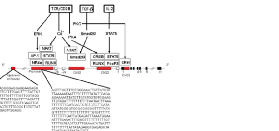
Fig 1. Molecules involved inFOXP3induction and stable expression and sequences analyzed. Enhancer (5’upstream enhancer, conserved non-coding sequences, CNS1, CNS2 and CNS3 serving as enhancer regions) and promoter region are shown (modified after36,37,68]. Promoter and enhancer regions are bound by several transcription factors and signals. A restriction in differentiation of nTregs is caused by protein inhibitor of the activated signal transducer and activator of transcriptionSTAT1which binds to the FOXP3promoter and recruits DNA methyltransferase [68]. CNS1, an intronic enhancer (enhancer 1) is responsive to Tumor-growth-factor-beta (TGFβ) by Smad2/3 binding sites, close to the NFAT site, essential for differentiation of induced iTregs. CNS2, the T cell receptor (TCR)-responsive enhancer (enhancer 2) contains CpG islands and binding sites for transcription factors,CREBandSTAT5. The unstable iTreg phenotype is associated with high methylation of the CNS2 region of Treg-specific demethylated regions (TSDRs). TSDR is a key factor in stability of Tregs [36]. Analyzed CpG regions were located in the 5’
upstream enhancer and the promoter of theFOXP3gene. Analyzed sequences are shown. The analyzed CpGs are located in the promoter region ofFOXP3which do not contain any CpG island. TheFOXP3 enhancer lies in a CpG island. Abbreviations: EKR: extracellular signal regulated kinase, PKA:
phosphokinase A, NFAT: Nuclear factor activated T cells, NR4a: Orphan nuclear receptor, RUNX: Runt-related transcription factor, CREB: CAMP responsible element binding protein 1, cRel: Proto-oncogene C-Rel, IL-2: interleukin-2, TCR: T cell receptor.
the criteria of DSM-IV [1]. Individuals with mental retardation, neurological or neurodegener-ative disorders impairing psychiatric evaluation as well as with severe somatic disorders were not included in this analysis. Medication with antidepressants and comorbidity with depres-sion was recorded. Both cases and controls were of Caucasian ethnicity. Smokers were defined by the consumption of more than two cigarettes/day.
The study was approved of by the ethics committee of the University of Muenster, Ger-many, written informed consent was obtained from all participating subjects, and the study was conducted according to the ethical principles of the Helsinki Declaration.
Quantification of TRECs and relative telomere length
DNA was extracted from separated whole EDTA blood using the FlexiGene DNA Kit (QIA-GEN, Hilden, Germany) according to the manufacturer's instructions. Signal-joint TREC con-centrations were determined by PCR as described in detail previously [43,44]. Recombination-activating gene 2 (RAG2) was used as a reference gene to normalize the quantity of DNA used for real-time quantitative polymerase chain reaction (RQ-PCR).
Determination of relative telomere length (RTL) was performed by calculating the ratio of a quantitative PCR reaction product from the same sample using specific primers for telomeres and a single copy gene as described previously [45–47].
Bisulfite pyrosequencing
Assays quantifying the methylation levels of CpGs in the target regions, i.e.FOXP3promoter
(human build hg 19 Chromosome X, 49121152–49121485 bp, len: 333) andFOXP35’
upstream enhancer (CpG human build hg 19 Chromosome X, 49126597–49126750 bp, len: 153) (both ensemble releaser 15 February 2014), were designed with the PyroMark Assay Design software (Qiagen, Hilden, Germany). Primers and sequences to analyze are listed in
Table 2.FOXP3is located on the X-chromosome (Xp11.23), resulting in hemizygosity in male
subjects and random inactivation of the second X-chromosome in females, subsequently meth-ylation data was stratified according to sex. Bisulfite conversion of DNA was performed using the EpiTect 96 Bisulfite Kit (Qiagen, Hilden, Germany) according to the manufacturer’s proto-col. PCR amplifications were performed on a Tetrad 2 cycler (BioRad, Munich, Germany) with an initial denaturation step at 95°C for 5 min, 40 cycles of 95°C for 30 s, primer-specific anneal-ing temperature of 60°C for 30 s, 72°C for 45 s, and a final extension step at 72°C for 10 min. The reaction mixture consisted of 2.5μl 10x PCR buffer with MgCl2, 0.5μl 10 mM dNTP mix,
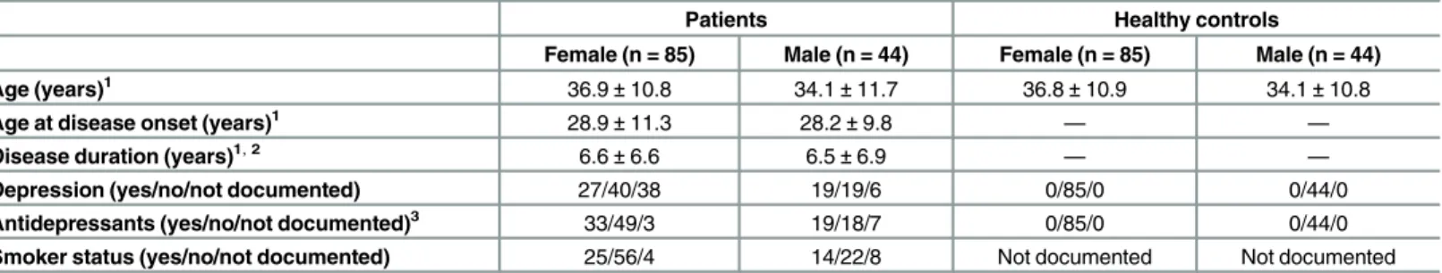
1.25μl of each forward and reverse primer (final concentration 0,5μM), 0.2μl (final Table 1. Demographics.
Patients Healthy controls
Female (n = 85) Male (n = 44) Female (n = 85) Male (n = 44)
Age (years)1 36.9±10.8 34.1±11.7 36.8±10.9 34.1±10.8
Age at disease onset (years)1 28.9±11.3 28.2±9.8 — —
Disease duration (years)1,2 6.6±6.6 6.5±6.9 — —
Depression (yes/no/not documented) 27/40/38 19/19/6 0/85/0 0/44/0
Antidepressants (yes/no/not documented)3 33/49/3 19/18/7 0/85/0 0/44/0
Smoker status (yes/no/not documented) 25/56/4 14/22/8 Not documented Not documented
1Values are given in mean±standard deviation. 2
Disease duration significantly correlated with age (R = 0.434; p = 0.007) in male patients and near to significance in female patients (R = 0.224; p = 0.056).
3Antidepressants: SSRIs: N = 46, Tricyclic Antidepressants: N = 2, NaSSA: N = 1, Melatonergic: N = 1, SSRI plus antipsychotics (off-label) N = 2.
concentration 1 U) Taq DNA polymerase (Roche Diagnostics, Mannheim, Germany), 18.3μl
PCR-grade water, and 1μl template DNA (75 ng). PCR products were visualized by
electro-phoresis on 1.5% agarose gel.
Bisulfite pyrosequencing was performed on a PyroMarkTMQ96 MD Pyrosequencing Sys-tem with the PyroMark Gold Q96 CDT Reagent Kit (Qiagen, Hilden, Germany). Our experi-mental methodology relied on simultaneous treatment of control and study samples in order to avoid batch effect and technical variability which is estimated around 1–2% of this assay. Bisulfite conversion, PCR amplifications and pyrosequencing of control and study samples were performed together. The sequences analyzed by bisulfite pyrosequencing are listed in
Table 3. Data analysis was done with the Pyro Q-CpG software (Qiagen, Hilden, Germany).
Statistical analysis
Shapiro-Wilk test was used to test for normal distribution, before applying Student’s t-test for normally-distributed and non-parametric Mann-Whitney-U for not normally-distributed independent variables. Correlations between variables were identified by Spearman's rank cor-relation coefficient. A generalized linear multivariate regression model was generated by step-wise regression to infer the influence of gender, depression, medication with antidepressants and smoking, adjusted for the chronological age (age at blood withdrawal) of the patient. Step-down exclusion was performed by excluding variables with the standardized regression coeffi-cient closest to 0 until a significant regression model was generated. Disease duration was excluded from the regression model due to strong correlation with age (Table 4). A p-value
0.05 was considered statistically significant. Given the explorative nature of the study, no
Table 2. Primers used for bisulfite pyrosequencing.
Gene Primer Sequence (5’-3’) Number of CpGs
FOXP3 forward Biotin-AGTTTGGTTTGTGGGAAATTGTT
reverse ACCCTATTATCTCATTAATACCTCTCA
Sequence 1 ATAAAAACAAAATTATTTTTAATA 1
Sequence 2 AAATTATTAAAAAAAAAAAATCTAC 4
FOXP3Enhancer forward ATGAAGGGGAGGAGGAAG
reverse Biotin-CCTCCAACTCCACCATAAC
Sequence 1 GAGGAAGAGGAGGTT 4
Sequence 2 GGGTTTTATTTGGTTTTTATATT 7
doi:10.1371/journal.pone.0157930.t002
Table 3. Sequences analyzed by bisulfite pyrosequencing.
Gene Sequence analyzed*
FOXP3CpG number Sequence 1 CRTAACAATTTCCCACAAACCAAACTA
. 1
FOXP3CpG number Sequence 2 RACTTCCACACCRTACAACRTAATTTTTCTTCTCRATATA
1 . . . 2 . . . 3 . . . 4
FOXP3Enhancer CpG number Sequence 1 TGTTTYGAGTTTTTATYGTTGTGTTTYGTTTTYGT
. . . 1 . . . 2 . . . 3 . . . 4
FOXP3Enhancer CpG number Sequence 2 TTYGTTGTYGTTYGYGTYGGGTYGTTTGGAGYGT
. . 1 . . . 2 . . . 3 . 4 . . 5 . . . . 6 . . . 7
*analyzed CpG sites are underlined and consecutively numbered
correction for multiple testing was performed. All statistical analyses were performed with SPSS Version 23.0 (Chicago, IL, USA).
Results
Lower TREC counts in panic disorder patients
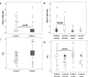
Patients (mean 0.49 ± 0.81) showed significantly lower TRECs/1,000 cells (mean 0.99 ± 1.90) compared to healthy controls (HC) (p = 0.004), which was due to significantly lower TRECs in female patients (Fig 2). In patients, TRECs negatively correlated with age (R = -0.245;
p = 0.025). An association of lower TREC counts with age was shown for both, female (R = -0.378; p = 0.019) and male patients (R = -0.578; p = 0.024). Regression analysis (R2= 0.051, p = 0.045) including age (p = 0.086) and gender (p = 0.468) revealed that having panic disorder (p = 0.016) was an independent factor for lower TRECs. Slightly lower TRECs were seen in patients treated with antidepressants compared to those without any medication (p = 0.024). Performing regression analysis (R2= 0.109, p = 0.019) in patients only, age (p = 0.031) was an
Table 4. Methylation ofFOXP3Promoter and Enhancer regions.
Females p-value1 Males p-value2
Patients Controls Patients Controls
Promoter sequence 1 69.06±4.94 65.54±9.19 0.008** 68.32±5.39 68.30±4.60 0.899
Position CpG 1 69.06±4.94 65.54±9.19 0.008** 68.32±5.39 68.30±4.60 0.899
Promoter sequence 2 (all 4 CpGs) 79.09±2.12 78.37±1.84 0.022* 70.61±3.47 70.52±3.08 0.802
Position CpG 1 90.21±2.12 90.20±1.86 0.621 82.04±3.41 82.28±2.90 0.856
Position CpG 2 75.76±2.36 74.89±1.89 0.054 68.81±3.31 68.74±3.58 0.941
Position CpG 3 75.05±3.88 74.13±2.98 0.011** 69.13±3.57 68.73±3.25 0.675
Position CpG 4 75.01±3.31 74.27±2.65 0.140 62.67±5.50 62.34±3.88 0.676
Promoter sequences 1+2 (all 5 CpGs) 77.01±2.40 75.80±2.56 0.005** 70.16±3.77 70.08±3.24 0.761 Enhancer sequence 1 (all 4 CpGs) 39.69±3.25 39.81±2.21 0.246 2.09±0.91 1.93±0.59 0.356
Position CpG 1 40.03±2.85 40.42±3.41 0.376 2.00±1.59 2.05±2.04 0.997
Position CpG 2 37.48±3.18 38.38±2.85 0.291 2.68±0.81 2.58±0.83 0.316
Position CpG 3 40.89±3.79 42.09±2.71 0.202 1.87±0.39 1.85±0.54 0.838
Position CpG 4 36.36±8.99 37.93±6.12 0.636 1.79±1.42 1.28±0.63 0.598
Enhancer sequence 2 (all 7 CpGs) 38.00±2.68 38.18±1.77 0.749 1.31±0.44 1.37±0.69 0.899
Position CpG 1 37.74±4.05 37.62±3.31 0.652 1.79±1.38 2.38±2.58 0.517
Position CpG 2 41.00±3.39 40.18±3.56 0.364 2.53±1.18 2.73±1.47 0.707
Position CpG 3 37.42±3.59 38.71±3.22 0.093 0.94±0.78 0.84±0.62 0.624
Position CpG 4 30.56±4.61 30.65±3.31 0.699 0.67±0.57 0.71±0.49 0.440
Position CpG 5 44.51±3.25 44.80±2.63 0.988 0.79±0.94 0.68±0.75 0.366
Position CpG 6 40.19±3.46 40.70±2.47 0.792 0.67±0.41 0.77±0.92 0.943
Position CpG 7 34.66±4.02 34.57±3.06 0.823 1.81±0.96 1.52±0.70 0.188
Enhancer sequences 1+2 (all 11 CpGs) 38.26±2.48 38.73±1.64 0.577 1.60±0.46 1.59±0.55 0.825
Values are given in mean percentages methylation±standard deviation. For all positions higher methylation was found in females compared to males in
patients (p<0.0001) and controls (p<0.0001) except for CpGs in promoter sequence 1 in position 1 (female patients versus male patients: p = 0.401; female
controls versus male controls: p = 0.159).
1
Comparison between female patients and female controls *= significant at p0.05
**= significant at p0.01.
2
Comparison between male patients and male controls: not significant.
independent factor for lower TRECs, whereas gender (p = 0.162), smoking (p = 0.727), and comorbid depression (p = 0.925) or antidepressants (p = 0.483) had no significant association.
FOXP3
promoter and enhancer methylation
Significantly higherFOXP3promoter methylation across all five CpGs as well as particularly at
sequence 1, CpG 1, and sequence 2, CpGs 2 and 3 was discerned in female patients compared to HC (Table 4, exemplarily shown inFig 3). Methylation at sequence 2, CpGs 2 and 3 highly correlated with each other (R = 0.698; p = 0.0001), but not with sequence 1, CpG 1. Regression analysis (R2= 0.059; p = 0.060) revealed that having panic disorder was an independent factor for higherFOXP3methylation across all five CpGs (p = 0.020) in female patients. Lower
meth-ylation across all five CpGs was seen in patients on antidepressants (mean methmeth-ylation 75.94 ± 2.63%) compared to those without any medication (77.78 ± 1.98%) (p = 0.014), with high significance atFOXP3promoter sequence 2, CpG 4 (antidepressants: 73.58 ± 2.39%; no
Fig 2. T cell receptor excision circles (TRECs).T cell receptor excision circles (TRECs) per 1,000 peripheral blood mononuclear cells (PBMCs) were significantly lower including all patients compared to controls (A) and in female patients with panic disorder compared to healthy female controls (B) (Mann-Whitney U test). Relative telomere lengths (RTLs) are shown in patients and compared to controls (C) and in female patients with panic disorder compared to healthy female controls (D) (Mann-Whitney U test). Circles (outliers) and asterisks (extreme values) represent individuals coded with numbers.
doi:10.1371/journal.pone.0157930.g002
Fig 3. Methylations at specific CpG sites in a representative female patient and a healthy female control person.Methylations at one specific CpG site inFOXP3promoter sequence 1 (position 1), and at four independent CpG sites inFOXP3promoter sequence 2 (positions 1 to 4) of a representative female patient (A, C) and a healthy female control person (B, D) were quantified in a single pyrosequencing run. Position-specific information in the context of the analyzed sequence presents broad-sequence methylation patterns (% methylation). The built-in quality control sites (highlighted in yellow) consisting of cytosines converted to thymines demonstrate full bisulfite conversion of the treated DNA.
antidepressants: 76.06 ± 3.66%; p = 0.004). Using a regression model for methylation across all five CpGs (R2= 0.276; p = 0.024) in female patients, antidepressants (p = 0.004) and age (p = 0.027) (depression p = 0.921; smoking p = 0.273) showed the strongest associations, with lowerFOXP3promoter methylation correlating with antidepressant medication and higher
age. No significant difference between female patients and female HC could be demonstrated forFOXP3enhancer methylation (Table 4). Categorization of female patients into smokers,
into patients with depression and into patients on antidepressants did not reveal any differ-ences inFOXP3enhancer methylation.
In males,FOXP3promoter and enhancer methylation were not different between patients
and HC (Table 4). Smoking male patients had significantly higher methylation atFOXP3
enhancer, sequence 2, CpG 5 (mean methylation 0.98 ± 0.73%) compared to non-smoking male patients (0.78 ± 1.19%) (p = 0.030). In male patients, significantly higher methylation was determined atFOXP3enhancer, sequence 2, CpG 4 (0.84 ± 0.70%) and 5 (1.16 ± 1.33%) in the
case of depression compared to male patients without depression (position 4 (0.51 ± 0.37%) and 5 (0.51 ± 0.33%); p = 0.039 and p = 0.006, respectively). Similarly, higher methylation was seen atFOXP3enhancer, sequence 2, CpG 5 in male patients on antidepressants
(1.15 ± 1.35%) compared to those without antidepressants (0.53 ± 0.31%) (p = 0.013). Methyla-tion atFOXP3enhancer, sequence 2, CpG 4 highly correlated with methylation atFOXP3
enhancer, sequence 2, position 5 (R = 0.675; p = 0.0001). Multiple regression analysis (R² = 0.345; p = 0.032) showed that methylation atFOXP3enhancer, sequence 2, CpG 5 was weakly
influenced by smoking (p = 0.067) and older age (p = 0.073) but not by depression (p = 0.267) or antidepressants (p = 0.868).
Relative telomere lengths
Relative telomere lengths (RTLs) were not different between patients and HC. However, within the patient group, smokers had significantly shorter telomeres (0.91 ± 0.30) compared to non-smokers (1.07 ± 0.37) (p = 0.018) and females (0.96 ± 0.34) had shorter telomeres than males (1.10 ± 0.32) (p = 0.017), although age and distribution of females were not significantly differ-ent between smokers and non-smokers. Stratifying for female patidiffer-ents aged35 years, differ-ence in RTLs was significant between smokers (0.84 ± 0.32) and non-smokers (1.12 ± 0.34) (p = 0.010). In patients, regression analysis (R² = 0.150; p = 0.008) identified smoking (p = 0.004), female gender (p = 0.011) and age (p = 0.041) as factors associated with shorter telomeres, whereas depression (p = 0.106) and antidepressants (p = 0.446) were less important. Interestingly, hypermethylation atFOXP3promoter, sequence 2, CpGs 2 (R = -0.495;
p = 0.0001) and 3 (R = -0.309; p = 0.035) correlated with shorter telomeres in female patients.
Discussion
The present study demonstrated significant lower TRECs in both female and male panic disor-der patients as well as significant hypermethylation of theFOXP3promoter region in female
patients with panic disorder as compared to healthy controls. No difference in relative telomere length was discerned between patients and controls, but significantly shorter telomeres in females, smokers and individuals aged35 years of the patient group.
an impaired thymic function and/or highly proliferating peripheral T cells contributing to the lower TREC numbers and the strong influence of age on TRECs as seen in the patient group. As TRECs are not only a marker for thymic function but also for peripheral replication with dilution of TRECs, relative telomere lengths were evaluated to estimate replicative activity of peripheral lymphocytes [20]. Although no association of telomere lengths were identified with the categorical diagnosis of panic disorder, telomere lengths were shorter in females and smok-ers within the panic disorder sample and associated with higher age. In contrast to T cell-spe-cific TRECs, telomeres were measured in samples from total leukocytes and may be greatly influenced by other subpopulations than naive T cells. This may also allow speculation for an effect of smoking habits, steroids or female hormones or other environmental factors for con-founding the telomere results [45].
Diminished thymic function has been associated with inflammatory diseases [13,14,17] due to compensatory proliferative mechanisms in the periphery and increased Th1 and Th17 responses in relation to a dysfunctional Treg activity. Additionally, lower output of thymic-dependent naturally occurring Tregs may result in lower suppression of inflammatory responses in the periphery. Our study revealed hypermethylation of theFOXP3promotor
region—potentially resulting in reduced immunosuppressive Treg function—in female but not in male patients with panic disorders, corroborates the idea of a prematurely aged immune sys-tem in this particular patient subgroup. Although a strong sex bias of autoimmunity, with most autoimmune diseases predominantly affecting females, is well known [51,52], the underlying mechanisms are not well understood. The absence of a second (inactivated) X chromosome in males, sex hormones, and sex-specific differences in gene regulation due to internal and exter-nal (i.e. environmental) factors, all can influence the susceptibility to disease. Particularly, hor-mone factors may explain the higher differences regarding methylation status of CpG regions within theFOXP3promoter [53,54] and lower TRECs in females [55].
Interestingly, besides older age, antidepressants were found to be associated with a relative demethylation at specific CpGs within theFOXP3promoter region. An association between
age andFOXP3hypomethylation with increased immunosuppressive Treg function has been
suggested by a recent study in mice [56]. Likewise,FOXP3demethylation could constitute a
molecular correlate of beneficial effects of antidepressants on the immune system in panic dis-order. This notion is supported by first therapy-epigenetic studies showing dynamic methyla-tion changes after successful antidepressant or even psychotherapeutic treatment in depression or anxiety disorders [57–59]. However, it has to be noted that the CpGs observed to be rela-tively demethylated in association with antidepressants were different from CpGs associated with panic disorder in the present sample. In males, methylation was higher at specific CpGs in theFOXP3enhancer region in smokers as well as in patients with comorbid depression
com-pared to non-smoking patients or patients without depression, respectively. Antidepressants were associated with higher methylation at the same CpG position. However, given only minor
FOXP3enhancer methylation in general (seeTable 4) in male subjects and an underpowered
sample size of the male subsample, these results are to be considered with caution.
In addition to the present cross-sectional study design, other factors influencing inflamma-tion, such as fat tissue mass in obese patients, comorbid diabetes or latent infections (e. g. Cyto-megalovirus infections) contributing to an immune-risk-phenotype [13,60], may limit the interpretation of our results. Also, it is unclear whether the findings from this study may be interpreted as primary or secondary events to panic disorder, although a stress-induced, and thus secondary neuroendocrinological effect of panic disorder on the immune system particu-larly in older females, may be a reasonable explanation for reduced thymic activity and hyper-methylation ofFOXP3. This interpretation is also supported by mouse models [61–64] and
because of limitations in available samples many groups showed that demethylation of pro-moter and enhancer regions ofFOXP3corresponds to expression of FoxP3 protein in
periph-eral lymphocytes [28,65]. The biological relevance of 5’upstream enhancer was demonstrated by the ability of methotrexate treatment to restore defective Treg function through demethyla-tion in rheumatoid arthritis patients [65]. Differences in the methylation ofFOXP3promotor
are quite small just above the background noise of the method and may be unable to explain all alterations found in patients with panic disorder. However, at least small differences in pro-moter methylation may influence the accessibility of the gene and, thus, the ability to provide stableFOXP3expression as shown by others [65]. Although the transactivation activity of
FOXP3 promoter appears to be weak, a weak transactivation activity may help prevent promis-cuous FOXP3 induction [66].
One limitation of our study is that lymphocytes were not separated into CD25highCD4+ T cells, defining mainly nTregs, and in naive and other T cell subpopulations, as a differentiated methylation pattern at theFOXP3enhancer region was found on activated and only transiently
FoxP3-expressing T cells with impermanent change of methylation status [34]. This may explain that we were not able to find significant differences in the methylation of CpG regions of the analyzed enhancer regions and only small differences in the promoter region as several T cell subpopulations with different methylation levels at theFOXP3promotor regions may
con-tribute to the methylation results. Another important limitation to mention is the fact that bisulfite sequencing cannot discriminate between 5-methylcytosine and 5-hydroxymethylcyto-sine. Therefore, the output from bisulfite sequencing cannot solely be interpreted as showing only 5-methylcytosines, but it could also include the 5-hydroxymethylcytosines. 5-hydroxy-methylcytosine has been postulated to play an important role in the process of demethylation [67], where 5-hydroxymethylcytosine facilitates passive demethylation and in turn promotes gene transcription.
Regarding the RTL data, it has to be taken into account that several factors could influence the outcome such as current inflammatory state as measurable by C-reactive protein levels, anti-inflammatory medications, paternal age, menopause, exercise, diet or childhood trauma. However, unfortunately, these data were not available and thus, have not been corrected for in the present study. Mechanisms and directions of interaction between anxiety and immune function as well as their relation to an increased risk of mortality and morbidity, also consider-ing early as well as recent life events need to be further evaluated in longitudinal studies.
In summary, the present study noted reduced TRECs in panic disorder patients compared to controls as well asFOXP3hypermethylation in patients with panic disorder potentially
reflecting impaired thymus and immunosuppressive Treg function. From the present results, we expect that female and older patients with panic disorder may show particularly strong effects regarding immunosenescence and its role in development of age-associated diseases, such as cardiovascular events, autoimmune disorders, cancer and infectious rates accounting for the known increased morbidity and mortality of anxiety disorders. Targeted prevention and early treatment of anxiety disorders could therefore aid in mitigating their detrimental effects on the immune system and thereby in lowering the risk of diseases associated with age such as coronary heart disease, cardiovascular death and cancer.
Acknowledgments
Author Contributions
Conceived and designed the experiments: MP GA NEH KD. Performed the experiments: DH CAS GKP JL NEH GA TH. Analyzed the data: MP DH CAS GKP JL GA NEH TH. Contrib-uted reagents/materials/analysis tools: KPL PZ VA KD. Wrote the paper: MP GA NEH KD.
References
1. Wittchen HU, Jacobi F, Rehm J, Gustavsson A, Svensson M, Jönsson B, et al. The size and burden of mental disorders and other disorders of the brain in Europe 2010. Eur Neuropsychopharmacol. 2011; 21: 655–679. doi:10.1016/j.euroneuro.2011.07.018PMID:21896369
2. Murray CJ, Abraham J, Ali MK, Alvarado M, Atkinson C, Baddour LM, et al.: The State of US Health, 1990–2010: Burden of Diseases, Injuries, and Risk Factors. JAMA. 2013; 310: 591–608. doi:10.1001/ jama.2013.13805PMID:23842577
3. McEwan BS, Stellar E. Stress and the individual. Mechanisms leading to disease. Arch Intern Med. 1993; 153: 2093–2101. PMID:8379800
4. Albert CM, Chae CU, Rexrode KM, Manson JE, Kawachi I. Phobic anxiety and risk of coronary heart disease and sudden cardiac death among women. Circulation. 2005; 111: 480–487. PMID:15687137
5. Knottnerus A, Pop VJ. Anxiety predicted premature all-cause and cardiovascular death in a 10-year fol-low-up of middle-aged women. J Clin Epidemiol. 2010; 62(4): 452–456.
6. Roest AM, Martens EJ, de Jonge P, Denollet J. Anxiety and risk of incident coronary heart disease: a meta-analysis. J Am Coll Cardiol. 2010; 56: 38–46. doi:10.1016/j.jacc.2010.03.034PMID:20620715
7. Vogelzangs N, Seldenrijk A, Beekman AT, van Hout HP, de Jonge P, Penninx BW. Cardiovascular dis-ease in persons with depressive and anxiety disorders. J Affect Disord. 2010; 125: 241–248. doi:10. 1016/j.jad.2010.02.112PMID:20223521
8. Irie M, Asami S, Nagata S, Ikeda M, Miyata M, Kasai H. Psychosocial factors as a potential trigger of oxidative DNA damage in human leukocytes. Atherosclerosis. 2006; 185: 320–326.
9. Pitsavos C, Panagiotakos DB, Papageorgiou C, Tsetsekou E, Soldatos C. Anxiety in relation to inflam-mation and coagulation markers, among healthy adults: the ATTICA study. Atherosclerosis. 2006; 185: 320–326. PMID:16005881
10. Brennan AM, Fargnoli JL, Williams CJ, Li T, Willett W, Kawachi I, et al. Phobic anxiety is associated with higher serum concentrations of adipokines and cytokines in women with diabetes. Diabetes Care. 2009; 32: 926–931. doi:10.2337/dc08-1979PMID:19223611
11. Hoge EA, Brandstetter K, Moshier S, Pollack MH, Wong KK, Simon NM. Broad spectrum of cytokine abnormalities in panic disorder and posttraumatic stress disorder. Depress Anxiety. 2009; 26: 447–55. doi:10.1002/da.20564PMID:19319993
12. Vogelzangs N, Beekman AT, de Jonge P, Penninx BW. Anxiety disorders and inflammation in a large adult cohort. Transl Psychiatry. 2013; 23: e249.
13. Prelog M. Aging of the immunsystem: a risk factor for autoimmunity? Autoimm Rev. 2006; 5: 136–139. 14. Prelog M, Schwarzenbrunner N, Sailer-Höck M, Kern H, Klein-Franke A, Ausserlechner MJ, et al. Pre-mature aging of the immune system in children with juvenile idiopathic arthritis. Arthritis Rheum. 2008; 58: 2153–2162. doi:10.1002/art.23599PMID:18576332
15. Appay V, Sauce D, Prelog M. The role of the thymus in immunosenescence: lessons from the study of thymectomized individuals. Aging. 2010; 2: 78–81. PMID:20354268
16. Prelog M. Immune-aging and autoimmune disease in children. Curr Immunol Rev. 2011; 7: 116–123. 17. Mayerl C, Prelog M. Immunosenescence and juvenile idiopathic arthritis. Autoimm Rev. 2012; 11: 297–
300.
18. Douek DC, McFarland RD, Keiser PH, Gage EA, Massey JM, Haynes BF, et al. Changes in thymic function with age and during the treatment of HIV infection. Nature 1998; 396: 690–695. PMID: 9872319
19. Hazenberg MD, Verschuren MC, Hamann D, Miedema F, van Dongen JJ. T cell receptor excision cir-cles as markers for recent thymic emigrants: basic aspects, technical approach, and guidelines for interpretation. J Mol Med (Berl) 2001; 79: 631–640.
20. Harris JM, Hazenberg MD, Poulin JF, Higuera-Alhino D, Schmidt D, Gotway M, et al. Multiparameter evaluation of human thymic function: interpretations and caveats. Clin Immunol. 2005; 115: 138–46. PMID:15885636
22. Armanios M, Blackburn EH. The telomere syndromes. Nat Rev Genet. 2012; 13: 693–704. doi:10. 1038/nrg3246PMID:22965356
23. Kiecolt-Glaser JK, Jaremka LM, Derry HM, Glaser R. Telomere lengths: a marker of disease suscepti-bility? Brain Behav Immun. 2013; 34: 29–30. doi:10.1016/j.bbi.2013.08.004PMID:23954395
24. Shalev I, Moffitt TE, Braithwaite AW, Danese A, Fleming NI, Goldman-Mellor S, et al. Internalizing disor-ders and leukocyte telomere erosion: a prospective study of depression, generalized anxiety disorder and post-traumatic stress disorder. Mol Psychiatry. 2014: 1–8.
25. Okereke OI, Prescott J, Wong JY, Han J, Rexrode KM, De Vivo I. High phobic anxiety is related to lower leukocyte telomere length in women. PLoS One. 2012; 7: e40516. doi:10.1371/journal.pone. 0040516PMID:22808180
26. Kananen L, Surakka I, Pirkola S, Suvisaari J, Lönnqvist J, Peltonen L, et al. Childhood adversities are associated with shorter telomere length at adult age both in individuals with an anxiety disorder and controls. PLoS One. 2010; 5: e10826. doi:10.1371/journal.pone.0010826PMID:20520834
27. Hoen PW, Rosmalen JG, Schoevers RA, Huzen J, van der Harst P, de Jonge P. Association between anxiety but not depressive disorders and leukocyte telomere length after 2 years of follow-up in a popu-lation-based sample. Psychol Med. 2013; 43: 689–697. doi:10.1017/S0033291712001766PMID: 22877856
28. Kennedy A, Schmidt EM, Cribbs AP, Penn H, Amjadi P, Syed K, Read JE, Green P, Gregory B, Bren-nan FM. A novel upstream enhancer of FOXP3, sensitive to methylation-induced silencing, exhibits dysregulated methylation in rheumatoid arthritis Treg cells. Eur J Immunol. 2014; 44: 2968–2978. doi: 10.1002/eji.201444453PMID:25042153
29. Chalan P, van den Berg A, Kroesen BJ, Brouwer L, Boots A. Rheumatoid arthritis, immunosenescence and the hallmarks of aging. Curr Aging Sci. 2015; 8: 131–146. PMID:26212057
30. Lal G, Bromberg JS. Epigenetic mechanisms of regulation of Foxp3 expression. Blood 2009; 114:3727–3735. doi:10.1182/blood-2009-05-219584PMID:19641188
31. Janson PCJ, Winderdal ME, Marits P, Thörn M, Ohlsson R, Winqvist O. FOXP3 promoter demethyla-tion reveals the committed Treg populademethyla-tion in humans. Plos One 2008; 3:e1612. doi:10.1371/journal. pone.0001612PMID:18286169
32. Miyara M, Yoshioka Y, Kitoh A, Shima T, Wing K, Niwa A et al. Functional delineation and differentiation dynamics of human CD4+ T cells expressing the FoxP3 transcription factor. Immunity 2009; 30:899. doi:10.1016/j.immuni.2009.03.019PMID:19464196
33. Lal G, Zhang N, van der Touw W, Ding Y, Ju W, Bottinger EP, Reid SP, Levy DE, Bromberg JS. Epige-netic regulation of Foxp3 expression in regulatory T cells by DNA methylation. J Immunol. 2009; 182: 259–273. PMID:19109157
34. Baron U, Floess S, Wieczorek G, Baumann K, Grützkau A, Dong J, Thiel A, Boeld TJ, Hoffmann P, Edinger M, Türbachova I, Hamann A, Olek S, Huehn J. DNA methylation in the human FOXP3 locus discriminates regulatory T cells from activated FOXP3+ conventional T cells. Eur J Immunol. 2007; 37: 2378–2389. PMID:17694575
35. Morikawa H, Sakaguchi S. Genetic and epigenetic basis of Treg cell development and function: from a FoxP3-centered view to an epigenome-defined view of natural Treg cells. Immunol Rev. 2014; 259: 192–205. doi:10.1111/imr.12174PMID:24712467
36. Takahashi R, Yoshimura A. SOCS1 and regulation of regulatory T cells plasticity. J Immunol Res. 2014;e943149.
37. Masahide T, Greene MI. Cooperative regulatory events and Foxp3 expression. Nature Immunol. 2011; 12: 14–16.
38. Jia L, Zhu L, Wang JZ, Wang XJ, Chen JZ, Song L, et al. Methylation of FOXP3 in regulatory T cells is related to the severity of coronary artery disease. Atherosclerosis 2013; 228: 346–352. doi:10.1016/j. atherosclerosis.2013.01.027PMID:23566804
39. Kim SJ, Lee H, Lee G, Oh SJ, Shin MK, Shim I, et al. CD4+CD25+ regulatory T cell depletion modulates anxiety and depression-like behaviors in mice. PLoS One. 2012; 7: e42054. doi:10.1371/journal.pone. 0042054PMID:22860054
40. Schmidt D, Reber SO, Botteron C, Barth T, Peterlik D, Uschold N, et al. Chronic psychosocial stress promotes systemic immune activation and the development of inflammatroy Th cell responses. Brain Behav Immun. 2010; 24: 1097–1104. doi:10.1016/j.bbi.2010.04.014PMID:20451603
42. Ferreira TB, Kasahara TM, Barros PO, Vieira MM, Bittencourt VC, Hygino J, et al. Dopamine up-regu-lates Th17 phenotype from individuals with generalized anxiety disorder. J Neuroimmunol. 2011; 238: 58–66. doi:10.1016/j.jneuroim.2011.06.009PMID:21872345
43. Kimmig S, Przybylski GK, Schmidt CA, Laurisch K, Möwes B, Radbruch A, et al. Two subsets of naïve
T helper cells with distinct T cell receptor excision circle content in human adult peripheral blood. J Exp Med. 2002; 195: 789–794. PMID:11901204
44. Thiel A, Alexander T, Schmidt CA, Przybylski GK, Kimmig S, Kohler S, et al. Direct assessment of hthy-mic reactivation after autologous stem cell transplantation. Acta Haematol. 2008; 119: 22–27. doi:10. 1159/000117824PMID:18292651
45. Almanzar G, Eberle G, Lassacher A, Specht C, Koppelstaetter C, Heinz-Erian P, et al. Maternal ciga-rette smoking and its effect on neonatal lymphocyte subpopulations and replication. BMC Pediatr. 2013; 13: 57. doi:10.1186/1471-2431-13-57PMID:23597118
46. Cawthon RM. Telomere measurement by quantitative PCR. Nucleic Acids Res. 2002; 30: e47. PMID: 12000852
47. Koppelstaetter C, Jennings P, Hochegger K, Perco P, Ischia R, Karkoszka H, et al. Effect of tissue fixa-tives on telomere length determination by quantitative PCR. Mech Ageing Dev. 2005; 126: 1331–1333. PMID:16182339
48. Hong M, Zheng J, Ding ZY, Chen JH, Yu L, Niu Y, et al. Imbalance between Th17 and Treg cells may play an important role in the development of chronic unpredictable mild-stress-induced depression in mice. Neuroimmunomodulation. 2013; 20: 39–50. doi:10.1159/000343100PMID:23172104
49. La Via MF, Munno I, Lydiard RB, Workman EW, Hubbard JR, Michel Y, et al. The influence of stress intrusion on immunodepression in generalized anxiety disorder patients and controls. Psychosom Med. 1996; 58: 138–142. PMID:8849630
50. Goodwin RD. Association between infection early in life and mental disorders among youth in the com-munity: a cross-sectional study. BMC Public Health. 2011; 11: 878. doi:10.1186/1471-2458-11-878 PMID:22103993
51. Dai R, Ahmed SA. Ther Clin Risk Manag. 2014; 10: 151–163. doi:10.2147/TCRM.S33517PMID: 24623979
52. Rubtsova K, Marrack P, Rubtsov AV. Sexual dimorphism in autoimmunity. J Clin Invest. 2015; 125: 2187–2193. doi:10.1172/JCI78082PMID:25915581
53. Rainbow DB, Yang X, Burren O, Pekalski ML, Smyth DJ, Klarqvist MDR, Penkett CJ, Brugger K, Martin H, Todd JA, Wallace C, Wicker LS. Epigenetic analysis of regulatory T cells using multiplex bisulfite sequencing. Eur J Immunol. 2015; 45: 3200–3203. doi:10.1002/eji.201545646PMID:26420295
54. Nielsen CH, Larsen A, Nielsen AL. DNA methylation alterations in response to prenatal exposure of maternal cigarette smoking: A persistent epigenetic impact on health from maternal lifestyle? Arch Toxi-col 2016; 90: 231–245. doi:10.1007/s00204-014-1426-0PMID:25480659
55. Geenen Van, Poulin JF, Dion ML, Martens H, Castermans E, Hansenne I, Moutschen M, Sékaly RP, Cheynier R. Quantification of T cell receptor rearrangement excision circles to estimate thymic function: an important new tool for endocrine-immune physiology. J Endocrinol 2003; 176: 305–311. PMID: 12630915
56. Garg SK, Delaney C, Toubai T, Ghosh A, Reddy P, Banerjee R, et al. Aging is associated with increased regulatory T cell function. Aging Cell. 2014; 13: 441–448. doi:10.1111/acel.12191PMID: 24325345
57. Dalton VS, Kolshus E, McLoughlin DM. Epigenetics and depression: return of the repressed. J Affect Disord. 2014; 155: 1–12. doi:10.1016/j.jad.2013.10.028PMID:24238955
58. Duclot F, Kabbaj M. Epigenetic mechanisms underlying the role of brain-derived neurotropic factor in depression and response to antidepressants. J Exp Biol. 2015; 218: 21–31. doi:10.1242/jeb.107086 PMID:25568448
59. Roberts S, Lester KJ, Hudson JL, Rapee RM, Creswell C, Cooper PJ, et al. Serotonin tranporter meth-ylation and response to cognitive behaviour therapy in children with anxiety disorders. Transl Psychia-try. 2014; 4: e444. doi:10.1038/tp.2014.83PMID:25226553
60. Wikby A, Johansson B, Olsson J, Löfgren S, Nilsson BO, Ferguson F. Expansions of peripheral blood CD8 T-lymphocyte subpopulations and an association with cytomegalovirus seropositivity in the elderly: the Swedish NONA immune study. Exp Gerontol. 2002; 37: 445–453. PMID:11772532
61. Manfro GG, Alexandre Netto C, Pollack M, Mezzomo KM, Preffer F, Kradin R. Stress regulates the lym-phocyte homing receptor. Arq Neuropsiquiatr. 2003; 61: 20–24. PMID:12715014
63. Gimsa U, Kanitz E, Otten W, Tuchscherer M, Tuchscherer A, Ibrahim SM. Tumour necrosis factor receptor deficiency alters anxiety-like behavioural and neuroendocrine stress responses of mice. Cyto-kine. 2012; 59: 72–78. doi:10.1016/j.cyto.2012.04.001PMID:22561136
64. Harpaz I, Abutbul S, Nemirovsky A, Gal R, Cohen H, Monsonego A. Chronic exposure to stress predis-poses to higher autoimmune susceptibility in C57BL/6 mice: Glucocorticoids as a double-edged sword. Eur J Immunol. 2013; 43: 758–769. doi:10.1002/eji.201242613PMID:23255172
65. Cribbs AP, Kennedy A, Penn H, Amjadi P, Green P, Read JE, Brennan F, Gregory B, Williams RO. Methotrexate restores regulatory T cell function through demethylation of FoxP3 upstream enhancer in patients with rheumatoid arthritis. Arthr Rheumat 2015; 67: 1182–1192.
66. Li X, Zheng Y. Regulatory T cell identity: formation and maintenance. Trends Immunol. 2015; 36: 344–
353. doi:10.1016/j.it.2015.04.006PMID:25981968
67. Guo JU, Su Y, Zhong C, Ming GL, Song H. Emerging roles of TET proteins and 5-hydroxymethylcyto-sines in active DNA demethylation and beyond. Cell Cycle 2011; 10: 2662–2668. PMID:21811096