Incidence and primary causes of unplanned
extubation in a neonatal intensive care unit
Incidência e principais causas de extubação não planejada em
unidade de terapia intensiva neonatal
INTRODUCTION
Tracheal intubation is frequently performed in neonatal intensive care units (NICUs) and is likely one of the most painful and stress-inducing procedures for newborns.(1,2) Tracheal intubation is often performed in an emergency due to the aggravation of respiratory problems, such as apnea, obstruction of the endotracheal tube (ETT), and accidental (AE) and/or unplanned (UE) extubation.(2,3) Patient maintenance using artiicial airways is currently a safe practice, but it is not free from complications.(3-5) AE and/or UE are the most frequent adverse events in the NICU.(6-8)
UE is deined as any unexpected extubation that occurs at unplanned times due to patient agitation or as a result of patient handling by the healthcare staf.(9,10) However, variations in this deinition are apparent. Some authors(4,6) report the premature removal of the intratracheal cannula associated with AE, and Poliana Cardoso Ribeiro de Oliveira1, Laura Alves
Cabral1, Renata de Carvalho Schettino1, Simone
Nascimento Santos Ribeiro1
1. Hospital Sofia Feldman - HSF - Belo Horizonte (MG), Brazil.
ABSTRACT
Objective: his study established the incidence and primary causes of unplanned extubation in newborns in the neonatal intensive care units of the Hospital Soia Feldman, Belo Horizonte (Minas Gerais).
Methods: his retrospective study was conducted between July 1, 2009 and April 30, 2010. Unplanned extubations and their primary causes were assessed using an adverse events form. he following variables were assessed: gender, corrected age, present weight, duration of mechanical ventilation time, and motives/causes of the event on the day of the unplanned extubation event.
Results: Fifty-four unplanned extubations occurred, which corresponded to an incidence of 1.0 event/100 days of mechanical ventilation. his rate was higher
among newborns with a corrected age of 30 to 36 weeks and weight < 1,000 g. he primary causes of unplanned extubations included patient agitation, inappropriate handling of patients during the performance of procedures, and inappropriate ixation and positioning of the endotracheal tube.
Conclusion: he incidence of unplanned extubation in the investigated neonatal intensive care units was low during the study period compared to previously reported data. Nevertheless, the assessment of the quality of procedures, the continuous follow-up of newborns, and the monitoring of the causes of extubation are required to further reduce this incidence.
Keywords: Airway extubation; Physical therapy modalities; Incidence; Infant, newborn; Intensive care units, neonatal
This study was conducted at the neonatal intensive care unit of Hospital Sofia Feldman - HSF - Belo Horizonte (MG), Brazil.
Conflicts of interest: None. Submitted of February 27, 2012 Accepted on August 15, 2012
Corresponding author:
Laura Alves Cabral Sofia Feldman Hospital
Rua Antônio Bandeira, 1.060 - Tupi
other authors(8,9) suggest that UE involves the premature removal of the intratracheal cannula by the patient’s own actions (i.e., self-extubation or deliberate spontaneous extubation).
he evaluation of UE includes physical and clinical signs, such as tube displacement, the presence of vocalization, sudden unexplainable air escape, gastric distension, cyanosis or a reduction in peripheral oxygen saturation, and the absence of respiratory movements and/or air entry to the lungs.(1,9) he risk of UE is higher in the NICU compared to the pediatric ICU due to the younger patient age, less use of sedation, absence of physical restraints, performance of a greater number of procedures, and the handling routine of the NICU multiprofessional staf, particularly in the case of preterm very low-weight newborns.(4,8,11,12)
Infants and newborns, particularly very low-weight newborns, exhibit a higher risk of UE due to the shorter length of the trachea and cognitive immaturity.(6,8,9) The potential complications of UE include respiratory failure, risks associated with re-intubation, increased duration of mechanical ventilation (MV) and hospitalization, hypoxia, pneumothorax, secondary pneumonia, bronchopulmonary dysplasia, upper airway trauma, and delayed psychomotor development. However, UE is rarely associated with increased mortality.(5-8)
he frequency of UE in intubated pediatric patients varies between 0.6 and 13.3%, which corresponds to a rate of 0.11 to 1.26 events per 100 days of MV.(3,8,13) his frequency varies in neonatal patients between 11.5 and 19.2%, which corresponds to a rate of 1.98 to 3.0 events per 100 days of MV.(3,8,14) However, few studies have assessed the incidence and primary causes of UE in the NICU.(3,8,9,13,14)
he State Health Secretary of Minas Gerais(15) reported that the proportion of premature live (gestational age < 37 weeks) and low birth weight (< 2,500 g) newborns in the state of Minas Gerais (MG) was 7.6% (N = 19,681) and 9.6% (N = 24,986), respectively, in 2008. he Hospital Soia Feldman (HSF) in Belo Horizonte (MG) assisted 9,559 live newborns in that year; 1,130 (11.8%) of these newborns exhibited low birth weight, and 1,010 (10.6%) newborns were premature.(15) hese data demonstrate the importance and impact of HSF in the care of high-risk pregnant women and newborns in the state of Minas Gerais.
herefore, the present study assessed the incidence and primary causes of UE events that were recorded by the physical therapy staf of the NICUs at HSF.
METHODS
his retrospective and descriptive study was conducted in the NICUs of HSF between July 1, 2009 and April 30, 2010. he institutional Research Ethics Committee approved this study (ruling 15/2010), which complied with the Declaration of Helsinki (informed consent was waived by the CEP).
Newborns who were admitted to the NICUs of HSF, subjected to MV using orotracheal intubation, and exhibited UE adverse events were included in the study. Newborns who were subjected to MV using tracheostomy and exhibited decannulation adverse events were excluded.
HSF is a reference hospital for mother-child care in Belo Horizonte, and the NICU includes 41 beds.
UE events and primary associated causes were assessed using an adverse events form, which is routinely used by the physical therapy staf of the NICUs at HSF. his form includes the following data: gender, corrected age, weight, duration of MV, time, and motives/causes of the event for UE occurrence. he on-duty physician completed one adverse events form per UE occurrence. he total number of hospitalized patients and the total number of patients under MV (both intubated and tracheostomized) were recorded daily.
UE was deined as any unexpected extubation that occurred at unplanned times,(4,6) due to patient agitation, or as a result of patient handling by the NICUs staf for the purpose of the present study.
he following causes of UE were included in the data collection form: patient agitation (disorganized motions of the upper and lower limbs with or without weeping); inappropriate patient handling by the staf during the performance of procedures; inappropriate ETT ixation (wet, dirty, and/or loose ixation); inappropriate positioning of the ETT; inappropriate positioning of the newborns in beds (unaligned head and trunk, lack of restraints around the body); traction of the ETT by the MV circuit; and other causes, e.g., change in ETT ixation (inappropriately performed procedure), vomiting, and non-reported (UE was recorded in the form but its cause was not described).
the following equation: number of UE x 100/number of days under MV.(4)
he number of days the patients were maintained with an artiicial airway was termed “patient-days with artiicial airway”. he number of UE events per 100 days of MV/intubation was analyzed monthly.(4)
Statistical analysis was performed usingtheStatistical Package For Social Sciences software (SPSS, Chicago, IL, USA), version 13.0.
RESULTS
A total of 13,034 newborns were admitted to the NICUs during the investigated period. A total of 6,729 of these newborns were subjected to MV, and orotracheal tubes were used in 5,389 of these newborns.
A total of 58 adverse event forms were completed during the investigated period. hree of these forms were excluded because they were iled due to an accidental decannulation of tracheostomized newborns. One adverse event form of an UE event was also excluded because it was completed inappropriately. A total of 54 forms were included in the analysis.
Fifty-four UE events occurred during the investigated period, which corresponded to an incidence of 1.0 UE per 100 days of intubation. he highest index was 1.79 events per 100 days of MV/intubation, which occurred in January 2010. he lowest index was 0.37, which occurred in April 2010.
hirty-one newborns were male (57.5%). he highest UE index (70.4%) was observed in newborns with corrected ages of 30 to 36 weeks (mean 34 ± 1.03 weeks) (Table 1). he highest index of UE (40.7%) was observed in newborns with a present weight < 1,000 g (Table 1).
he average number of hospitalized newborns was 43.4 per day. he average number of newborns under MV was 22.4 per day. he average number of tracheostomized newborns was 4.4 per day, and the average number of intubated (non-tracheostomized) newborns was 17.9 per day.
he mean duration of MV prior to UE was 11.2 days (range 1 to 39 days). Most UE (50%) occurred during the irst 7 days of MV (Table 1).
he following clinical signs suggested the occurrence of UE: audible weeping in 22 cases (26.8%), exteriorization of tubes in 22 cases (26.8%), cyanosis in 19 cases (23.2%), worsening of the respiratory pattern in 10 cases (12.2%), gastric contents in the ETT in six cases (7.3%), and bradycardia in three cases (3.7%).
An average of 1.51 clinical signs were suggestive of UE per event.
he following primary causes of UE were identiied and analyzed in the present study: patient agitation in 30.8% of cases (24); inappropriate handling of patients during the performance of procedures (e.g., blood collection, change of dressings, lumbar puncture, radiographs, and placement of newborns in the beds) in 17.9% of cases (14); inappropriate ETT ixation in 17.9% of cases (14); inappropriate ETT position in 16.6% of cases (13); inappropriate positioning of newborns in beds in 9% of cases (7); other causes such as change in ETT ixation and vomiting in 3.9% of cases (3); not reported in 2.6% of cases (2); and inappropriate MV circuit position in 1.3% of cases (1). An average of 1.44 causes per event was demonstrated.
Analysis of the time of UE occurrence revealed that the frequency was highest in the morning and corresponded to 46.3% of the cases (25) followed by 31.5% of the cases at night (17) and 22.2% of the cases in the afternoon (12).
DISCUSSION
he incidence of UE in the NICUs at HSF was 1.0 per 100 days of MV/intubation, which is lower than the previously reported values for newborns(3,8,14) of 1.98
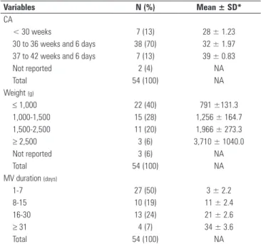
Table 1 - Distribution of the occurrence of unplanned extubations according to the mean corrected age, weight, and duration of mechanical ventilation
Variables N (%) Mean ± SD*
CA
< 30 weeks 7 (13) 28 ± 1.23
30 to 36 weeks and 6 days 38 (70) 32 ± 1.97
37 to 42 weeks and 6 days 7 (13) 39 ± 0.83
Not reported 2 (4) NA
Total 54 (100) NA
Weight (g)
≤ 1,000 22 (40) 791 ±131.3
1,000-1,500 15 (28) 1,256 ± 164.7
1,500-2,500 11 (20) 1,966 ± 273.3
≥ 2,500 3 (6) 3,710 ± 1040.0
Not reported 3 (6) NA
Total 54 (100) NA
MV duration (days)
1-7 27 (50) 3 ± 2.2
8-15 10 (19) 11 ± 2.4
16-30 13 (24) 21 ± 2.6
≥ 31 4 (7) 34 ± 3.6
Total 54 (100) NA
to 3.00 events per 100 days of MV/intubation. his diference may be attributed to the professional training programs that are frequently performed at HSF to promote continued staf education and improve the care of newborns in the NICUs. However, the inappropriate or lack of form completion may have caused some underreporting and an underestimation of the actual incidence of UE.
he implementation of 24-hour physical therapy assistance at HSF may have also contributed to the lower UE rate. his strategy allows for systematic assessments of both newborns and the ETT positioning and ixation by the physical therapy staf, which reduces the frequency of UE. However, further reductions in the incidence of UE remains a challenge for the investigated NICUs due the short-term (e.g., laryngospasm, bronchospasm, laryngeal edema, aspiration pneumonia, hypotension, hypoxia, and death), medium-term (e.g., increased duration of MV, and higher incidence of MV-related pneumonia), and long-term (e.g., longer hospitalization and complications associated with airway damage) iatrogenic potential of UE.(8,16-18)
he highest incidence of UE occurred in January 2010, and the lowest incidence was observed in April 2010. hese indings may be related to the number of newborns under MV per day in the NICUs. he highest rate of UE also occurred within the irst 7 days of MV, which may be explained by the numerous procedures that the newborns underwent during their irst days of life, such as radiographs, blood sample collections, and lumbar puncture. he reduced use of continuous sedation at this institution might have increased the frequency of UE during this period.
he highest UE rates were observed at gestational ages of 30 to 36 weeks (6/7) and weights ≤ 1,000 g. hese results may be due to the shorter length of the trachea in these newborns and their positioning during procedures, including spine lexion during lumbar puncture and changes in position that were performed by the newborns themselves (e.g., spinal hyperextension in the supine position).(6,8,19,20)
Todres et al.(21) observed 7- to 28-mm ETT movements in preterm newborns, which may also contribute to the higher index of UE in this population. Other factors are associated with UE in newborns, such as the use of ETT without balloons, cognitive immaturity, and the lack of physical restraints.(8,14,22)
Electronic devices, such as pulse oximeters and ventilator alarms (tidal volume, minute ventilation, and pressure peaks), are of paramount importance in the
diagnosis of UE(8,13). However, diagnosis in the present and previous studies(4,9) was established using clinical signs (primarily audible crying), the exteriorization of ETT, and a decrease in peripheral oxygen saturation (cyanosis). herefore, the importance of adequate patient monitoring and the continued training of the NICU multiprofessional staf in the detection of UE must be emphasized because vital signs monitors may emit belated or no signals or perform “false” readings when their sensitivity is low.(4,13)
Age, the amount of secretions, agitation, duration of intubation, ETT ixation and movement, and the route of intubation enhance the risk for UE, especially in neonates.(4,11,12,23) herefore, the results of the present study are consistent with previous studies because the primary causes of UE related to patient agitation and the inappropriate manipulation and ixation of the ETT. hese factors may also be associated with other factors, such as the inappropriate positioning of the MV circuit, agitation, and vomiting.
he orotracheal route of intubation is used at HSF, and ixation of the ETT is performed using sutures following a modiication of Gregory’s original technique.(4,13,24) A physician records the length of the external or proximal part of the tube after intubation and ETT ixation in the patients’ clinical records. An on-duty physician performs and replaces ETT ixation whenever the tube becomes loose, wet, and/or dirty, according to established institutional protocols.(25)
he ixation and positioning procedures for ETT that are performed by the on-duty physician must be reassessed because a signiicant fraction of the causes of UE were related to the inappropriate ixation and positioning of the ETT, and these mistakes are avoidable. A periodic training program for the physical therapy staf should be implemented.
occurrence of UE that is associated with each of the identified risk factors.
he limbs of the newborns who underwent UE were not restrained, and most of these newborns were not continuously sedated. However, some of newborns who sufered UE received a bolus of fentanyl and/ or midazolam when prescribed. hese factors may have allowed the newborns to move their upper limbs towards the ETT to cause some of the UE events. he use and efects of chemical and physical restraints for the prevention of UE are controversial.(4,8,26) herefore, the multiprofessional staf at HSF NICUs must discuss analgesia, sedation, and the criteria for the weaning and early extubation of newborns under MV more thoroughly to ensure that these patients do not remain unnecessarily awake under MV, which may lead to UE and its related complications.
he assessment and quality of continued patient care must be further improved in NICUs to reduce the incidence of UE. Clinical signs that are indicative of possible UE should especially be monitored, and procedures to control the causes of UE should be implemented.(6,16,17) he implementation of programs to continuously improve the quality of care may play an important role in the reduction of the incidence of UE.(26,27) hese programs must not be performed in an isolated manner, but they must include the full participation of all staf. he results of the present study demonstrated that each UE event was associated with at least one of the procedures that are performed by diferent members of the NICU teams.
he present study is clinically relevant because it contributes to the ongoing discussion of UE in the neonatal setting. Data on UE are scarce, and controversies in the deinition and discussion of UE and AE remain.(8,9,13) he present study also provides data to improve the performance and frequency of the procedures that are performed by the full multiprofessional NICU staf at HSF, particularly physical therapy. he present study also contributes to improvements in UE reports to ensure that all events and the number of hospitalized patients in the NICU is systematically documented on a daily basis and the number of intubated and tracheostomized patients who are subjected to MV is distinguished. These improvements in UE reporting facilitate for the establishment of UE and the performance of further studies.
Some limitations of the present study relate to the loss of data and missing information on the need for re-intubation after an UE event and the subjection
of patients to an extubation procedure. his result emphasized the need for appropriate records of UE at HSF. Importantly, no instance of UE was directly related with death.
Newborns undergoing weaning from MV (i.e., less sedation and greater agitation) are at risk for UE, which reinforces the need for the accurate recording of the abovementioned data. A further limitation of the present study is that the risk factors for UE in the NICUs of HSF were not established because the clinical staf did not record daily data on the number of newborns under MV (per orotracheal route) who did not experience UE during the investigated period. his study emphasizes the need to record this information, but further studies to identify the risk factors for UE are required.
CONCLUSION
he incidence of UE at HSF was lower than previous reports. However, programs to improve the quality of care and continued education must be revised to further reduce the rate of these events and their possible complications.
ACKNOWLEDGMENTS
We thank the HSF “Integrality Incubator” Inter-institutional Technical Scientiic Strengthening Program for its support.
RESUMO
Objetivo: Determinar a incidência e as principais causas de extubação não planejada em recém-nascidos nas unidades de te-rapia intensiva neonatais do Hospital Soia Feldman, de Belo Horizonte (MG).
Métodos: Estudo retrospectivo, realizado durante o perío-do de 1o de julho de 2009 a 30 de abril de 2010. Os eventos
de extubação não planejada e as principais causas associadas a estes foram avaliados por meio de uma icha de eventos ad-versos. Foram analisadas as seguintes variáveis: gênero, idade gestacional corrigida, peso atual, tempo em ventilação mecâ-nica, horário e motivos/causas do evento no dia da extubação não programada.
inade-quada do paciente durante execução de procedimentos; ixação inadequada e posicionamento do tubo endotraqueal.
Conclusão: A incidência de extubação não planejada nas unidades de terapia intensiva neonatais pôde ser considerada baixa, de acordo com o período avaliado, quando comparada aos dados relatados na literatura. Contudo, uma avaliação da
qualidade dos procedimentos e um acompanhamento contínuo desses recém-nascidos, assim como a monitoração das causas, são necessários para reduzir, ainda mais, tal incidência.
Descritores: Extubação; Modalidades de isioterapia; Incidência; Recém-nascido; Unidades de terapia intensiva neonatal
REFERENCES
1. Franck LS, Vaughan B, Wallace J. Extubation and reintubation in the NICU: identifying opportunities to improve care. Pediatri Nurs. 1992;18(3):267-70. 2. Eusébio M, Fernandes E. O uso de pré-medicação na intubação traqueal
não emergente do recém-nascido em Portugal. Acta Pediatr Port. 2008;39(1):3-7.
3. Veldman A, Trautschold T, Weiss K, Fischer D, Bauer K. Characteristics and outcome of unplanned extubation in ventilated preterm and term newborns on a neonatal intensive care unit. Paediatr Anaesth. 2006;16(9):968-73. 4. Piva JP, Garcia PC. Ventilação mecânica em pediatria. In: Piva JP, Carvalho
P, Garcia PC. Terapia intensiva em pediatria. 4a ed. Rio de Janeiro: Medsi; 1997. p. 235-60.
5. Hermeto F, Martins BM, Ramos JR, Bhering CA, Sant’Anna GM. Incidence and main risk factors associated with extubation failure in newborns with birth weight < 1,250 grams. J Pediatr (Rio J). 2009;85(5):397-402. 6. Ream RS, Mackey K, Leet T, Green MC, Andreone TL, Loftis LL, et al.
Association of nursing workload and unplanned extubations in a pediatric intensive care unit. Pediatr Crit Care Med. 2007;8(4):366-71.
7. Turner BS. Maintaining the artificial airway: current concepts. Pediatr Nurs. 1990;16(5):487-93.
8. Da Silva PSL. Extubação não-planejada. In: Barbosa AP, Carvalho WB, Johnston C. Desmame e extubação em pediatria e neonatologia. Rio de Janeiro: Atheneu; 2010. p. 165-71.
9. Carvalho FL, Mezzacappa MA, Calil R, Machado HC. Incidence and risk factors of accidental extubation in a neonatal intensive care unit. J Pediatr (Rio J). 2010;86(3):189-95.
10. Kleiber C, Hummel PA. Factors related to spontaneous endotracheal extubation in the neonate. Pediatr Nurs. 1989;15(4):347-51.
11. Scott PH, Eigen H, Moye LA, Georgitis J, Laughlin JJ. Predictability and consequences of spontaneous extubation in a pediatric ICU. Crit Care Med. 1985;13(4):228-32.
12. Braun MS. Prevention of accidental extubation in newborns. Am J Dis Child. 1988;142(1):1240-3.
13. Piva JP, Amantéa S, Luchese S, Giugno K, Maia TR, Einloft L. Extubação acidental em uma unidade de terapia intensiva. J Pediatr (Rio J). 1995;71(2):72-6. 14. Loughead JL, Brennan RA, DeJuilio P, Camposeo V, Wengert J, Cooke D.
Reducing accidental extubation in neonates. Jt Comm J Qual Patient Saf. 2008;34(3):164-70, 125.
15. Secretaria de Estado de Saúde de Minas Gerais. Subsecretaria de Vigilância em Saúde. Análise de Situação de Saúde - Minas Gerais 2010. Belo Horizonte: Secretaria de Estado de Saúde de Minas Gerais; 2010. [citado 2012 Ago 19]. Disponível em: http://www.saude.mg.gov. br/publicacoes/estatistica-e-informacao-em-saude/analises-de-situacao-de-saude/publicacao_subsec_saude_FINAL.pdf.
16. Little LA, Koenig JC Jr, Newth CJ. Factors affecting accidental extubations in neonatal and pediatric intensive care patients. Crit Care Med. 1990;18(2):163-5.
17. Nicolau CM, Lahóz AL. Fisioterapia respiratória em terapia intensiva pediátrica e neonatal: uma revisão baseada em evidências. Pediatria (São Paulo). 2007;29(3):216-21.
18. Nieves J. Avoiding spontaneous extubation of nasotracheal or oral tracheal tubes. Pediatr Nurs. 1986;12(3):215-8.
19. Erenberg A, Nowak AJ. Appliance for stabilizing orogastric and orotracheal tubes in infants. Crit Care Med. 1984;12(8):669-71.
20. Frank BS, Lewis RJ. Experience with intubated patients does not affect the accidental extubation rate in pediatric intensive care units and intensive care nurseries. Pediatr Pulmonol. 1997;23(6):424-8.
21. Todres ID, deBros F, Kramer SS, Moylan FM, Shannon DC. Endotracheal tube displacement in the newborn infant. J Pediatr. 1976;89(1):126-7. 22. Goldsmith JP, Karotkin EH. Introduction to assisted ventilation. In:
Goldsmith JP, Karotkin EH. Assisted ventilation of the neonate. 4th ed. Philadelphia: Saunders; 2003. p. 1-14.
23. Kapadia FN, Bajan KB, Raje KV. Airway accidents in intubated intensive care unit patients: an epidemiological study. Crit Care Med. 2000;28(3):659-64. 24. Rivera R, Tibballs J. Complications of endotracheal intubation
and mechanical ventilation in infants and children. Crit Care Med. 1992;20(2):193-9.
25. Oliveira CR, Gontijo FO, Cabral LA, David RB, Schettino RC, Ribeiro SN, Araújo VC, Azevedo VM, Lopes TC. Instrução de trabalho técnico-assistencial da equipe de fisioterapia do Hospital Sofia Feldman. 3a ed. Belo Horizonte: HSF; 2010.
26. da Silva PS, de Aguiar VE, Neto HM, de Carvalho WB. Unplanned extubation in a pediatric intensive care unit: impact of a quality improvement programme. Anaesthesia. 2008;63(11):1209-16.