Glass Coatings on Titanium Substrates
*e-mail: [email protected]
Pulsed Laser Deposition of SiO
2- P
2O
5- CaO - MgO
Glass Coatings on Titanium Substrates
Ednan Joannia,b*, Marta Carmona Ferroc, Cezarina Cela Mardarea,
Andrei Ionut Mardarea, José Ramiro Afonso Fernandesa,b,
Sandra Cristina de Almeida Pinac
aINESC-Porto, Unidade de Optoelectrónica e Sistemas Electrónicos,
Rua do Campo Alegre, 687, 4169-007 Porto, Portugal
bUniversidade de Trás-os-Montes e Alto Douro, Departamento de Física,
5001-911 Vila Real, Portugal
cUniversidade de Aveiro, Departamento de Engenharia Cerâmica e do Vidro,
3810-193 Aveiro, Portugal
Received: September 16, 2003; Revised: April 5, 2004
Thin films of bioactive glass-ceramic have been deposited on titanium substrates by the Pulsed Laser Deposition (PLD) technique under different experimental conditions. The effect of param-eters such as deposition pressure and temperature of heat treatments was studied. The microstruc-ture and the crystalline phases of the coatings were characterized using SEM, EDX and XRD analysis; the phases present were titanium oxides, calcium magnesium silicates and phosphates. The adhesion of the as-deposited films has been examined by scratch tests. The interfacial adhe-sion of the coatings was better when the deposition was performed at low pressure. Samples were immersed in simulated body fluid (SBF), and a calcium-phosphate precipitate was observed on the surface of less crystallized samples, suggesting that there is some relationship between surface reactivity and crystallinity.
Keywords:coatings, pulsed laser deposition, glass-ceramics, biomaterials
1. Introduction
Many studies have established hydroxyapatite (HAp) as a material of choice for applications involving bone repair and bone-implant contact. On the other hand, new glasses and glass-ceramics based on the SiO2 - CaO - P2O5 system with different amounts of minor constituents exhibit an even greater degree of osteoinductive behaviour1. Other
advan-tages of these bioactive glasses include the flexibility in-herent to the glass processing methods, and the possibility to manipulate the composition and thermal history in order to adjust the amount and nature of the crystalline phases present in the final material2,3.
Bioceramics and bioactive glasses are well known for their bioactivity, but their poor mechanical properties con-stitute a limitation for load-bearing applications. One solu-tion for this problem is to cover metals (such as Ti, Ti al-loys, Co - Cr alloys and stainless steel), which form the structural part of the implant, with a ceramic or a vitreous
layer4-6. The most promising metallic system for implant
applications was found to be Ti and its alloys because of their biocompatibility and mechanical properties7,8.
Plasma-spraying is the most common method used for coating Ti-based implants, because of its simplicity and the ability to form relatively thick layers (typically 50-200 µm) of HAp or glasses9-11. However the poor mechanical
prop-erties are the major drawback of this kind of coating; the adhesion between the sandblasted titanium surface and the ceramic layer is low because it is almost exclusively due to mechanical interlocking12. Alternative techniques such as
electrolytic deposition, sol-gel, powder sintering, ion beam deposition, RF sputtering and others have been explored to improve bond strength and other properties of the coatings, but these methods are still very much open to new develop-ments13,14.
cur-rently being applied in a growing number of fields, mainly due to its ability to deposit high quality films from a variety of materials. A unique characteristic of the PLD technique is the production of dense films that preserve the stoichi-ometry of the target and have a high degree of adhesion to the substrate15,16. By controlling the composition of the gas
in the vacuum chamber (O2, Ar and H2O) and the substrate
temperature, both amorphous and crystalline HAp films, α
and β tricalcium phosphates and tetracalcium phosphates could be made from the same HAp target17-19.
In the present study a series of samples were prepared by depositing a bioactive glass on Ti substrates using the PLD technique under different experimental conditions. The effect of parameters such as deposition pressure and tem-perature of heat treatments was studied. Coatings were char-acterized in terms of morphology, crystallinity, adhesion strength and in vitro behaviour. The relationship between crystallinity and in vitro behaviour in SBF is discussed.
2. Experimental
2.1. Deposition and heat treatments
The targets for pulsed laser deposition (PLD) were fab-ricated by melting and casting a glass with the molar com-position 31 SiO2; 26.7 MgO; 31.7 CaO; 10.6 P2O5. This glass, when heated for 1 h at 870 °C, shows the presence of a single Mg-substituted whitlockite phase, 3(Ca,Mg)O.P2O5, but in samples heat-treated at 970 °C enstatite, (Mg,Ca)O.SiO2, also precipitates. This glass-ceramic is machineable and moderately bioactive20,21. Commercially
available titanium disks (99.6 wt. (%) Ti) were used as substrates. The substrate surface was prepared for deposi-tion by polishing, washing with acetone, immersing in a
solution with 15%HNO3, 1%HF and 84% H2O (vol%) for
3 min, followed by ultrasonic cleaning in deionised water. The deposition was performed using a KrF laser (Lambda Physik LPX 300) with a wavelength of 248 nm and a 10 Hz pulse repetition rate. The laser fluence was 5.58 J/cm2 and the number of pulses was 18000. The vacuum
chamber was always pumped to a base pressure of at least 10-7mbar, and for some depositions the pressure was
in-creased by the introduction of oxygen (10-2 mbar). The
coat-ings were deposited at room temperature. The distance be-tween the target and the substrate was fixed at 4 cm. After deposition, some of the coatings were heat-treated for 1h at 870 and 970 °C, in air.
2.2. Characterization
The surface morphology of the films was examined us-ing scannus-ing electron microscopy (Hitachi H9000-NA), and energy dispersive X-ray (EDX) analysis was used for el-emental analysis. In order to assess the crystallinity of the as-deposited and heat-treated coatings, X-ray diffraction
(Rigaku PMG-VH) measurements were performed with a scanning speed of 0.1°/min.
The adhesion strength of the coatings was evaluated using scratch tests performed on a CSem-Revetest scratcher with a Rockwell C diamond stylus (hemispherical tip, 200 µm), under linearly increasing load (from 1 to 20 N) with a specimen traversal rate of 10 mm/min. The samples were inspected by SEM and optical microscopy. The adhe-sion was evaluated by taking into consideration any coating losses, whether by flaking or by massive loss in the scratch channel22.
In order to test their in vitro behaviour in a simulated plasma, the as-deposited coatings and the heat-treated coat-ings were soaked in 10 mL SBF at 37 °C and pH 7.4 for 7 days, with daily solution renewal. The amounts of Si, Ca, P and Mg ions in solution were analysed by inductive cou-pled plasma spectrometry (ICP). Morphological characteri-sation by SEM was carried out before and after SBF im-mersion.
3. Results and Discussion
3.1. Morphology and structure
Figure 1 shows the typical surface morphologies of the coatings as deposited. They consist of a continuous matrix full of spheres ranging in size between 50 nm and 3 µm. This morphology is typical for glass and metal films made by PLD. The spheres are droplets of molten material from the target and they cannot be avoided by manipulating the deposition parameters (temperature, pressure or laser fluence). Even when the substrate is placed off-axis, the glass films made by PLD still present this kind of surface topography23. In the films made at low pressure, 10-7 mbar
(Fig. 1a), the packing of the spheres is very dense, while in the films made at high pressure, 10-2 mbar (Fig. 1b), the
surface is rougher and the spheres are weakly connected among themselves. The packing density of the spheres is related to the loss of kinetic energy of the particles during the transport of material between the target and the substrate due to the collisions with the O2 molecules in the chamber atmosphere.
The coatings had thicknesses ranging between 1 and
5 µm, but it was not easy to have an exact measurement
because of the roughness of the substrate, the morphology of the coatings and the difficulty in fracturing the titanium substrates without destroying the coatings. In order to ob-serve the cross sections of the films, some coatings were repeated on silicon substrates using the same experimental conditions. Fig. 2 shows the cross section of one of the coat-ings made at low pressure (10-7 mbar). The film is very
Glass Coatings on Titanium Substrates
droplets of molten glass in relation to the thickness of the film. For this particular application the surface roughness due to the spheres is actually favourable because it helps the osteointegration of the implants24.
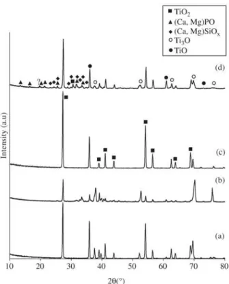
Figure 3 shows the XRD patterns of the titanium substrates heat-treated at 870 °C and 970 °C, for 1 h in air, and substrates with coatings prepared at 10-7 mbar and
heat-treated at the same temperatures. The heat-heat-treated coatings are crystallized even for the lower heat treatment tempera-ture and exhibit peaks from titanium oxides, as well as peaks that can be attributed to calcium magnesium phosphates and calcium magnesium silicates, whereas the diffractograms for the heat-treated substrates show only the peaks charac-teristic of Ti3O and TiO2. With the increase of the heat
treat-Figure 1. SEM micrographs of typical film surfaces made at: a)
low pressure (10-7 mbar); b) high pressure (10-2 mbar).
(a)
Figure 2. Cross section of a film made at low pressure (10-7mbar)
on a silicon substrate.
ment temperature the peak intensities increase, indicating a higher degree of crystallinity in the films, and a series of small peaks appear at low diffraction angles, all of them
(b)
Figure 3. X-ray diffraction patterns of samples heat-treated for 1 h
corresponding also to the phosphates and silicates with the exception of one peak which could not be identified. These peaks are not present in the diffractogram for the Ti substrates heat-treated under the same conditions. Previous DTA studies on samples from the same glass showed two exothermic peaks at 829 °C and 936 °C, corresponding to the onset of crystallization for these two phases25.
The morphology of the heat-treated coatings is shown in Figs. 4a and 4b. In the case of the heat treatment at 870 °C, the coatings appeared to begin their crystallization, while at 970 °C an extensive oxidation of the metallic substrate occurred and two different morphologies, thin plates and agglomerates were observed. The EDS analysis showed that the agglomerates in the sample heat-treated at 870 °C are richer in Ca and Mg than the surrounding matrix. In the sample heated at 970 °C, the molar concentration of Ti in the plates is higher than the concentration of Ca and Mg, whereas the opposite happens in the agglomerates.
3.2. Adhesion
The inherent properties of the coating and the substrate, namely the coating morphology and the substrate surface roughness, make the measurement of the adhesion strength very difficult. Due to these characteristics, the scratch test does not give reliable information of the load that is repre-sentative of the coating adhesion; it can only provide quali-tative results, useful for comparative studies.
The interfacial adhesion of the glass coatings is better for the lower deposition pressure (10-7 mbar), than for the
higher deposition pressure (10-2 mbar) as observed in Fig. 5a
and 5b, respectively. This was expected from the surface
morphology of the films shown in Fig. 1. The films made at low pressure are denser while in the films made at high pres-sure the cohesion between the glass spheres is weaker and as a result a lower adhesion was obtained.
3.3. Surface reactivity in SBF
After soaking in SBF, the as-deposited coatings made at low pressure showed signs of surface dissolution (Fig. 6a). This type of behaviour was also observed by Wolke et al.26
for amorphous calcium phosphate coatings (4 µm thick)
deposited by RF magnetron sputtering, but the authors re-ported a good durability for much thinner (0.1µm thick) heat-treated coatings even after 4 weeks immersion in SBF. Although the coatings deposited by PLD and sputtering are thinner than the ones made by plasma spraying, they are denser, more homogeneous and more adherent, conse-quently having improved mechanical properties18,26,27. For
the coatings heat-treated for 1 h at 870 °C a granular precipi-tate was deposited (Fig. 6b). EDS analysis showed that this precipitate was rich in Ca and P. In the case of the coatings heat-treated for 1h at 970°C no changes were observed.
The growth of the Ca-P precipitate was only observed on the coatings heat-treated for 1 h at 870 °C. As shown in Fig. 3, the samples heat-treated at 870 °C are less crystal-lised than the ones at 970 °C. It is likely that calcium phos-phate nuclei formed in SBF grow faster on samples with a residual glassy phase, since super saturation of the SBF solution will be favoured by dissolution of the glass matrix. The presence of a certain amount of amorphous phase is thought to be essential in promoting dissolution of the ma-terial and further growth of the apatite layer in a simulated
Figure 4. Scanning electron micrographs of heat-treated coatings: a) 870 °C, 1h; b) 970 °C, 1 h.
Glass Coatings on Titanium Substrates
plasma26,28. In the case of the coatings heat-treated at 970 °C,
the Ca-P precipitate did not appear after immersion in SBF probably because the coatings were almost fully crystal-lised, as suggested by Figs. 3d and 4b.
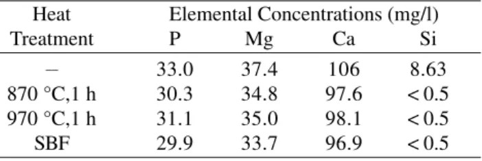
The elemental concentrations of Si, Ca, P and Mg, ob-tained by ICP confirmed that dissolution in SBF of the as-deposited coatings was more severe than in the samples heat-treated in both conditions, as shown in Table 1. In contrast with the as-deposited coatings, the heat-treated coatings dissolved only slightly; after the test, the Ca and P
concen-(a) (b)
Figure 5. SEM picture of a scratch test performed on as-deposited coatings grown at: a) low pressure (10-7 mbar); b) high pressure
(10-2 mbar).
Figure 6. SEM micrograph of coating surface after 7 days in SBF with renewal: a) as-deposited coating; b) heat-treated coating at 870 °C.
(a) (b)
trations in solution were similar to the original ion concen-tration in the SBF.
4. Conclusions
treat-ments. Titanium oxides, calcium magnesium silicates and phosphates were present in the heat-treated coatings. Scratch tests provided indication that the interfacial adhe-sion of the coatings was better when the deposition was performed at low pressure.
The coatings heat-treated for 1 h at 870 °C induced the formation of a Ca - P precipitate when immersed in a renewed SBF solution; this did not occur for the amor-phous coatings and for the coatings heat-treated at 970 °C for 1 h in air. These results suggest that, similarly to what happens with bulk bioactive glasses and glass-ceramics, the behaviour of the coatings in simulated plasma is de-pendent on the amount of glassy phase present. Work is now in progress to clarify the mechanisms of apatite for-mation on the surface of the coatings and to confirm if the
in vitro bioactivity observed in bulk samples is maintained in the films deposited by the PLD technique.
References
1. Hench, L.L.; Wilson, J. Bioceramics: Mater. and Appl., p. 11, 1995.
2. Brink, M.; Turunen, T.; Happonen, R.P.; Yli-Urpo, A.
J. Biomed. Mater. Res., v. 37, p. 114, 1997.
3. Peitl Filho, O.; Latorre, G.P; Hench, L.L. J. Biomed. Mater. Res., v. 30, p. 509, 1996.
4. De Groot, K.; Geesink, R.; Klein, C.P.A.T.; Serekian, P. J. Biomed. Mater. Res., v. 21, p. 1375, 1987.
5. Tomisia, A.T.; Moya, J.S.; Guitian, F. Bioceramics:
Mater. and Appl., p. 303, 1995.
6. Verne, E.; Ferraris, M.; Ventrella, A.; Paracchini, L.; Krajewski, A.; Ravaglioli, A. J. Eur. Ceram. Soc., v. 18, p. 363, 1998.
7. Niinomi, M. Mater. Sci. And Eng. A, v. 243, p. 231,
1998.
8. Van Noort, R. J. Mater. Sci., v. 22, p. 3801, 1987. 9. Gottschling, S.; Kohl, R.; Engel, A.; Oel, H.J.
Bioceramics: Mater. And Appl., p. 201, 1995. 10. Oku, T.; Suganuma, K.; Wallenberg, L.R.; Tomsia,
A.P.; Gomez-Vega, J.M.; Saiz, E. J. Mater. Sci.: Mater. In Med., v. 12, p. 413, 2001.
11. Sun, L.; Berndt, C.C.; Khor, K.A.; Cheang, H.N.; Gross, K.A. J. Biomed. Mater. Res., v. 62, n. 2, p. 228, 2002. 12. Ravaglioli, A.; Krajewski, A.; Bioceramics – Mater.,
Propert., Appl., Chapman & Hall, p. 198, 1992. 13. Mardare, C.C.; Mardare, A.I.; Fernandes, J.R.A.;
Joanni, E.; Pina, S.C.A.; Fernandes, M.H.V.; Correia, R.N. J. Eur. Ceram. Soc., v. 23, p. 1027, 2003. 14. Weng, W.; Baptista, J.L. J. Mater. Sci.: Mater. In Med.,
v. 9, p. 159, 1998.
15. Kim, M.C.; Choi, J.W.; Yoon, S.J.; Yoon, K.H.; Kim, H.J. Jpn. J. Appl. Phys., v. 41, p. 3817, 2002. 16. Mardare, A.I.; Mardare, C.C.; Joanni, E.; Fernandes,
J.R.A.; Vilarinho, P.M.; Kholkin, A.L. Ferroelectrics, n. 293, p. 177, 2003.
17. Cotell, C.M. Pulsed Laser Deposition Of Thin Films, John Wiley And Sons, Inc., New York, p. 549, 1994. 18. Garcia-Sanz, F.J.; Mayor, M.B.; Arias, J.L.; Pou, J.;
Leon, B.; Perez-Amor, M.; J. Mater. Sci.: Mater. In
Med., v. 8, p. 861, 1997.
19. Arias, J.L.; Mayor, M.B.; Garcia-Sanz, F.J.; Pou, J.;
Leon, B.; Perez-Amor, M.; Knowles, J.C. J. Mater.
Sci.: Mater. In Med., v. 8, p. 873, 1997.
20. Oliveira, J.M.; Fernandes M.H.; Correia, R.N.
Biomaterials, v. 16, n. 11, p. 849, 1995.
21. Serro, A.P.; Fernandes, A.C.; Saramago, B.; Fernandes, M.H.V. J. Biomed. Mater. Res. n. 61, p. 99, 2002. 22. Sekler, J.; Steinmann, P.A.; Hintermann, H.E. Surf. And
Coat. Tech., v. 36, p. 519, 1988.
23. D’Alessio, L.; Ferro, D.; Marotta, V.; Santagata, A.; Teghil, R.; Zaccagnino, M. Appl. Surf. Sci., n. 183, p. 10, 2001.
24. Gonzáles, P.; Serra, J.; Liste, S.; Chiussi, S.; León, B.; Pérez-Amor, M.; Martínes-Fernández, J.; de Arellano-López, A.R.; Valera-Feria, F.M. Biomaterials, v. 24, p. 4827, 2003.
25. Oliveira, A.L.; Oliveira, J.M.; Correia, R.N.; Fernandes, M.H. Glastechn. Berichte Glass Sci. and Techn., v. 67C, p. 367, 1994.
26. Wolke, J.G.C.; De Groot, K.; Jansen, J.A. J. Mater. Sci., v. 33, p. 3371, 1998.
27. Ding, S.-J. Biomaterials, v. 24, p. 4233, 2003. 28. Li, P.; Yang, Q.; Zhang, F.; Kokubo, T. J. Mater. Sci.:
Mater. In Med., v. 3, p. 452, 1992.
Table 1. Elemental concentration of the SBF after immersion of
coatings.
Heat Elemental Concentrations (mg/l)
Treatment P Mg Ca Si
__ 33.0 37.4 106 8.63
870 °C,1 h 30.3 34.8 97.6 < 0.5
970 °C,1 h 31.1 35.0 98.1 < 0.5