Materials Research, Vol. 10, No. 2, 161-163, 2007 © 2007
*e-mail: [email protected]
Quantitative Analysis of Lead in Polysulfide-based Impression Material
Aparecida Silva Bragaa, Sebastião Roberto Silva Bragab, Alma Blásida Concepción Elizaur Benitez Catirsea*,
Luis GeraldoVazc, Francisco de Assis Mollo Juniorc
a
Department of Dental Materials and Prosthetics,
Ribeirão Preto School of Dentistry, University of São Paulo – USP,
Av do Café, s/n, 14040-904 Monte Alegre, Ribeirão Preto - SP, Brazil
b
Department of Biological Science, Araraquara School of Pharmaceutical Sciences,
Júlio de Mesquita Filho University, Araraquara - SP, Brazil
c
Department of Dental Materials and Prosthesis, Araraquara School of Dentistry,
Julio de Mesquita Filho University - Araraquara, SP, Brazil
Received: January 4, 2007; Revised: May 31, 2007
Permlastic® is a polysulfide-based impression material widely used by dentists in Brazil. It is composed of
a base paste and a catalyzer containing lead dioxide. The high toxicity of lead to humans is ground for much concern, since it can attack various systems and organs. The present study involved a quantitative analysis of the concentration of lead in the material Permlastic®. The lead was determined by plasma-induced optical emission
spectrometry (Varian model Vista). The percentages of lead found in the two analyzed lots were 38.1 and 40.8%. The lead concentrations in the material under study were high, but the product’s packaging contained no information about these concentrations.
Keywords: Permlastic®, impression materials, polysulfides, lead
1. Introduction
Permlastic® is a polysulfide-based impression material for complete
and partial dentures, inlays, onlays, crowns and fixed partial and/or removable dentures. The molding material is supplied in two tubes, one containing the base paste and the other a catalyzer. The base paste contains polysulfide polymer, fillers and plasticizers. According to the manufacturer, the polysulfide polymer has a low molecular weight, comprising terminal and pendent mercaptan groups. The lead dioxide catalyzes the condensation of the terminal and the pendent groups with -SH groups in other molecules, lengthening and cross-linking the chain. In the process, the material is transformed from a paste into rubber.
The presence of lead is a cause of concern due to its toxicity. Although the environment contains many sources of this metal, hu-man exposure to lead compounds occurs mainly through food, air, drinking water, ink and sand1. Lead poisoning has proved to be one
of the most difficult environmental health problems to control. Part of this difficulty is due to the lack of clear manifestations at an early stage of the process, which may remain asymptomatic until a severe encephalopathic crisis occurs.
Dental treatments are a source of exposure, because when pa-tients are subjected to dental procedures, they are exposed to the lead contained in some of the impression materials, as in the case of polysulfides.
Because this material comes in packaging without information about its lead content and given the toxicity of this metal, research is necessary to warn dentists about the safety measures to be applied in its use.
The present study involved a quantitative analysis of the lead content in the material Permlastic®.
2. Material and Method
The quantitative analysis of lead was carried out by the Labora-tory of Chemical Analyses at the Federal University of São Carlos
Center for Materials Characterization and Development – CCDM (Laboratório de Análise Química do Centro de Caracterização e Desenvolvimento dos Materiais da Universidade Federal de São Carlos – CCDM). Lots 3-1252 and 4-1083 of Permlastic®
catalyzer paste were analyzed. This material is produced by Kerr Corporation, USA.
The lead was determined in a Varian, model VISTA plasma-induced optical emission spectrometer. The samples were dried in a sand bath at 200 °C, then ground and digested in an acid medium (HNO3 + H2O2).
3. Results and Discussion
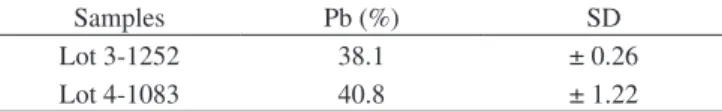
The results obtained correspond to the sample’s dry mass and are expressed as weight percentage, as shown in Table 1.
In the present study, the lead concentrations presented by the two lots of Permlastic® material were high (38.1 and 40.8% of Pb).
Previ-ous researches in which the cytotoxicity and local tissue imitation of elastic impression materials were tested revealed that the most severe reactions were caused by materials containing lead peroxide2-4.
In addition to local imitation, once it is absorbed in the body, lead may cause systemic reactions that are well documented in the literature5-7.
The efficiency of lead absorption depends on the path of exposure, age and nutritional status. Human adults absorb from 10 to 15% of the lead ingested, while children may absorb over 50%1.
The lead absorbed by the body is distributed mainly in the blood, soft tissue and bone, with bone containing close to 95% of the total lead in the body. The half-life of lead ranges from 25 to 28 days in the blood, and 20 years in the bones8,9. Approximately 75% of the
162 Braga et al. Materials Research
Nonspecific signs and symptoms of lead intoxication include a metallic taste, constipation, insomnia, irritability, muscle and joint pain, tremors, colic, and purplish-blue gums. Specific effects occur in target organs and systems, such as the nervous, hematological, cardiovascular and reproductive systems, as well as the kidneys and fetus8,12. It should be kept in mind that the estimated fatal dose is
40 mg.kg-1 13, at which point a blood lead concentration of more than
20 µg.100 mL-1 may be associated with acute symptoms.
Lead causes anemia by reducing hemoglobin and shortening of the life of erythrocytes14, due to the fragility of the blood cell
mem-brane and the reduction in the enzymes involved in heme synthesis. This reduction is evidenced in blood lead levels of 50 µg.dL-1 in adults
and approximately 40 µg.dL-1 in children8.
Effects on the central nervous system, subencephalopathy and damage to the peripheral nerves occur at blood levels of 30-50 µg.dL-1
in adults15. Desres et al.16 reported a neuromuscular effect in children
with blood lead concentrations of less than 10 µg.dL-1.
Cognitive and neuropsychological deficiencies have been ob-served in workers with blood lead levels of 41-80 µg.dL-1 17 and
ir-reversible cerebral damage occurs at blood levels exceeding or equal to 100 µg.dL-1 in adults and 80-100 µg.dL-1 in children8.
Another manifestation characteristic of lead toxicity is renal disease (nephropathy). Two types of nephropathy have been observed in humans. Acute nephropathy, which occurs during the initial stages of excessive exposure, is characterized by reversible functional and morphological alterations in the proximal tubular epithelial cells. Chronic nephropathy occurs after prolonged exposure to lead, present-ing morphological and functional changes that include reduction of the glomerular filtration speed and tubular dysfunction5,18. Exposure to
lead is also linked with hypertension in the general population and in subjects occupationally exposed to the metal19,20 even in blood levels
as low as 7 µg.dL-1, and may predispose those individuals to heart
at-tacks21. Farmand et al.22 demonstrated that the oxidative stress caused
by lead as a result of the imbalance in the oxidant/antioxidant process plays an important role in the pathogenesis of hypertension.
Research on pregnant women’s exposure to lead has provided clear evidence of the adverse effects of this metal on reproduction, particularly abortions and stillbirths8,23. Studies have also shown that
pregnancy may also be affected by paternal exposure to lead24,25.
Available data on the possible genotoxic and carcinogenic action of lead are conflicting, according to Minozzo et al.26. Researches on
the human carcinogenicity of exposure to lead that demonstrated a high prevalence of death by stomach and lung cancer have been in-adequate due to the exposure of the research subjects to other metals, thereby leading to ambiguities in the interpretation of the results18,27.
However, the database on the carcinogenicity of lead compounds in laboratory animals is very large, with the kidney ranking as the first anatomical tumor induction site in rats and mice28. Tumors of the
pituitary, supra-renal and thyroid glands, in addition to lung tumors and cerebral gliomas, have also been observed in animals exposed to lead29.
In view of the toxic effects of lead, both local and systemic, the manufacturers of dental materials should be warned about the urgent need for printing clearly stated information about the product’s lead
content on the packaging. In the case of Permlastic®, the
concen-tration of this metal is not stated in the descriptive leaflet, but the manufacturer clearly discloses this information on the packaging, stating that the product contains lead, a chemical substance known to cause cancer, as is divulged in the State of California. Furthermore, the manufacturer declares that this material is not for sale in the USA or Canada, and warns that the material must not be ingested and that accidental contact with the skin and eyes must be avoided.
It should be noted that the buccal mucosa, and especially the sub-lingual portion, has a higher absorption potential than the skin due to the high permeability and vascularization of its tissue.
Although this product has been approved for use in Brazil by ANVISA – National Agency for Sanitary Vigilance (under Permit No. 10258190069), in vivo studies are crucial in view of this mate-rial’s high lead content, as well as its long biological half-life and the various sources of exposure to this metal, in order to ensure its safe use.
4. Conclusions
Based on our quantitative analysis, we found that the lead content in Permlastic® dental impression material is high.
References
1. Valverde M, Fortoul TI, Diaz Barriga F, Mejia J; Del Castillo ER. Ge-notoxicity induceced in Cd – 1 mice by inhaled lead: differential organ response. Mutagenesis. 2002; 17(1):55-61.
2. Gettleman L, Nathanson D, Shklar G, Brathwaite WJr, Darmiento L, Levine P, et al. Preliminary evaluation of toxicity and radiopacity of lead-containing elastic impression materials. Journal of the American
Dental Association. 1978; 96(6):987-93.
3. Spranley TJ, Gettleman L, Zirmmerman KL. Acute tissue irritation of polysulfide rubber impression materials. Journal of Dental Research.
1983; 62(5):548-51.
4. Sydiskis RJ, Gerhardt, DE. Cytotoxicity of impression materials. Journal of Prosthetic Dentistry. 1993; 69(4):431-5.
5. Cooper WC, Wong O, Kheifets L. Mortality among employees of lead battery plants and lead-producing plants, 1947-1980. Scandinavian Journal of Work, Environment & Health. 1985; 11(5):331-45. 6. Bellinger DC. Lead. Pediatrics. 2004;113(4 Suppl):1016-22.
7. Carmouche JJ, Puzas JE, Zhang X, Tiyapatanaputi P, Cory-Slechta DA, Gelein R, et al. Lead exposure inhibits fracture healing and is associated with increased chondrogenesis, delay in cartilage mineralization, and a decrease in osteoprogenitor frequency. Environmental Health Perspec-tives. 2005; 113(6):749-55.
8. U.S Environmental Protection Agency (EPA). Air Quality Criteria Lead. V. III. Enviromental Criteria and Assessment Office, Research Triangle Park, NC. EPA – 600/8-83/028cF, 1986, Avaliable form NTIS, Springfield, VA; PB87-142378.
9. ATSDR (Agency for Toxic Substances and Disease Registry). Toxico-logical Profile for Lead. Update. Prepared by Clemente International Corporation Under Contract, n. 205-88-0608 for ATSDR, U.S. Public Health Service, Atlanta, GA, 1993.
10. Jensen, AA. Metabolism and toxicokinetics. In: Grandjean PG, Grandjean EC, editors. Biological effects of organolead compounds. Boca Raton: CRC Press; 1983. p. 97-115.
11. U.S. Environmental Protection Agency (EPA). Evaluation of the Potential Carcinogenicity of Lead and Lead Compounds. Office of Health and Environmental Assessment. EPA/600/8-89/045A; 1989.
12. Costa LG, Aschner M, Vitalone A, Syeversen T, Soldin OP. Developmental neuropathology of environmental agents. Annual Review of Pharmacology
and Toxicology. 2004; 44:87-110.
13. Mack, RB. A hard day,s kineght, zinc, phosphide poisoning. North
Carolina Medical Journal. 1989; 50(1):17-18.
Table 1. Lead concentration of the material Permlastic® in weight percentage and standard deviation.
Samples Pb (%) SD
Lot 3-1252 38.1 ± 0.26
Vol. 10, No. 2, 2007 Quantitative Analysis of Lead in Polysulfide-based Impression Material 163
14. Kempe DS, Lang PA, Eisele K, Klarl BA, Wieder T, Huber SM, et al. Stimulation of erythrocyte phosphatidylserine exposure by lead ions. American Journal of Physiology Cell Physiology. 2005, 288(2): C396-402.
15. Ehle AL, McKee DC. Neuropsychological effect of lead in occupationally exposed workers: a critical review. Critical Reviews in Toxicology. 1990; 20(4):237-55.
16. Desres C, Beuter A, Richer F, Poitras K, Veilleux A, Ayotte P, et al. Neuromotor functions in Inuit preschool children exposed to Pb, PCBs, and Hg. Neurotoxicology and Teratology. 2005; 27(2):245-57.
17. Stollery BT, Broadbent DE, Banks HA, Lee, WR. Short term prospec-tive study of cogniprospec-tive functioning in lead workes. British Journal of
Industrial Medicine. 1991; 48(11):739-49.
18. Selevan SG, Landrigan PJ, Stern F.B, Jones J H. Mortality of lead smelter workers. American Journal of Epidemiology. 1985; 122(4):673-83.
19. Pocock SJ, Shaper AG, Ashby D, Delves HT, Clayton BE. The relationship between blood lead, blood pressure, stroke, and heart attacks in middle-aged British men Environmental Health Perspectives. 1988; 78:23-30.
20. Ni Z, Hou S, Barton CH, Vaziri ND. Lead exposure raises superoxide and hydrogen peroxide in human endothelial and vascular smooth muscle cells. Kidney International. 2004; 66(6):2329-36.
21. U.S Environmental Protection Agency (EPA). Air Quality Criteria Lead: Supplement to the 1986. Addendum. Environmental Criteria and Assessment Office. Research Triangle Park, N. C. EPA – 600/8 – 89/049F, 1990.
22. Farmand F, Eddaie A, Roberts CK, Sindhu RK. Lead-induced dysregu-lation of superoxide dismutases, catalase, glutathione peroxidase, and guanylate cyclase. Environmental Research. 2005; 98(1):33-9.
23. Baghurst PA, Robertson, EF, Mc Michael, AJ. The Port Pirie cohort study: Lead effects on pregnancy outcome and early childhood development.
Neurotoxicology. 1987; 8(3):395-401.
24. Lindbohm ML, Sallmen M, Anttila A, Taskinen H, Hemminki D. Paternal occupational lead exposure and spontaneous abortion. Scandinavian Journal of Work, Environment & Health. 1991; 17(2):95-103.
25. Shiau CY, Wang JD, Chen PC. Decreased fecundity among male lead workers. Occupational and Environmental Medicine. 2004; 61(11):915-23.
26. Minozzo R, Deimling LI, Gigante LP, Santos-Mello R. Micronuclei in peripheral blood lymphocytes of workers exposed to lead. Mutation
Research. 2004; 565(1):53-60.
27. Gerhardsson L, Chettle DR, Snglyst GF. Kidney effects in long term exposed lead smelter workers. British Journal of Industrial Medicine.
1992; 49(3):186-92.
28. Nogueira E. Rat renal carcinogenesis after chronic simultaneous exposure to lead acetate and N-nitrosodiethylamine. Virchows Archiv. B. 1987; 53(6):365-74.
29. Zawirska B, Medras K. The role of the kidneys in disorders of porphyrin metabolism during carcinogenesis induced with lead acetate. Archivum