An electronic pressure-meter
nociception paw test for rats
Departamento de Farmacologia, Faculdade de Medicina de Ribeirão Preto, Universidade de São Paulo, Ribeirão Preto, SP, Brasil
G.G. Vivancos*, W.A. Verri Jr.*, T.M. Cunha*, I.R.S. Schivo, C.A. Parada, F.Q. Cunha and S.H. Ferreira
Abstract
The objective of the present investigation was to compare the sensitivity of an electronic nociceptive mechanical paw test with classical mechanical tests to quantify the intensity variation of inflammatory nociception. The electronic pressure-meter test con-sists of inducing the hindpaw flexion reflex by poking the plantar region with a polypropylene pipette tip adapted to a hand-held force transducer. This method was compared with the classical von Frey filaments test and with the rat paw constant pressure test, a modification of the Randall and Selitto test developed by our group. When comparing the three methods, the electronic pres-sure-meter and the rat paw constant pressure test, but not the von Frey filaments test, detected time vs treatment interactions in pros-taglandin E2 (PGE2)-induced hypernociception. Both methods also detected the PGE2-induced hypernociception in dose- (50-400 ng/ paw) and time- (1-4 h) dependent manners, and time vs treatment interactions induced by carrageenin (25-400 µg/paw). Furthermore, the electronic pressure-meter test was more sensitive at early times, whereas the constant pressure test was more sensitive at later times. Moreover, the electronic pressure-meter test detected the dose-dependent antinociceptive effect of local indomethacin (30-300 µg/paw) and dipyrone (80-320 µg/paw) on carrageenin- (200 µg/paw) and PGE2- (100 ng/paw) induced hypernociception, re-spectively, and also detected the ineffectiveness of indomethacin (300 µg) on the effect of PGE2. Our results show that the electronic pressure-meter provides a sensitive, objective and quantitative mechanical nociceptive test that could be useful to characterize new nociceptive inflammatory mediators and also to evaluate new peripheral analgesic substances.
Correspondence
S.H. Ferreira
Departamento de Farmacologia FMRP, USP
Av. Bandeirantes, 3900 14049-900 Ribeirão Preto, SP Brasil
Fax: +55-16-633-0021 E-mail: [email protected]
Research supported by CNPq, CAPES, FAPESP and PRONEX.
*These authors contributed equally
to this study.
Received June 2, 2003 Accepted November 25, 2003
Key words
•Hyperalgesia
•Nociception
•Dipyrone
•Indomethacin
•von Frey filaments
•Constant pressure test
Introduction
The objective of the present investigation was to compare the sensitivity of an elec-tronic nociceptive mechanical paw test (elec-tronic pressure-meter) with the classical von
this test presents some disadvantages such as the large number of attempts required to evaluate the nociceptive threshold and the problems with the standardization of the filaments (6). The electronic pressure-meter has been previously used in humans (7) and rats (8). It is an adaptation of the classical von Frey filaments test in which the pressure intensity is recorded automatically after paw removal.
The Randall and Selitto nociceptive test (9) and the writhing test have been used extensively for the development of nonste-roidal anti-inflammatory drugs. The modifi-cation of the Randall and Selitto nociceptive test developed by our group uses a small constant pressure and a different behavioral response (freezing reaction) as an end-point (2). This test was instrumental in the discov-ery of various seminal findings, which were later confirmed by other nociceptive meth-ods such as formalin-induced flinches (10,11), chemically induced writhing (12,13) and the classical Randall and Selitto method (14). In fact, the modified Randall and Selitto test was used in several pioneering studies of inflammatory nociceptor sensitization (hy-pernociception): the participation of the cAMP/Ca2+ pathway in the mechanism of
hypernociception (15), the peripheral effect of opiates (16), the cytokine cascade in-volved in the onset of inflammatory hypernociception (17-24), the peripheral memory of nociceptor sensitization (25,26), and the spinal retrograde sensitization of primary sensory neurons (27).
Compared with chemical tests (acetic acid writhing test and formalin test), me-chanical tests have an important practical advantage by allowing the dissociation be-tween the nociceptor sensitization (by an injection of a phlogogen or of a specific inflammatory mediator) and the overt behav-ioral end-point induced by the mechanical stimulus (28).
In order to evaluate the sensitivity of the electronic pressure-meter test, we sensitized
or induced an inflammatory response in the paws with different doses of prostaglandin E2 (PGE2) or with carrageenin, respectively.
PGE2-induced hypernociception was
com-pared to the von Frey filaments test and our modification of the Randall and Selitto test. Carrageenin-induced hypernociception was compared to the modification of the Randall and Selitto test. We also determined if the pressure-meter test was able to detect the qualitative difference previously observed with the constant pressure test in the effect of indomethacin and dipyrone on carragee-nin- and PGE2-induced hypernociception.
Material and Methods
Animals
The experiments were performed on male Wistar rats weighing 180 to 200 g (Univer-sity of São Paulo, Ribeirão Preto, SP, Brazil) housed in the animal care facility of the School of Medicine of Ribeirão Preto and taken to the testing room at least 1 h before the experiments. Food and water were avail-able ad libitum. All behavioral testing was performed between 9:00 am and 4:00 pm and the animals were used only once. Ani-mal care and handling procedures were in accordance with the guidelines of the Inter-national Association for the Study of Pain (IASP) on the use of animals in pain re-search. All efforts were made to minimize the number of animals used and their dis-comfort.
von Frey filaments and electronic pressure-meter paw tests for rats
or urination movements and not resting over the paws. In these experiments, either a series of von Frey filaments (Stoelting, Chi-cago, IL, USA) with logarithmically increas-ing stiffness (-2.35 to 2.65 log of force, g) or the pressure-meter, which consisted of a hand-held force transducer fitted with a 0.7 mm2 polypropylene tip (electronic von Frey
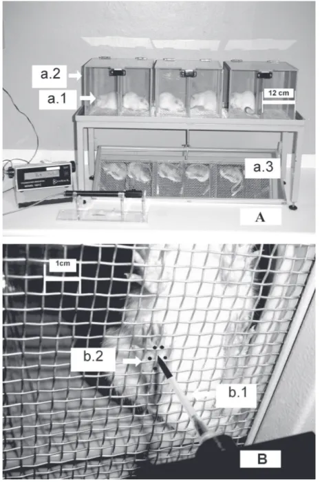
anesthesiometer, IITC Inc., Life Science Instruments, Woodland Hills, CA, USA), were used. The investigator was trained to apply the filaments or the polypropylene tip perpendicularly to one of the five distal foot-pads with a gradual increase in pressure. A tilted mirror below the grid provided a clear view of the animal’s hindpaw (Figure 1, panel B). The tests consisted of poking the hindpaw to provoke a flexion reflex followed by a clear flinch response after paw with-drawal. Each von Frey filament was applied for approximately 3-4 s to induce the end-point reflex. Testing was initiated with the filament handle marked 5.46, which corre-sponds to 1.46 log of force (g), which is in the middle of the filament series. The response to this filament defines if a series of a weaker or a stronger filament will be tested. The weakest filament able to elicit a response was consid-ered to be the mechanical threshold (g).
The results are reported as ∆ log of force (g) which was calculated by subtracting the value of the measurements (log of force) after treatment from that of the first measure-ment (before treatmeasure-ment). With the electronic pressure-meter, the intensity of the stimulus was automatically recorded when the paw was withdrawn. The equipment was cali-brated to determine the pressure linearly until 80 g. The stimulation of the paw was re-peated until the animal presented three similar measurements (the difference between the highest and the lowest measurement should be less than 10 g). The animals were tested before and after the treatments and the re-sults are reported as the ∆ withdrawal thresh-old (g), which was calculated by subtracting the value of the measurements after the
treatments from that of the first measure-ment (before treatmeasure-ment).
Rat paw constant pressure test
Paw sensitivity was also measured using
Figure 1. Apparatus for the electronic pressure-meter test and the area to which the polypropylene tip should be applied. Panel A: Rats (a.1) were placed in acrylic cages (a.2;
12 x 20 x 17 cm high) with a wire grid floor. A tilted mirror (a.3) below the grid provided a clear view of the animal’s hindpaw. Panel B: A 0.7 mm2 polypropylene tip (b.1) fitted to a
the rat paw constant pressure test, which is a modification of the Randall and Selitto test (2). In this method, a constant pressure of 20 mmHg was applied via a syringe piston moved by compressed air to an area of 15 mm2 of the dorsal surface of the rat paw, and
discontinued (reaction time) when the ani-mal exhibited a typical freezing reaction. The freezing reaction was indicated by brief ap-nea, concomitant with a retraction of the head and forepaws and a reduction in the escape movements that animals may make in order to escape from the position imposed by the hands of the experimenter. Usually, ap-nea was associated with successive waves of muscular tremor. For each animal, the la-tency to the onset of the freezing reaction (from the time of first pressure application) was measured before and after administra-tion of the agents. The results are reported as the ∆ reaction time which was calculated by subtracting the value of the measurements during the experiment from that of the first measurement (before treatment).
Drugs
Dipyrone and PGE2 were purchased from
Sigma (St. Louis, MO, USA). Carrageenin
was obtained from FMC Corporation (Phila-delphia, PA, USA) and indomethacin from Prodome Química e Farmacêutica (São Paulo, SP, Brazil).
Carrageenin and dipyrone were diluted in sterile saline. A stock solution of PGE2 was
prepared in 10% ethanol, and further dilu-tions were made in saline; the final concen-tration of ethanol was 1%. Indomethacin was diluted in Tris-HCl buffer, pH 8.0, which was administered alone to the control groups.
Drug administration
Drugs were injected subcutaneously in a 50-µl volume into the plantar region of rats. A 26-G hypodermic needle was inserted into the skin of the second footpad (to avoid back flow) and the tip of the needle was placed among the five distal footpads, at the same site where filaments or the tip of the pres-sure-meter were applied.
Statistical analysis
Two-way analysis of variance (ANOVA) was used to compare the groups and doses at all times. The factors analyzed were treat-ments, time and time vs treatment
interac-Intensity of hypernociception (∆
withdrawal threshold, g)
20
10
0 A
* *
*
* * *
* * * *
* *
*
*
*
* *
* *
*
* * *
C B
ns
ns
ns
ns ns
Intensity of hypernociception
(
∆
log force, g)
0.50
0.00
Intensity of hypernociception
(
∆
reaction time, s)
20
10
0
Saline PGE2 50 ng PGE2 100 ng PGE2 200 ng PGE2 400 ng
1 2 3 4
Time (h) 1 2Time (h)3 4 1 2Time (h)3 4
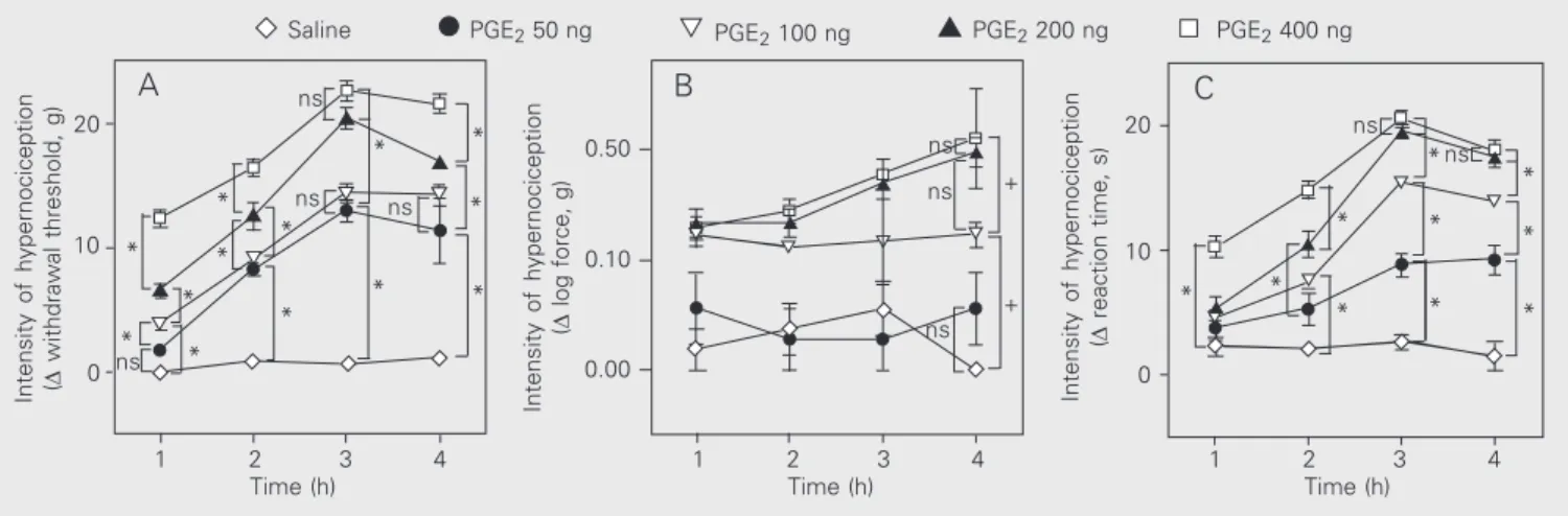
Figure 2. Dose-response curve for the hypernociception induced by intraplantar prostaglandin E2 (PGE2) in rats. Saline was injected in the control group.
The animals were tested with the electronic pressure-meter paw test (A), von Frey filaments (B) and the rat paw constant pressure test (C). The results are reported as the mean ± SEM of 4-6 animals per group. *P < 0.05 (one-way ANOVA followed by the Tukey test) indicates significantly different time points (A and C). +P < 0.05 (one-way ANOVA followed by the Tukey test) indicates a significant difference between curves (B). ns
indicates no statistically significant differences. ns
ns
0.10
+
tion. When there was a significant time vs treatment interaction, one-way ANOVA fol-lowed by the Tukey test was performed for each time in order to distinguish dose effects. For nonsignificant time vs treatment interac-tion curves, the mean of repeated measures at different times for each animal was calcu-lated and one-way ANOVA followed by the Tukey test was used to compare the doses. These same statistical tests were used for dose-response curves for a single time point. Results of statistical tests with P < 0.05 were considered to be significant.
Results
Comparison of the mechanical
hypernociception induced by intraplantar injections of PGE2 using the electronic pressure-meter test, the von Frey filaments test and the rat paw constant pressure test
Figure 2 compares the sensitivity of the electronic pressure-meter, the von Frey fila-ments and the rat paw constant pressure tests in detecting the hypernociception in-duced by intraplantar injection of PGE2 (50,
100, 200 and 400 ng). Panels A, B and C in Figure 2 show a dose-dependent hypernoci-ceptive effect after intraplantar injection of PGE2 determined by these three mechanical
nociceptive methods. Statistical analysis (ANOVA) indicated that time course inter-acted with treatments when the electronic pressure-meter and rat paw constant pres-sure tests were used (panels A and C, respec-tively). Both methods also detected hyperno-ciception in a time- and dose-dependent manner. However, the von Frey filaments (panel B) detected significant differences only between curves.
Comparison of the hypernociception induced by intraplantar injections of carrageenin using the electronic pressure-meter test and the rat paw constant pressure test
Figure 3 compares the use of the elec-tronic pressure-meter and rat paw constant pressure tests to detect the hypernociception induced by carrageenin (25, 50, 100, 200 and 400 µg). Panels A and B in the figure show a dose-dependent hypernociceptive effect detected by the electronic pressure-meter and rat paw constant pressure tests, respectively. Statistical analysis (ANOVA) indicated that time course interacted with treatments when both methods were used. The electronic pressure-meter was more sen-sitive than the rat paw constant pressure test at early times, whereas the rat paw constant pressure test was more sensitive at later
Intensity of hypernociception (∆
withdrawal threshold, g)
40
30
20
10
0
-10
0.5 2 3 4
Time (h)
0.5 1 2 3 4
Time (h)
Intensity of hypernociception
(
∆
reaction time, s)
20
10
0
* *
* *
*
* * *
* *
1
* *
*
*
*
* *
*
*
*
*
Saline Cg 50 µg Cg 100 µg Cg 200 µg Cg 400 µg
A B
Cg 25 µg
times. Furthermore, the electronic pressure-meter test distinguished between the control curve and the curves of two different doses of carrageenin (200 and 400 µg), while the rat paw constant pressure test detected dif-ferences between the control and only three different doses of carrageenin (100, 200 and 400 µg) injected in the animals.
Dose-dependent antinociceptive effects of indomethacin on carrageenin-induced and of dipyrone but not of indomethacin on PGE2-induced mechanical hypernociception quantified by the electronic pressure-meter test
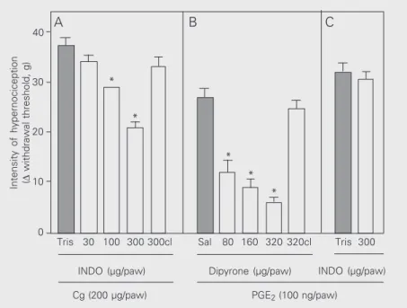
Subcutaneous administration of indo-methacin (30, 100 and 300 µg/paw) partially blocked the hypernociception induced by intraplantar injection of carrageenin (200 µg)
in a dose-dependent manner (Figure 4, panel A). The ineffectiveness of indomethacin at its maximum dose (300 µg) on PGE2
-in-duced hypernociception (100 ng; Figure 4, panel C) was detected by the electronic pressure-meter. On the other hand, the ef-fectiveness of dipyrone (80, 160 and 320 mg/paw) in blocking PGE2-induced
hyper-nociception was detected in a dose-depend-ent manner. In the contralateral paw, the maximum dose of indomethacin (300 µg/ paw) or dipyrone (320 µg) had no effect, excluding a systemic effect (Figure 4, panels A and B, respectively).
Discussion
In the present study, we have used hyper-nociception (increased hyper-nociception) to de-scribe the behavioral response induced by the application of the von Frey filaments test, the electronic pressure-meter test and the constant pressure test. The terms allodynia and hyperalgesia describe distinct nocicep-tive symptoms in man (29,30). The me-chanical tests have been used to measure increased experimental nociceptor sensitiv-ity referred to either as allodynia or hyperal-gesia by different investigators. In fact, thus far there is no demonstration that these symptoms describe different second mes-senger events in the inflammatory response. The use of the terms hypersensitivity or hyperexcitability was also avoided because they have specific meaning in immunology and electrophysiology, respectively.
Our results showed the applicability of the electronic pressure-meter test to detect nociceptor hypernociception in rats when its measurements were compared with those obtained with von Frey filaments and with the constant pressure test (our modification of the Randall and Selitto test; Ref. 2). This commercial instrument (electronic von Frey anesthesiometer) is similar to that success-fully used to quantify neuropathic allodynia (8). One of the advantages of this electronic
Figure 4. Effect of indomethacin on carrageenin- (Cg) and prostaglanadin E2- (PGE2) induced
hypernociception and of dipyrone on PGE2-induced hypernociception in rats. In panels A
and B, animals were pretreated locally with indomethacin (INDO, 30, 100 and 300 µg in A
and 300 µg in C) or Tris-HCl buffer, pH 8.0 (Tris), 30 min before injection of the
hypernociceptive agent. In panel B, dipyrone (80, 160 and 320 µg) or saline (Sal) was injected subcutaneously into the rat paw 2 h after PGE2 administration. Dipyrone (320 µg)
was also injected into the contralateral (cl) paw to evaluate a possible systemic effect of the drug. Animals were tested 3 h after injection of the hypernociceptive agents. The results are reported as the mean ± SEM of 5-6 animals. *P < 0.05 compared to the respective control (one-way ANOVA followed by the Tukey test).
Intensity of hypernociception (∆
withdrawal threshold, g)
40
*
*
* *
*
A B C
Tris 30 100 300 300cl Sal 80 160 320 320cl
INDO (µg/paw) Dipyrone (µg/paw) INDO (µg/paw) Tris 300 30
20
10
0
method over the classical von Frey filaments lies in a decrease in the number of attempts required to evaluate the nociceptive thresh-old and in the elimination of problems con-cerning the standardization of the filaments (6).
The PGE2-induced hypernociception
could be detected by the electronic pressure-meter test, von Frey filaments test and the rat paw constant pressure test. However, the von Frey filaments test did not reveal differ-ences in time vs treatment interaction, which were detected by the electronic pressure-meter test and the rat paw constant pressure test. Also, the last two methods distinguished the influence of different doses on the PGE2
-induced hypernociception, but the electronic pressure-meter was more sensitive than the rat paw constant pressure test at early times, whereas the rat paw constant pressure test was more sensitive at later times.
When hypernociception was induced by carrageenin it could be detected by the elec-tronic pressure-meter test and the rat paw constant pressure test. Moreover, both meth-ods detected time vs treatment interactions in a dose- and time-dependent manner, and again, the electronic pressure-meter test was more sensitive than the rat paw constant pressure test at early times, whereas the rat paw constant pressure test was more sensi-tive at later times in quantifying hypernoci-ceptive inflammatory stimuli.
Although the rat paw constant pressure test seems more discriminative in detecting differences in the effects of PGE2 or
carra-geenin at later times, which are near the hypernociceptive peak, this method has a much more subjective end-point which may limit its usefulness. On this basis, it would be preferable to apply a less subjective end-point method such as the electronic pres-sure-meter test. These apparent discrepan-cies might indicate that the different tests detect the hypernociception of different sets of primary sensory neurons, which have a different time course of initiation and
dura-tion of hypernocicepdura-tion. In fact, using the electronic pressure-meter test, we observed that sensitization of the skin in the plantar region of the rat paw differs temporally and biochemically from that of the profound intraplantar tissues (31).
automatically recorded (7,38). It is reason-able to assume that the electronic pressure-meter test has methodological characteris-tics similar to those of the other mechanical nociceptive tests, whose predictivity for de-velopment of nonsteroidal anti-inflammatory drugs should be better than that of chemical methods (28). The electronic pressure-meter test also has advantages over other mechani-cal tests, such as the Randall and Selitto and rat paw constant pressure tests, since it is not necessary to restrain the animals, avoiding the stress component. Furthermore, depend-ing on the number of attempts required for an experiment, the mechanical stimulus applied by the Randall and Selitto method may be harmful to the animal inducing edema per se, a fact not observed with the electronic
pres-sure-meter test.
In conclusion, we described the elec-tronic pressure-meter test, which is a useful tool to characterize new nociceptive media-tors and also to evaluate new classes of peripheral analgesics that are COX inhibitors or directly block ongoing nociceptor hypernociception.
Acknowledgments
The authors wish to express their appre-ciation to Sérgio Roberto Rosa for excellent technical support. The authors also want to express gratitude to Prof. Francisco Silveira Guimarães for useful advice about statistical analysis and to Luiz Fernando Ferrari for help with the photos.
References
1. von Frey M (1896). Untersuchunger über die Sinnesfunctionen der menschlichen Haut. Bandes der Abhandlungen der mathematisch-physischen Classe der Königl. Sächsischen Gesellschaft der Wissenschaften, 23: 175-266.
2. Ferreira SH, Lorenzetti BB & Correa FMA (1978). Central and peripheral antialgesic action of aspirin-like drugs. European Journal of Pharmacology, 53: 39-48.
3. Kim S & Chung J (1992). An experimental model for peripheral neuropathy produced by segmental spinal nerve ligation in the rat.
Pain, 50: 355-363.
4. Omote K, Kawamata T, Nakayama Y, Kawamata M, Hazama K & Namik IA (2001). The effects of peripheral administration of a novel selective antagonist for prostaglandin E receptor subtype EP(1), ONO-8711, in a rat model of postoperative pain. Anesthesia and Analgesia, 92: 233-238.
5. Sousa AM & Prado WA (2001). The dual effect of a nitric oxide donor in nociception. Brain Research, 897: 9-19.
6. Cunha TM, Verri Jr WA, Vivancos GG, Moreira IF, Reis S, Cunha FQ & Ferreira SH (2004). An electronic pressure-meter nociception paw test for mice. Brazilian Journal of Medical and Biological Research, 37: 401-407.
7. Jensen K, Andsersen HO, Olesen J & Lindblom U (1986). Pres-sure-pain threshold in human temporal region. Evaluation of a new pressure algometer. Pain, 25: 313-323.
8. Möller KÄ, Johansson B & Berg OG (1998). Assessing mechanical allodynia in the rat paw with a new electronic algometer. Journal of Neuroscience Methods, 84: 41-47.
9. Randall L & Selitto JJ (1957). A method for measurement of analge-sic activity on inflamed tissues. Archives Internationales de Pharma-codynamie et de Thérapie, 111: 409-419.
10. Vinegar R, Truax JF & Selph JL (1976). Quantitative comparison of the analgesic and anti-inflammatory activities of aspirin, phenacetin
and acetaminophen in rodents. European Journal of Pharmacolo-gy, 37: 23-30.
11. Granados-Soto V, Alonso-Lopez R, Asomoza-Espinosa R, Rufino MO, Gomes-Lopes LD & Ferreira SH (2001). Participation of COX, IL-1 beta and TNF alpha in formalin-induced inflammatory pain.
Proceedings of the Western Pharmacology Society, 44: 15-17. 12. Duarte IDG, Nakamura M & Ferreira SH (1988). Participation of the
sympathetic system in acetic acid-induced writhing in mice. Brazil-ian Journal of Medical and Biological Research, 21: 341-343. 13. Miranda HF, Lopez J, Sierralta F, Correa A & Pinardi G (2001). NSAID
antinociception measured in a chemical and a thermal assay in mice.
Pain Research & Management, 6: 190-196.
14. Aley KO, Green PG & Levine JD (1995). Opioid and adenosine peripheral antinociception are subject to tolerance and withdrawal.
Journal of Neurosciences,15: 8031-8038.
15. Ferreira SH & Nakamura M (1979). I - Prostaglandin hyperalgesia: a cAMP/Ca2+ dependent process. Prostaglandins, 18: 179-190.
16. Ferreira SH, Duarte ID & Lorenzetti BB (1991). Molecular base of acetylcholine and morphine analgesia. Agents and Actions Supple-ments, 32: 101-106.
17. Nakamura M & Ferreira SH (1987). A peripheral sympathetic compo-nent in inflammatory hyperalgesia. European Journal of Pharmacolo-gy, 135: 145-153.
18. Ferreira SH, Lorenzetti BB, Bristow AF & Poole S (1988). Interleukin-1ß as a potent hyperalgesic agent antagonized by a tripeptide analogue. Nature, 334: 689-700.
19. Cunha FQ, Lorenzetti BB, Poole S & Ferreira SH (1991). Interleukin-8 as a mediator of sympathetic pain. British Journal of Pharmacolo-gy,104: 765-767.
21. Cunha JM, Cunha FQ, Poole S & Ferreira SH (2000). Cytokine-mediated inflammatory hyperalgesia limited by interleukin-1 recep-tor antagonist. British Journal of Pharmacology, 130: 1418-1424. 22. Ferreira SH, Lorenzetti BB, Cunha FQ & Poole S (1993). Bradykinin
release of TNF-α plays a key role in the development of inflamma-tory hyperalgesia. Agents and Actions, 38: C7-C9.
23. Ferreira SH, Lorenzetti BB & Poole S (1993). Bradykinin initiates cytokine-mediated inflammatory hyperalgesia. British Journal of Pharmacology, 110: 1227-1231.
24. Lorenzetti BB, Veiga FH, Canetti CA, Poole S, Cunha FQ & Ferreira SH (2002). Cytokine-induced neutrophil chemoattractant 1 (CINC-1) mediates the sympathetic component of inflammatory mechanical hypersensitivity in rats. European Cytokine Network, 13: 456-461.
25. Ferreira SH, Lorenzetti BB & de Campos DI (1990). Induction, blockade and restoration of a persistent hypersensitive state. Pain, 42: 365-371.
26. Sachs D, Cunha FQ, Poole S & Ferreira SH (2002). Tumour necrosis factor-alpha, interleukin-1beta and interleukin-8 induce persistent mechanical nociceptor hypersensitivity. Pain, 96: 89-97.
27. Ferreira SH & Lorenzetti BB (1994). Glutamate spinal retrograde sensitization of primary sensory neurons associated with nocicep-tion. Neuropharmacology, 33: 1479-1485.
28. Le Bars D, Gozariu M & Cadden SW (2001). Animal models of nociception. Pharmacological Reviews, 53: 597-652.
29. Onttonen T & Pertovaara A (2000). The mechanical antihyperalgesic effect of intrathecally administered MPV-2426, a novel alpha2-adrenoceptor agonist, in a rat model of postoperative pain. Anesthe-siology, 92: 1740-1745.
30. Vrinten DH, Gispen, WH, Groen GJ & Adan RA (2000). Antagonism
of the melanocortin system reduces cold and mechanical allodynia in mononeurophatic rats. Journal of Neurosciences, 20: 8131-8137. 31. Vivancos GG, Parada CA & Ferreira SH (2003). Opposite nocicep-tive effects of the arginine/NO/cGMP pathway stimulation in dermal and subcutaneous tissues. British Journal of Pharmacology, 138: 1351-1357.
32. Vane JR (1971). Inhibition of prostaglandin synthesis as a mechan-ism of action for aspirin-like drugs. Nature New Biology, 231: 232-235.
33. Lorenzetti BB & Ferreira SH (1985). Mode of action of dipyrone: direct antagonism of inflammatory hyperalgesia. European Journal of Pharmacology, 114: 375-381.
34. Lorenzetti BB & Ferreira SH (1996). Activation of the arginine-nitric oxide pathway in primary sensory neurons contributes to dipyrone-induced spinal and peripheral analgesia. Inflammation Research, 45: 308-311.
35. Aguirre-Bañuelos P & Granados-Soto V (1999). Evidence for a pe-ripheral mechanism of action for the potentiation of the antinocicep-tive effect of morphine by dipyrone. Journal of Pharmacology and Toxicology, 42: 79-85.
36. Collier HO, Dinnenen LC, Johnson CA & Schneider C (1968). The abdominal constriction response and its suppression by analgesic drugs in the mouse. British Journal of Pharmacology, 32: 295-310. 37. Dubuisson D & Dennis SG (1977). The formalin test: a quantitative
study of the analgesic effects of morphine, meperidine, and brain stem stimulation in rats and cats. Pain, 4: 161-174.