An electronic pressure-meter
nociception paw test for mice
Departamento de Farmacologia, Faculdade de Medicina de Ribeirão Preto, Universidade de São Paulo, Ribeirão Preto, SP, Brasil
T.M. Cunha*, W.A. Verri Jr.*, G.G. Vivancos*, I.F. Moreira, S. Reis, C.A. Parada, F.Q. Cunha and S.H. Ferreira
Abstract
The aim of the present investigation was to describe and validate an electronic mechanical test for quantification of the intensity of inflammatory nociception in mice. The electronic pressure-meter test consists of inducing the animal hindpaw flexion reflex by poking the plantar region with a polypropylene pipette tip adapted to a hand-held force transducer. This method was compared to the classical von Frey filaments test in which pressure intensity is automatically recorded after the nociceptive hindpaw flexion reflex. The electronic pressure-meter and the von Frey filaments were used to detect time versus treatment interactions of carrageenin-induced hypernociception. In two separate experiments, the elec-tronic pressure-meter was more sensitive than the von Frey fila-ments for the detection of the increase in nociception (hypernoci-ception) induced by small doses of carrageenin (30 µg). The elec-tronic pressure-meter detected the antinociceptive effect of non-steroidal drugs in a dose-dependent manner. Indomethacin admin-istered intraperitoneally (1.8-15 mg/kg) or intraplantarly (30-300 µg/ paw) prevented the hypersensitive effect of carrageenin (100 µg/ paw). The electronic pressure-meter also detected the hypernoci-ceptive effect of prostaglandin E2 (PGE2; 10-100 ng) in a dose-dependent manner. The hypernociceptive effect of PGE2 (100 ng) was blocked by dipyrone (160 and 320 µg/paw) but not by intraplantar administration of indomethacin (300 µg/paw). The present results validate the use of the electronic pressure-meter as more sensitive than the von Frey filaments in mice. Furthermore, it is an objective and quantitative nociceptive test for the evaluation of the peripheral antinociceptive effect of anti-inflammatory anal-gesic drugs, which inhibit prostaglandin synthesis (indomethacin) or directly block the ongoing hypernociception (dipyrone). Correspondence
S.H. Ferreira
Departamento de Farmacologia FMRP, USP
Av. Bandeirantes, 3900 14049-900 Ribeirão Preto, SP Brasil
Fax: +55-16-633-0021 E-mail: [email protected]
Research supported by CNPq, CAPES, FAPESP and PRONEX.
*These authors contributed equally to this study.
Received June 2, 2003 Accepted December 8, 2003
Key words •Hyperalgesia
•Nociception
•Indomethacin
•Dipyrone
•von Frey filaments
Introduction
The type and site of application of a nociceptive stimulus, the sensitivity to anal-gesic drugs, as well as a typical innate behav-ioral response (end-point) characterize noci-ceptive tests. There is a growing consensus
are also used for the generation of transgenic and knockout animals for different molecules, including immune and inflammatory media-tors, and also for their specific receptors (1). The most popular test for the investigation of nociception in this animal species is the writhing test (2), quantified by the frequency of abdominal constrictions induced by in-flammatory stimuli (i.e., acetic acid and zy-mosan). The tail flick and the formalin paw tests have also been extensively used (2). Although the writhing test is quite sensitive for detection of analgesic compounds, its therapeutic predictivity is inferior to the clas-sical Randall and Selitto nociceptive test in rats (2). However, with the tests (except for the classical Randall and Selitto test), it is rather difficult to determine if an analgesic is causing antinociception by blocking the sen-sitization or by the activation of the nocicep-tors, phenomena that characterize inflam-matory pain, which has a quite different mechanism. Sensitization of the nociceptors (hypernociception) is a metabotropic event involving at least the stimulation of the cAMP/PKA/Ca2+ or the phospholipase/PKC/
Ca2+ pathways (3-8).
In recent years, the classical von Frey filaments test (9) has become popular among the mechanical tests used for rats and mice (10,11). This mechanical test has an advan-tage over the writhing test and formalin test in mice because it distinguishes between the two components of inflammatory pain, i.e., sensitization and activation of the nocicep-tor.
In the present study, we standardized and evaluated an electronic version of the von Frey filaments test for mice. An electronic pressure-meter test has been described for use in humans and rats (12,13). We induced an inflammatory process and sensitized the paws of mice with different doses of carra-geenin and prostaglandin E2 (PGE2),
respec-tively, to compare the sensitivity of the von Frey filaments and the electronic pressure-meter tests. We also used paws treated with
carrageenin or with PGE2 tocompare the
sensitivity of the tests in detecting the anal-gesic action of indomethacin, a cyclooxy-genase (COX) inhibitor or dipyrone, a di-rect-acting anti-hyperalgesic drug (14-16).
Material and Methods
Animals
The experiments were performed on 25-30-g Swiss mice (University of São Paulo, Ribeirão Preto, SP, Brazil) housed in the animal care facility of the School of Medi-cine of Ribeirão Preto and taken to the test-ing room at least 1 h before the experiment. Food and water were available ad libitum.
All behavioral testing was performed be-tween 9:00 am and 4:00 pm. The mice were used only once. Animal care and handling procedures were in accordance with the In-ternational Association for Study of Pain (IASP) guidelines on the use of animals in pain research. Efforts were made to mini-mize the number of animals used and their suffering.
von Frey filaments and electronic pressure-meter paw tests for mice
In a quiet room, mice were placed in acrylic cages (12 x 10 x 17 cm high) with a wire grid floor 15-30 min before testing. During this adaptation period, the paws were poked 2-3 times. Before paw stimulation, the animals were quiet, without exploratory movements or defecation and not resting on their paws. In these experiments, we used either a series of von Frey filaments (Stoelting, Chicago, IL, USA) with logarithmically incremental stiff-ness (-1.17 to 0.74 log of force, g) or a pressure-meter which consisted of a hand-held force transducer fitted with a 0.5 mm2
investiga-tor was trained to apply the filaments or the polypropylene tip perpendicularly to the central area of the hindpaw with a gradual increase in pressure. A tilted mirror below the grid provid-ed a clear view of the animal’s hindpaw. The test consisted of poking a hindpaw to provoke a flexion reflex followed by a clear flinch response after paw withdrawal. Each one of the von Frey filaments was applied for ap-proximately 3-4 s to induce the end-point reflex. Testing was initiated with the filament handle marked 4.31, a value corresponding to 0.31 log of force (g) which is in the middle of the filament series. The response to this fila-ment defined if a series of weaker or stronger filaments would be tested. The weakest fila-ment able to elicit a response was taken to be the mechanical threshold (g).
The results are reported as the ∆ log of force (g) calculated by subtracting the value of the measurements (log of force) after treatment from that of the first measurement for the von Frey test (before treatment). In the electronic pressure-meter test the inten-sity of the stimulus was automatically re-corded when the paw was withdrawn. The maximal force applied was 18 g. The stimu-lation of the paw was repeated until the animal presented two similar measurements. If the results were inconsistent (great differ-ence in the baseline response compared to the other animals of the experiment), another animal was used. The results are reported as the ∆ withdrawal threshold (g) which was calculated by subtracting the values obtained after the treatments from the first measure-ment (before treatmeasure-ment).
Drugs
Dipyrone and PGE2 were purchased from
Sigma (St. Louis, MO, USA). Carrageenin was obtained from FMC Corporation (Phila-delphia, PA, USA), and indomethacin from Prodome Química e Farmacêutica (São Paulo, SP, Brazil).
Carrageenin and dipyrone were diluted
in sterile saline. A stock solution of PGE2
was prepared in 10% ethanol, and further dilutions were made in saline; the final con-centration of ethanol was 1%. Indomethacin was diluted in 0.1 M Tris-HCl buffer, pH 8.0. Tris-HCl buffer alone was used for the control groups.
Drug administration
For local administration, drugs were in-jected subcutaneously into the plantar re-gion of the hindpaws. A hypodermic 26-G needle was inserted into the skin of the sec-ond footpad (to avoid back flow) and the tip of the needle was introduced until the central area of the hindpaw, in the same place where filaments or the tip of the pressure-meter were applied. A volume of 25 µl was admin-istered. For systemic treatment, drugs were injected intraperitoneally in a volume of 200 µl. Doses were calculated based on animal weight.
Statistical analysis
Two-way analysis of variance (ANOVA) was used to compare the groups and doses over all times. The factors analyzed were treatments, time and time vs treatment
inter-action. When a significant time vs treatment
interaction was detected, one-way ANOVA followed by the Tukey test was performed for each time in order to distinguish dose effects. One-way ANOVA followed by Tukey test was also used for dose-response curves for a single time point. Results with P < 0.05 were considered to be significant.
Results
Comparison of the sensitivity for the
detection of carrageenin-induced mechanical hypernociception by the electronic pressure-meter and the von Frey filaments tests
carrageenin-induced hypernociception was detected by both the electronic pressure-meter (panel A; 10, 30, 100 and 300 µg/paw) and the classi-cal von Frey filaments (panel B; 30, 100 and 300 µg/paw) tests. There was a significant increase in paw hypernociception with a time versus treatment interaction (ANOVA). The hypernociception induced by a low dose of carrageenin (30 µg/paw) was detected by
the electronic pressure-meter but not by the von Frey filaments as a significant effect.
Detection of the local and systemic effects of indomethacin on the carrageenin-induced hypernociception by the electronic pressure-meter test
The systemic (panel A) and local (panel
Intensity of hypernociception (
∆
log force, g)
2
1
0 Intensity of hypernociception (∆
withdrawal threshold, g)
12
9
6
3
0
-3
0 0.5 1 3 5 0 0.5 1 3 5
Time after injection (h)
Saline 10 µg Cg 30 µg Cg 100 µg Cg 300 µg Cg
ns
ns
ns
* *
* * * *
*
* *
*
*
ns
ns ns
A B
Figure 1. Time course of the hypernociception induced by intraplantar injections of carrag-eenin (Cg) in mice detected with the electronic pressure-meter (A) and von Frey filaments (B). Saline was injected intra-plantarly in the control group. The results are reported as the mean ± SEM for 4-5 animals per group and are representa-tive of two separate experi-ments. *P < 0.05 indicates a significant difference between time-points indicated by the brackets. ns = nonsignificant (two-way ANOVA and one-way ANOVA followed by the Tukey test).
Intensity of hypernociception (
∆
withdrawal threshold, g)
8
6
4
2
0
Tris 1.8 5 15 Tris 30 100 100 cl 300 300 cl
Indomethacin (mg/kg) Indomethacin (µg/paw)
Carrageenin (100 µg/paw)
*
*
*
* *
*
Figure 2. Effect of indomethacin on carrageenin-induced hyper-nociception in mice measured with the electronic pressure-meter. Animals were pretreated with indomethacin 30 min be-fore intraplantar injection of car-rageenin (100 µg). Indomethacin was injected intraperitoneally (A, systemic, 1.8, 5 and 15 µg/kg) or subcutaneously into the mouse paw (B, local, 30, 100 and 300 µg/paw or contralateral (cl) paw, 100 and 300 µg/paw). Tris-HCl buffer (Tris) was injected in the control groups. The bars indicate the withdrawal threshold 3 h af-ter carrageenin administration. The results are reported as the mean ± SEM for 5 animals per group and are representative of two separate experiments. *P < 0.05 compared to control (Tris) (one-way ANOVA followed by the Tukey test).
Systemic Local
B) inhibitory effects of indomethacin on the hypernociception induced by carrageenin (100 µg) is shown in Figure 2. Both systemic (1.8, 5 and 15 mg/kg, intraperitoneally) and local (30, 100 and 300 µg/paw) administra-tion of indomethacin produced a dose-de-pendent (ANOVA) blockade of the hyper-nociceptive state induced by carrageenin (100 µg/paw). Although the local administration of indomethacin at a dose of 300 µg/paw had a systemic effect on carrageenin-induced hypernociception, the dose of 100 µg/paw had only a local effect since it was not able to inhibit the carrageenin-induced hypernoci-ception in the contralateral paw.
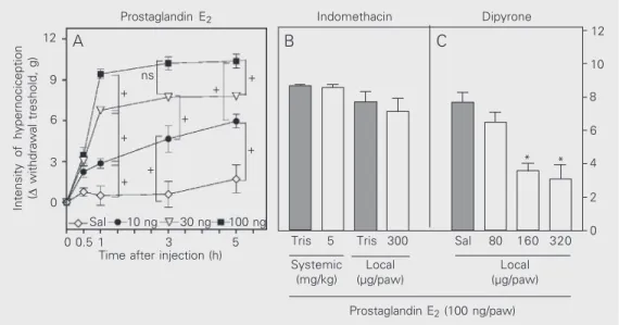
Dose-response curve for the hypernociception induced by intraplantar injection of PGE2 in
mice and its blockade by dipyrone but not by indomethacin
The electronic pressure-meter was able to detect the statistically significant intra-plantar effect of the three doses of PGE2 (10,
30 and 100 ng; Figure 3, ANOVA). Paw hypernociception tended to increase with time (particularly with small doses) and there was a significant interaction between time and the different treatments (Figure 3A, ANOVA). Dipyrone (80, 160 and 320 µg; Figure 3C), but not indomethacin (Figure 3B), inhibited the PGE2-induced (100 ng)
hypernociception in a dose-dependent man-ner (ANOVA).
Discussion
In the present study, we have used the word hypernociception (increased nocicep-tion) to describe the behavioral response induced by the application of von Frey fila-ments or the electronic pressure-meter. The terms allodynia and hyperalgesia describe distinct nociceptive symptoms in man (18,19). von Frey filaments have been used to measure increased experimental nocicep-tor sensitivity referred to as allodynia or hyperalgesia by different investigators. In
12
Intensity of hypernociception (∆
withdrawal treshold, g)
12
10
8
6
4
2
0 9
6
3
0
0 0.5 1 3 5 Time after injection (h) Sal 10 ng 30 ng 100 ng
Tris 5 Tris 300 Sal 80 160 320
Systemic (mg/kg)
Local (µg/paw)
Local (µg/paw)
Prostaglandin E2 (100 ng/paw)
A B C
Figure 3. Dose-response curve for the hypernociception induced by intraplantar injection of prostaglandin E2 (PGE2) in mice and its blockade by dipyrone but not by indomethacin measured with the electronic pressure-meter. Panel A shows the time course of the PGE2-induced hypernociception at the doses of 10, 30 and 100 ng/ paw. In Panel B, dipyrone (80, 160 and 320 µg/paw) or saline (Sal) was injected 2 h after PGE2 (100 ng). In Panel C, pretreatment with indomethacin (300 µg) or Tris-HCl buffer (Tris) was performed 30 min before injection of the hypersensitizing agent. Measurements were made 3 h after PGE2 in A and B. The results are reported as the mean ± SEM for 4-5 animals per group and are representative of two separate experiments. +P < 0.05 indicates a significant difference between points (A, two-way ANOVA and one-way ANOVA followed by the Tukey test); *P < 0.05 compared to control (Sal or Tris; B and C, one-way ANOVA followed by the Tukey test).
Prostaglandin E2 Indomethacin Dipyrone
ns
+ +
+
+ +
+
+ +
fact, so far there is no demonstration that these symptoms describe different second messenger events in the inflammatory re-sponse. The use of the terms hypersensitivity or hyperexcitability was also avoided be-cause they have specific meanings in immu-nology and electrophysiology, respectively. We evaluated the efficacy of the elec-tronic pressure-meter to detect nociceptor hypernociception in mice. This commer-cially available instrument (electronic von Frey) is similar to that successfully used to quantify neuropathic allodynia (13) and in-flammatory hypernociception in rats (17). The electronic method has several advan-tages over the classical von Frey filaments: a) reduction of the number of attempts required to evaluate the nociceptive threshold; b) elimination of the problems of filament stan-dardization; c) stimulation of areas of equal size (the area varies with the diameter of the von Frey filaments), d) the end-point is automatically recorded (12,20).
Carrageenin-induced hypernociception could be detected in the inflamed paws of the mice by poking the paws with the von Frey filaments and the electronic pressure-meter. The electronic pressure-meter detected hy-pernociception as early as 1 h after the injection of carrageenin at the lower doses, which was not detected by the von Frey filaments.
The usefulness of the electronic pres-sure-meter for the study of analgesics in mice was demonstrated with indomethacin and dipyrone. Indomethacin is a COX inhib-itor and dipyrone has specific antinocicep-tive effects on PGE2-induced
hypernocicep-tion, which are not shared by COX inhibi-tors. The activation of the arginine-NO-cGMP pathway contributes to dipyrone-in-duced spinal and peripheral analgesia (14) via opening of K+ ATP-sensitive channels (15).
The electronic pressure-meter detected the systemic and local antinociceptive effects of a standard COX inhibitor, indomethacin, in a dose-dependent manner. The electronic test differentiated among increasing doses of PGE2 although no time discrimination
be-tween 1 and 5 h after administration was observed. In general, hypernociception tended to be more intense at later times. Although indomethacin (as expected) showed no local effect on PGE2-induced hypernociception,
dipyrone showed a dose-dependent antinoci-ceptive effect. Thus, this technique is ca-pable of differentiating between drugs that prevent the development of tion (blockade of carrageenin hypernocicep-tion) and those which directly antagonize ongoing hypernociception, i.e., dipyrone.
A clear dissociation in time between the hypernociceptive and the nociceptive stimuli, an easily defined and recorded end-point induced by the nociceptive stimulus, and the ability to differentiate between different classes of peripheral analgesics are essential characteristics of a useful test for the devel-opment of new analgesics as well as for the investigation of inflammatory pain mechan-isms. In the present investigation, we showed that the electronic pressure-meter test in mice fulfills all of these basic conditions.
Acknowledgments
Referentes
1. Mogil JS & Grisel JE (1998). Transgenic studies of pain. Pain, 77:
107-128.
2. Le Bars D, Gozariu M & Cadden SW (2001). Animal models of nociception. Pharmacological Reviews, 53: 597-652.
3. Taiwo YO & Levine JD (1989). Contribution of guanine nucleotide regulatory proteins to prostaglandin hyperalgesia in the rat. Brain Research, 492: 400-403.
4. Cunha FQ, Teixeira MM & Ferreira SH (1999). Pharmacological modulation of secondary mediator systems - cyclic AMP and cyclic GMP - on inflammatory hyperalgesia. British Journal of Pharmacol-ogy, 127: 671-678.
5. Ferreira SH & Nakamura M (1979). I-Prostaglandin hyperalgesia: a cAMP/Ca2+ dependent process. Prostaglandin, 18: 179-190. 6. Lynn B & O’Shea NR (1998). Inhibition of forskolin-induced
sensiti-zation of frog skin nociceptors by the cyclic AMP-dependent pro-tein kinase A antagonist H-29. Brain Research, 780: 320-362. 7. Taniguchi K, Shinjo K, Mizutani M, Shimada K, Ishikawa T, Menniti
FS & Nagahisa A (1997). Anti-nociceptive activity of CP-101,606, NMDA receptor NR2B subunit antagonist. British Journal of Phar-macology, 122: 809-812.
8. Coderre TJ (1992). Contribution of protein kinase C to central sensi-tisation and persistent pain following tissue injury. Neuroscience Letters, 140: 181-184.
9. von Frey M (1896). Untersuchunger über die Sinnesfunctionen der menschlichen Haut. Bandes der Abhandlungen der mathematisch-physischen Classe der Königl. Sächsischen Gesellschaft der Wissenschaften, 23: 175-266.
10. Prado WA, Schiavon VF & Cunha FQ (2002). Dual effect of local application of nitric oxide donors in a model of incision pain in rats.
European Journal of Pharmacology, 441: 57-65.
11. Shimoyama M, Tanaka K, Hasue F & Shimoyama N (2002). A mouse
model of neuropathic cancer pain. Pain, 99: 167-174.
12. Jensen K, Andsersen HO, Olesen J & Lindblom U (1986). Pres-sure-pain threshold in human temporal region. Evaluation of a new pressure algometer. Pain, 25: 313-323.
13. Möller KÄ, Johansson B & Berg OG (1998). Assessing mechanical allodynia in the rat paw with a new electronic algometer. Journal of Neuroscience Methods, 84: 41-47.
14. Lorenzetti BB & Ferreira SH (1996). Activation of the arginine-nitric oxide pathway in primary sensory neurons contributes to dypirone-induced spinal and peripheral analgesia. Inflammation Research, 45: 308-311.
15. Alves D & Duarte I (2002). Involvement of ATP-sensitive K(+) chan-nels in the peripheral antinociceptive effect induced by dipyrone.
European Journal of Pharmacology, 24: 47-52.
16. Ferreira SH, Moncada S & Vane JR (1973). Prostaglandins and the mechanism of analgesia produced by aspirin-like drugs. British Jour-nal of Pharmacology, 49: 86-97.
17. Vivancos GG, Verri Jr WA, Cunha TM, Schivo IRS, Parada CA, Cunha FQ & Ferreira SH (2004). An electronic pressure-meter nociception paw test for rats. Brazilian Journal of Medical and Biological Re-search, 37: 391-399.
18. Onttonen T & Pertovaara A (2000). The mechanical antihyperalgesic effect of intrathecally administered MPV-2426, a novel alpha2-adrenoceptor agonist, in a rat model of postoperative pain. Anesthe-siology, 92: 1740-1745.
19. Vrinten DH, Gispen WH, Groen GJ & Adan RA (2000). Antagonism of the melanocortin system reduces cold and mechanical allodynia in mononeuropathic rats. Journal of Neuroscience, 20: 8131-8137.