○ ○ ○ ○ ○ ○ ○ ○ ○ ○ ○ ○ABSTRACT○ ○ ○ ○ ○ ○
Case Repor
t
○ ○ ○ ○ ○ ○ ○ ○ ○ ○ ○ ○ ○ ○ ○ ○ ○ ○ ○ ○ Introduction
Most duodenal diverticula are asympto-matic structures. About 75% of duodenal di-verticula are located in the second portion of the duodenum. They can be periampullary, when they originate within a range of 2 to 3 cm from Vater’s ampulla,1 or ampullary when the papilla ends at the bottom of the diver-ticulum.
The duodenal diverticula rarely produce signs of inflammation, obstruction, hemor-rhage or perforation. In some cases secondary biliary-pancreatic complications are found when a diverticulum originates from the re-gion of Vater’s papilla. We here describe an ampullary duodenal diverticulum case asso-ciated with cholangitis.
○ ○ ○ ○ ○ ○ ○ ○ ○ ○ ○ ○ ○ ○ ○ ○ ○ ○ ○ ○ CASE REPORT
The patient, a 74-year-old white woman, was admitted to the Taubaté University Hos-pital having had pain in the right upper quad-rant for 2 days, and jaundice and fever with chills (Charcot’s triad). She referred to similar episodes in the past. In giving her history, she revealed that she had been hypertensive for 35 years and was submitted to cholecystec-tomy 30 years earlier. She had had diabetes over the last 2 years and was taking captopril and insulin daily.
Biochemical evaluations showed: hematocrit 35%, hemoglobin concentration 10.9 g/dl, leukocyte count 30.0 x 10³/ml and total bilirubin 3.75 mg/dl, with conjugated bilirubin 3.66 and unconjugated bilirubin 0.9. Serum amylase was 1000 U/l, aspartate
transaminase 90 U/l, alkaline phosphatase 818 U/l, gamma-glutamyl transferase 1306 U/l and glycemia 113 mg/dl.
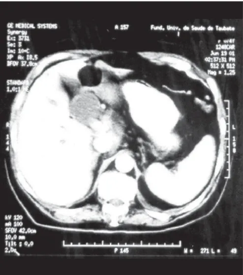
Abdomen ultrasonography was inconclu-sive. Computed tomography showed that the intra and extra-hepatic bile ducts were very much dilated and the pancreas head had in-creased volume and was heterogeneous (Fig-ure 1). Endoscopic retrograde cholangiopan-creatography revealed a patent duodenal pa-pilla opening at the bottom of a diverticulum, a common bile duct with an internal diam-eter of 3.5 cm and the absence of gallstones (Figure 2).
While these examinations were being car-ried out, the interned patient underwent clini-cal treatment with hypotensor drugs, insulin, parenteral hydration, third generation cepha-losporin (ceftriaxone) and symptomatic medi-cine. The treatment accomplished provided suggestive regression of the acute-phase symp-toms of the disease and consequent decrease in the surgical risk.
The surgery consisted of supra-umbilical median laparotomy to remove the previous scar. The liver was found to have normal size and a smooth surface. Sectioning of the com-mon bile duct was performed at the point of its largest diameter, with distal stump suture and choledochojejunal anastomosis using a Roux-en-Y loop. Choledochostomy was done to evaluate the intraductal pressure, and sub-sequent cholangiography through a T tube.
The postoperative period elapsed without incidents or complications. On the 8th post-operative day, cholangiography through the T tube revealed that the bile ducts were di-lated and tortuous, and the choledochojejunal
• Joaquim Mendes Castilho Netto
• Manlio Basilio Speranzini
Ampullary duodenal
diverticulum and cholangitis
Surgical Clinical Service, Hospital Universitário, Department of Medical
Sciences, Universidade de Taubaté, Taubaté, São Paulo, Brazil
CONTEXT: Ampullary duodenal diverticulum compli-cated by cholangitis is little known in clinical prac-tice, especially when there are no gallstones in the common bile duct or there is no biliary tree ectasia or hyperamylasemia. A case of this asso-ciation is presented, in which the surgical treat-ment was a biliary-enteric bypass.
CASE REPORT: A 74-year-old diabetic white woman was admitted to the Taubaté University Hospital, complaining of pain in the right upper quadrant, jaundice and fever with chills (Charcot’s triad). She had had cholecystectomy 30 years earlier. She underwent clinical treatment with parenteral hydration, insulin, antibiotics and symptomatic drugs. Imaging examinations were provided for diagnosis: ultrasound, computed tomography and endoscopic retrograde cholangiopancreato-graphy. The surgical treatment consisted of chole-dochojejunostomy utilizing a Roux-en-y loop. The postoperative period progressed without incidents, and a DISIDA scan demonstrated the presence of dynamic biliary excretion. The patient remained asymptomatic when seen at outpatient follow-up.
KEY WORDS: Duodenal diverticulum. Cholangitis. Choledochojejunostomy.
São Paulo Medical Journal — Revista Paulista de Medicina
174
anastomosis was pervious with Roux limb opacification. The patient was discharged one day after this. The drainage tube was removed on the 21st day after the operation. At a fol-low-up 13 months later, the patient under-went radioisotope scanning using Technetium-99m (diisopropylphenyl carboxymethyl iminodiacetic acid, or DISIDA), which showed good excretion into the Roux limb (Figure 3). Postoperative follow-up over ap-proximately two years was uneventful and she continued to present a satisfactory condition.
○ ○ ○ ○ ○ ○ ○ ○ ○ ○ ○ ○ ○ ○ ○ ○ ○ ○ ○ ○ DISCUSSION
The ampullary duodenal diverticulum is located at the choledochoduodenal junction and the periampullary diverticulum is at its side. Both types of diverticula are included in the list of possible etiological factors for acute pan-creatitis.2 According to Naranjo-Chavez et al.,3 the ampullary diverticulum is the possible cause of chronic pancreatitis, which would not oc-cur with a periampullary diverticulum. In our report, there was probable pancreatic disease characterized by hyperamylasemia, and the pancreatic head was enlarged with a heteroge-neous aspect. The studies of Kim et al.4 showed that the association between diverticula and gallstones was significant in patients with pri-mary gallstones but not in those with second-ary gallstones. The pathogenesis for the primsecond-ary stones was associated with the presence of bile stasis and bacterial infection. In our patient, endoscopic retrograde cholangiopancrea-tography showed ampullary diverticulum, pro-nounced dilation of the common bile duct, bile stasis and absence of gallstones.
Periampullary diverticula, and particu-larly the ampullary diverticula, hamper cath-eterization and bile duct examinations.5 Fur-thermore, giant duodenal diverticula are re-sponsible for the false-positive findings from magnetic resonance imaging, endoscopic ret-rograde cholangiopancreatography and hepatobiliary scintigraphy.6 In the case we re-port, the papilla was in the diverticulum and catheterization of the common bile duct was performed with difficulty, without direct visualization of the papilla. Oddi’s sphincter insufficiency, as verified by endoscopic ret-rograde cholangiopancreatography, has been reported in the presence of both ampullary and periampullary diverticula.7
The cholangitis manifested by our patient can be explained by the increasing bile con-tamination from bacterial proliferation that takes place in the presence of duodenal diver-ticula.8
Figure 3. DISIDA scan after biliary-enteric bypass, showing the presence of dynamic biliary excretion.
Figure 1. Computed tomography scan of the patient, showing common bile duct that is very much dilated.
Figure 2. Duodenal diverticulum and absence of gallstones inside the dilated common bile duct, demonstrated by endoscopic retrograde cholangiography.
São Paulo Medical Journal — Revista Paulista de Medicina
175
Provided that the finding of a diverticu-lum is only incidental, without signs of in-flammation, perforation, hemorrhage or ob-struction, no treatment is needed. In cases of biliary-pancreatic disease secondary to a di-verticulum, the initial resolution is by means of endoscopic papillotomy, which relieves the jaundice and cholangitis. This is, however, not always definitive due to recurrence over the long term. Surgical removal of the diver-ticulum, in association with papillosphincte-roplasty is the best treatment because it is a
simple procedure and avoids recurrence of symptoms in the biliary system.9
When diverticulectomy is technically dif-ficult or considered to have major risk in the presence of a dilated bile duct, the best proce-dure is choledochojejunal anastomosis using a Roux-en-Y loop. For patients with little dila-tion of the common bile duct or submitted to Billroth II gastrectomy, choledochoduodenal anastomosis may be the procedure of choice.
In the present report, the common bile duct was fully dilated and there were
manipu-lation risks regarding the biliary-pancreatic confluence. Thus, the most appropriate pro-cedure for the patient was to effect total drain-age from the common bile duct into the Roux limb of the jejunum, thereby separating the associated link between the diverticulum and cholangitis.
The good postoperative evolution, the DISIDA scan showing good excretion into the Roux limb, and the follow-up with absence of any digestive symptoms, demonstrate the success of the operation.
1. Lobo DN, Balfour TW, Iftikhar SY, Rowlands, BJ. Periampullary
diverticula and pancreaticobiliary disease. Br J Surg 1999;86(5):588-97.
2. Uomo G, Manes G, Raggozino A, Cavallera A, Rabitti, PG.
Periampullary extraluminal duodenal diverticula and acute pan-creatitis: an underestimated etiological association. Am J Gastroenterol 1996;91(6):1186-8.
3. Naranjo-Chavez J, Schwarz M, Leder G, Beger HG. Ampullary
but not periampullary duodenal diverticula are an etiologic fac-tor for chronic pancreatitis. Dig Surg 2000;17(4):358-63.
4. Kim MH, Myung SJ, Seo DW, et al. Association of
○ ○ ○ ○ ○ ○ ○ ○ ○ ○ ○ ○ ○ ○ ○ ○ ○ ○ ○ ○ ○ ○ ○ ○ ○ ○ ○ ○ ○ ○ ○ ○ ○ ○ ○ ○ ○ ○ ○ ○ ○ ○ ○ ○ ○ ○ ○ ○ ○ ○ ○ ○ ○ ○ ○ ○ ○ ○ ○ ○ ○ ○ ○ ○ REFERENCES
periampullary diverticula with primary choledocholithiasis but not with secondary choledocholithiasis. Endoscopy 1998;30(7):601-4.
5. Fogel EL, Sherman S, Lehman GA. Increased selective biliary
cannulation rates in the setting of periampullary diverticula: main pancreatic duct stent placement followed by pre-cut bil-iary sphincterotomy. Gastrointest Endosc 1998:47(5):396-400.
6. Gupta S, Rajagopal S, Chander R, Sawroop K, Bhatnagar A.
Gi-ant duodenal diverticulum: a cause of false-positive findings of magnetic resonance imaging, cholangiopancreatography and hepatobiliary scintigraphy. Clin Nucl Med 2000;25(12):1037-8.
7. Lotveit T, Osnes M, Aune S, Larsen S. Studies of the
choledocho-duodenal sphincter in patients with and without juxta-papil-lary duodenal diverticula. Scand J Gastroenterol 1980;15(7):875-80.
8. Skar V, Skar AG, Midtvedt T, Osnes M. Bacterial growth in the
duodenum and in the bile of patients with gallstone disease treated with endoscopic papillotomy (EPT). Endoscopy 1986;18(1):10-3.
9. Pinotti HW, Tacla M, Pontes JF, Bettarello A. Surgical
proce-dures upon juxta-ampullary duodenal diverticula. Surg Gynecol Obstet 1972;135(1):11-6.
Divertículo duodenal ampolar e colangite
CONTEXTO: Divertículo duodenal ampolar complicado com colangite é pouco comum na prática clínica, especialmente se há ausência de cálculos no colédoco, ectasia da árvore biliar e hiperamilasemia. Um caso desta associação é apresentado. O tratamento foi cirúrgico por meio de derivação biliodigestiva.
RELATO DE CASO: Mulher branca de 74 anos, diabética, colecistectomizada há 30 anos, foi internada no Hospital Universitário de Taubaté acometida de dor no hipocôndrio direito, icterícia e febre com calafrios (tríade de Charcot). Submeteu-se a tratamento
clí-○ ○ ○ ○ ○ ○ ○ ○ ○ ○ ○ ○ ○ ○ ○ ○ ○ ○ ○ ○ ○ ○ ○ ○ ○ ○ ○ ○ ○ ○ ○ ○ ○ ○ ○ ○ ○ ○ ○ ○ ○ ○ RESUMO
Joaquim Mendes Castilho Netto, MD. Titular collabo-rating professor, Discipline of General Surgery, Department of Medical Sciences, Universidade de Taubaté, Taubaté, São Paulo, Brazil.
Manlio Basilio Speranzini, MD. Titular collaborating professor, Discipline of General Surgery, Department of Medical Sciences, Universidade de Taubaté, Taubaté, São Paulo, Brazil.
Sources of funding: Not declared Conflict of interest: Not declared
Date of first submission: September 30, 2002 Last received: March 17, 2003
Accepted: May 19, 2003 Address for correspondence Joaquim Mendes Castilho Netto
Avenida Tiradentes, 205 — Centro Taubaté/SP — Brasil — CEP 12030-180 Tel./Fax (+55 12) 232-6175 E-mail: [email protected]
COPYRIGHT © 2003, Associação Paulista de Medicina ○ ○ ○ ○ ○ ○ ○ ○ ○ ○ ○ ○ ○ ○ ○ ○ ○ ○ ○ ○
Publishing information
nico por meio de hidratação parenteral, in-sulina, antibióticos e medicamentos sintomá-ticos, enquanto a tomografia computado-rizada e a colangiopancreatografia retrógrada endoscópica eram providenciadas para escla-recimento diagnóstico. O tratamento cirúr-gico constou de derivação coledocojejunal com alça em Y de Roux. O pós-operatório decorreu sem incidentes; a cintilografia feita com tecnécio-99m (DISIDA) mostrou fácil excreção do contraste para o jejuno e a paci-ente permanece assintomática em seguimen-to ambulaseguimen-torial.
PALAVRAS-CHAVE: Divertículo duodenal. Colangite. Derivação biliodigestiva.