○ ○ ○ ○ ○ ○ ○ ○ ○ ○ ○ ○ ABSTRACT○ ○ ○ ○ ○ ○ ○ ○ ○ ○ ○ ○ ○ ○ ○ ○ ○ ○ ○ ○ ○ ○ ○ ○ ○ ○ ○
Introduction
In 1964, Pfeiffer described an acroce-phalosyndactyly syndrome consisting of bicoronal craniosynostosis, midface deficiency, broad thumbs, broad big toes and partial and variable soft-tissue syndactyly of the hands and feet.1 Autosomal dominant inheritance with complete penetrance is characteristic, despite variable expressivity related to the presence or absence of syndactyly and its degree of severity. Based on the severity of the phenotype, Cohen proposed the division of Pfeiffer syn-drome (Online Mendelian Inheritance in Man classification: OMIM 101600) into 3 clinical subtypes.2 Classic Pfeiffer syndrome, desig-nated Type 1, involves individuals with mild manifestations, associated with normal neu-rological and intellectual development, gen-erally has good outcome and can be found dominantly inherited. Type 2 consists of clo-verleaf skull, Pfeiffer hands and feet, severe exorbitism, central nervous system involve-ment, elbow ankylosis or synostosis. Type 3 is similar to type 2 but without the cloverleaf skull. Types 2 and 3 have poor prognosis due to severe neurological compromise and vari-ous visceral anomalies, and they generally re-sult in early death. To date, all cases of types 2 and 3 have only had sporadic occurrence.
We report one case of Pfeiffer syndrome type 2, with particular emphasis on the clini-cal presentation, differential diagnosis and the importance of early diagnosis.
○ ○ ○ ○ ○ ○ ○ ○ ○ ○ ○ ○ ○ ○ ○ ○ ○ ○ ○ ○
Case report
The patient in our case was a boy, born in
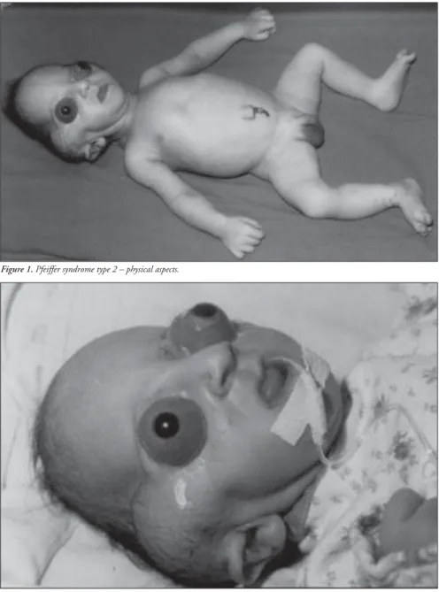
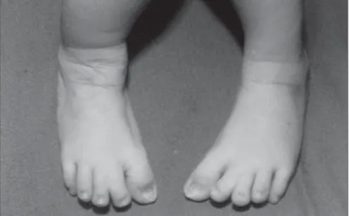
2001, as the third child of a healthy 34-year-old mother. The parents were not consanguin-eous, although the father was unknown. The mother had two other healthy sons from dif-ferent fathers. Labor began spontaneously and the infant was delivery vaginally at term with a birth weight of 2,975 g and Apgar score of 8/9. The propositus had a cloverleaf skull, se-vere exorbitism, choanal atresia, low-set and posteriorly-rotated ears, broad and medially-deviated halluces and partial cutaneous syn-dactyly of the second and third toes (Figures 1, 2 and 3). The ocular globes and eyelids were intact with shallow orbits that would have prevented the replacement of the eye. The ocular anterior structures were preserved, with-out iris or lens abnormalities. The baby de-veloped respiratory distress and died on the second day.
A computed tomography scan of the brain revealed a cloverleaf skull with cranio-encephalic asymmetry, bone deformities in the cranial base and orofacial aspect, asymmetric dilatation of the lateral ventricles, cerebral abnormalities and beaked nose (Figure 4). The orbital computed tomography showed a very shallow orbit with severe proptosis and pre-served optic nerve and extrinsic ocular mus-cles. Synostosis of the elbows was not detected on x-ray film.
○ ○ ○ ○ ○ ○ ○ ○ ○ ○ ○ ○ ○ ○ ○ ○ ○ ○ ○ ○
Discussion
Our patient had the clinical findings of Pffeifer Syndrome type 2 and despite this child’s bad outcome, a favorable prognosis can be achieved in some cases, especially with ag-gressive medical and surgical intervention.3 • Maria Kiyoko Oyamada
• Haideé Salgado Alonso Ferreira
• Marcelo Hoff
Pfeiffer syndrome
type 2 – case report
Hospital do Servidor Público Municipal de São Paulo, São Paulo, Brazil
OBJECTIVE: To report on a case of Pfeiffer Syndrome, with a discussion of the diagnostic characteristics and features of disease types and the differential diagnosis.
DESCRIPTION: The authors describe a newborn with cloverleaf skull, extreme bilateral exorbitism and choanal atresia, partial syndactyly of the second and third toes and broad medially-deviated big toes. The case reported was Pfeiffer Syndrome type 2, which usually has a poor prognosis.
COMMENTS: Pfeiffer Syndrome is a clinically vari-able disorder and consists of an autosomal domi-nantly-inherited osteochondrodysplasia with craniosynostosis. It has been divided into three types. Type 1 is commonly associated with nor-mal intelligence and generally good outcome. Types 2 and 3 generally have severe neurologi-cal compromise, poor prognosis, early death and sporadic occurrence. Potential for prolonged use-ful survival outcome can be achieved in some cases with early aggressive medical and surgical management according to recent literature.
KEY WORDS: Pfeiffer syndrome. Cloverleaf skull. Craniosynostosis. Syndactyly. Upper airway. Eye.
Case Repor
t
São Paulo Medical Journal — Revista Paulista de Medicina
177
For this, early clinical diagnosis is necessary. Besides the premature craniosynostosis of coronal sutures that gives the typical appear-ance of cloverleaf skull deformity, midfacial hypoplasia and hand and foot anomalies, ad-ditional anomalies may also include aque-ductal stenosis, hydrocephalus, cerebellar and brain stem herniation, low-set ears, external auditory canal stenosis or atresia and, unusu-ally, hydronephrosis, pelvic kidney and hypo-plastic gallbladder.2
The prognostic implications and genetic counseling required are distinct and depend on the subtype. Considerable clinical overlap may occur between the three subtypes. The absence of cloverleaf skull in type 3 can result in failure to diagnose the Pfeiffer Syndrome. Presence of classic Pfeiffer hands and feet in association with craniosynostosis are the ma-jor diagnostic clues in this type of syndrome. Although Pfeiffer Syndrome and Apert-type acrocephalosyndactyly (OMIM 101200) are noteworthy for some similarities, the two disorders appear to be nosologically and ge-netically distinct. Sometimes Pfeiffer Syn-drome has been confused with Saethre-Chotzen (OMIM 101400) and Jackson-Weiss syndromes (OMIM 123150), since broad toes may occur in both. The big toes of Saethre-Chotzen syndrome are more triangular, with a bulbous shape and in the valgus position. Broad big toes identical to those observed in Pfeiffer Syndrome may occur in some in-stances of Jackson-Weiss syndrome, although broad thumbs are never observed.2
Congenital airway anomalies clearly related to severity of midface hypoplasia have been re-ported in craniosynostosis syndromes, includ-ing Pfeiffer Syndrome types 1 and 2. Upper airway anomalies are an important source of morbidity and mortality in severe Pfeiffer Syn-drome, since chronic hypoventilation and hy-poxia are likely to be contributory factors in neurodevelopment deficits.
The trilobed skull deformity (cloverleaf skull) is a rare congenital anomaly that may be present as an isolated defect, but is usually part of an osteochondrodysplastic or dysostotic syndrome like Apert, Crouzon (OMIM 123500) or Pfeiffer syndrome. This anomaly has important prognostic implica-tions because of the limited brain growth and the eye exposure caused by shallow orbits. The pathogenesis of the cloverleaf skull is un-known, but the trilobed shape of the head, hydrocephalus and facial deformation have been attributed to intrauterine synostosis of cranial sutures.3 Abnormalities of the cranial base may have played a role in the respiratory
Figure 1. Pfeiffer syndrome type 2 – physical aspects.
Figure 2. Cloverleaf skull, exorbitism and low-set ears.
distress of the baby reported.
The ocular manifestations in Pfeiffer Syn-drome may be an inherent feature of the patho-logical process or may occur as a secondary complication. They can include hypertelorism, downslanting palpebral fissures, strabismus, anterior chamber dysgenesis including Peter’s anomaly (corneal clouding and variable iridolenticulocorneal adhesions), corneal scleralization, corectopia, atypical iris colo-bomata superiorly, retinal detachment and atrophic optic nerve heads. Disc edema, optic atrophy, and progressive optic nerve dysfunc-tion may accompany increased intracranial pressure even without evidence of hydrocepha-lus. Severe proptosis may cause corneal ulcer,
endophthalmitis and globe rupture.
Experience with corrective surgery is lim-ited. The aims of the surgery are: decompres-sion of the brain and remodeling of the skull, elongation and expansion of the bony orbits to accommodate the globes and enable eyelid closure, and opening of the nasopharyngeal airways by advancement of the naso-maxil-lary-zygomatic complex.3
Recent molecular genetic research has done much to advance our understanding of the molecular basis for some craniosynostosis syndromes. Pfeiffer Syndrome, either in fa-milial or sporadic cases, has been associated with fibroblast growth factor receptor 1 and 2 (FGFR1 locus 8p11.2-p11.1, OMIM
São Paulo Medical Journal — Revista Paulista de Medicina
178
1. Pfeiffer RA. Dominant erbliche akrocephalosyndactylie. Z.
Kinderheilk 1964;90:301-20.
2. Cohen MM. Pfeiffer syndrome update, clinical subtypes, and
guidelines for differential diagnosis. Am J Med Genet 1993;45(3):300-7.
3. Kroczek RA, Mühlbauer W, Zimmermann I. Cloverleaf skull
associated with Pfeiffer syndrome: pathology and management. Eur J Pediatr 1986;145(5):442-5.
4. Jabs EW. Toward understanding the pathogenesis of
craniosyn-ostosis through clinical and molecular correlates. Clin Genet
○ ○ ○ ○ ○ ○ ○ ○ ○ ○ ○ ○ ○ ○ ○ ○ ○ ○ ○ ○ ○ ○ ○ ○ ○ ○ ○ ○ ○ ○ ○ ○ ○ ○ ○ ○ ○ ○ ○ ○ ○ ○ ○ ○ ○ ○ ○ ○ ○ ○ ○ ○ ○ ○ ○ ○ ○ ○ ○ ○ ○ ○ ○ ○
REFERENCES
1998;53(2):79-86.
5. Rutland P, Pulleyn LJ, Reardon W, et al. Identical mutations in
the FGFR2 gene cause both Pfeiffer and Crouzon syndrome phenotypes. Nat Genet 1995;9(2):173-6.
6. Passos-Bueno MR, Sertie AL, Richieri-Costa A, et al.
Descrip-tion of a new mutaDescrip-tion and characterizaDescrip-tion of FGFR1, FGFR2, and FGFR3 mutations among Brazilian patients with syndromic craniosynostoses. Am J Med Genet 1998;78(3):237-41.
7. Okajima K, Robinson LK, Hart MA, et al. Ocular anterior
chamber dysgenesis in craniosynostosis syndromes with a
fibroblast growth factor receptor 2 mutation. Am J Med Genet 1999;85(2):160-70.
8. Glaser RL, Jiang W, Boyadjiev SA, et al. Paternal origin of
FGFR2 mutations in sporadic cases of Crouzon syndrome and Pfeiffer syndrome. Am J Hum Genet 2000; 66(3):768-77.
9. Benacerraf BR, Spiro R, Mitchell AG. Using
three-di-mensional ultrasound to detect craniosynostosis in a fe-tus with Pfeiffer syndrome. Ultrasound Obstet Gynecol 2000;16(4):391-4.
136350; and FGFR2 locus 10q26, OMIM 176943) gene mutations.4,5 Severe midface hypoplasia and exorbitism are more likely to be associated with FGFR2 gene mutation. Identical mutations in the fibroblast growth factor receptor gene have been identified in Crouzon, Jackson-Weiss and Pfeiffer Syn-drome types 2 and 3, thereby resulting in vari-able expression with distinct phenotypes.4,6 It has been demonstrated that the FGFR2 gene is also involved in the development of the an-terior chamber of the eye. Ser351Cys muta-tion has been identified in craniosynostosis patients with Peter’s anomaly and other ante-rior ocular chamber defects, and is likely to be associated with a severe phenotype and clinical course.7 Sporadic cases have been re-lated to advanced paternal age, and there has been speculation that older men are more sus-ceptible to a variety of germline mutations.8
The more severe types of Pfeiffer Syn-drome are due only to de novo mutations, but it is not possible to rule out the presence of mosaicism in one of the parents. Familial re-currence risk may be considered within the scope of genetic counseling.
Prenatal diagnosis can occasionally be made with three-dimensional ultrasound based on ultrasound findings like head shape, facial hand and foot abnormalities.9 Unfortunately, clini-cal management has major problems and ex-perience with corrective surgery is very limited, but the potential for a favorable outcome may be greater with prenatal diagnosis.
Electronic-Database Information
Online Mendelian Inheritance in Man (OMIM), available at URL http:// www.ncbi.nlm.nih.gov/Omim/
Figure 3. Broad big toes and cutaneous syndactyly between the second and third toes.
Figure 4. Computed tomography findings – dilatation of the lateral ventricles, cranial deformities and shallow orbit.
São Paulo Medical Journal — Revista Paulista de Medicina
179
Síndrome de Pfeiffer tipo 2 – relato de caso
OBJETIVO: Apresentar relato de um caso de síndrome de Pfeiffer tipo 2, discutindo as características diagnósticas que o diferenci-am dos outros tipos e os diagnósticos dife-renciais.
DESCRIÇÃO: Os autores descrevem recém-nas-cido com crânio em trevo, grave exorbitismo bilateral, atresia de coanas, sindactilia parcial cutânea de segundos e terceiros artelhos, hálux alargados e medialmente desviados, caracte-rizando o tipo 2 da síndrome de Pfeiffer. Esta síndrome, de manifestação clínica variável, é subdividida classicamente em três tipos
dis-○ ○ ○ ○ ○ ○ ○ ○ ○ ○ ○ ○ ○ ○ ○ ○ ○ ○ ○ ○ ○ ○ ○ ○ ○ ○ ○ ○ ○ ○ ○ ○ ○ ○ ○ ○ ○ ○ ○ ○ ○ ○
RESUMO
Maria Kiyoko Oyamada. Attending Doctor, Ophthalmol-ogy Clinic, Hospital das Clínicas, Faculdade de Medicina da Universidade de São Paulo, São Paulo, Brazil.
Haideé Salgado Alonso Ferreira. Doctor, Neonatology Clinic, Hospital do Servidor Público Municipal de São Paulo, São Paulo, Brazil.
Marcelo Hoff. Third-year resident doctor, Neonatology Clinic, Hospital do Servidor Público Municipal de São Paulo, São Paulo, Brazil.
Sources of funding: Not declared
Conflict of interest: None
Date of first submission: October 18, 2002
Last received: February 25, 2002
Accepted: May 19, 2003
Address for correspondence
Maria Kiyoko Oyamada
Rua Tabapuã, 41 — conjunto 32 — Itaim Bibi São Paulo/SP — Brasil — CEP 04533-010 Tel./Fax (+55 11) 3161-4403 E-mail: [email protected]
COPYRIGHT © 2003, Associação Paulista de Medicina
○ ○ ○ ○ ○ ○ ○ ○ ○ ○ ○ ○ ○ ○ ○ ○ ○ ○ ○ ○
Publishing information
tintos. O tipo 2 apresenta pior prognóstico. Chamam atenção para os recentes relatos de literatura, que reportam casos de melhor evo-lução, quando de abordagem clínica e cirúr-gica mais agressiva.
COMENTÁRIOS: A síndrome de Pfeiffer faz parte das cranioestenoses, constituindo ano-malia bastante rara, com herança autossômica dominante. O tipo 1 corresponde à forma menos grave e os tipos 2 e 3 podem ser de ocorrência esporádica e apresentam anoma-lias muito graves.
PALAVRAS-CHAVE: Síndrome de Pfeiffer. Crâ-nio. Trevo. Craniosinostose. Sindactilia. Vias aéreas superiores. Olho.