Postoperative abdominal
Postoperative abdominal
Postoperative abdominal
Postoperative abdominal
Postoperative abdominal CT
CT
CT
CT findings in patients submitted to
CT
findings in patients submitted to
findings in patients submitted to
findings in patients submitted to
findings in patients submitted to
Roux-en-y gastric bypass without ring
Roux-en-y gastric bypass without ring
Roux-en-y gastric bypass without ring
Roux-en-y gastric bypass without ring
Roux-en-y gastric bypass without ring
Achados tomográficos das alterações abdominais pós-operatórias dos pacientes
Achados tomográficos das alterações abdominais pós-operatórias dos pacientes
Achados tomográficos das alterações abdominais pós-operatórias dos pacientes
Achados tomográficos das alterações abdominais pós-operatórias dos pacientes
Achados tomográficos das alterações abdominais pós-operatórias dos pacientes
submetidos ao derivação gastrojejunal em Y-de-Roux sem anel
submetidos ao derivação gastrojejunal em Y-de-Roux sem anel
submetidos ao derivação gastrojejunal em Y-de-Roux sem anel
submetidos ao derivação gastrojejunal em Y-de-Roux sem anel
submetidos ao derivação gastrojejunal em Y-de-Roux sem anel
FERNANDA MARCONDES RIBAS1; PAULO AFONSO NUNES NASSIF,TCBC-PR2; CARMEN PAREDES MARCONDES RIBAS3; ULRICH ANDREAS DIETZ,
TCBC-OU3; FELIPE TUON3; EDUARDO WENDLER1; MARCELO SEIKI ENOKAWA4; KELLEN REGINA FERRI4
A B S T R A C T A B S T R A C T A B S T R A C T A B S T R A C T A B S T R A C T
Objective Objective Objective Objective
Objective: To evaluate by CT scan in patients undergoing laparoscopic Roux-en-Y gastric bypass without a ring for treatment of morbid obesity that looked for medical assistance after the operation. MethodsMethodsMethodsMethods: We studied 40 CT exams from patientsMethods attended at the radiology service with the intention to clarify abdominal complains. The patients were in post-bariatric surgical follow-up and were operated in the same hospital. We excluded patients who had undergone bariatric surgery by other surgical techniques, operated by another surgical team and the ones who did not agree with the administration of oral or intravenous iodinated contrast media and exceeding the weight limit of the examination table. ResultsResultsResultsResultsResults: The patients were aged from 23 to 70 years, 11 male and 29 female. There were no extra-abdominal changes, and 30 of the 40 patients had CT findings within normal limits. The presence of stenosis at the gastrojejunal anastomosis was found in one patient, internal hernias occurred in five, anastomotic leak in one and the presence of abscess occurred in three of patients. ConclusionConclusionConclusionConclusionConclusion: Total abdominal CT failed to inform the cause of the symptoms in 87.5% of patients seeking medical re-evaluation for symptoms of post-operative bariatric surgery.
Key words: Key words: Key words: Key words:
Key words: Patients. Tomography. Anastomosis, Roux-en-Y. Obesity, mobid. Bariatric surgery.
Work conducted at the Post-Graduation Program in Principles of Surgery of the Evangelic Faculty of Paraná, Evangelical University Hospital of Curitiba, Curitiba, Paraná State – PR, Brazil.
1. Master’s Degree, Post-Graduation Program in Principles of Surgery, Medical Research Institute, Evangelical School of Paraná / Evangelical University Hospital of Curitiba; 2. Permanent Professor, Post-Graduation Program in Principles of Surgery, Medical Research Institute, Evangelical School of Paraná / Coordinator, Metabolic and Bariatric Surgery Service, Evangelical University Hospital of Curitiba; 3. Permanent Professor, Post-Graduation Program in Principles of Surgery, Medical Research Institute, Evangelical School of Paraná / Evangelical University Hospital of Curitiba; 4. Scientific Initiation Scholarship Grantee, Evangelical School of Paraná / Evangelical University Hospital of Curitiba.
INTRODUCTION
INTRODUCTION
INTRODUCTION
INTRODUCTION
INTRODUCTION
D
ue to the multifactorial features of obesity, its treatment involves several types of approaches. The clinical standard treatment for morbid obesity continues to produce unsatisfactory results, with 95% of patients regaining their initial weight within two years 1.Obesity is associated with a series of related diseases, ultimately leading to early mortality 2,3.
Morbid obesity is considered when the person has body mass index (BMI) greater than 40 kg/m2 or greater than or equal to 35 kg/m2 in patients who have some comorbidity 4.
In response to the growing global epidemic of obesity, new treatments have been proposed and improved, emphasizing, among them, advances in surgery 2.
Due to the need for more effective intervention in the clinical management of severe obesity, the indications for bariatric operations have been growing 5.
The surgical treatment for severe obesity has been employed for nearly half a century. It began in the 1950s with operations that caused malabsorption, abandoned in the late 1970s due to their serious side effects. From then on, procedures that limit food intake began to dominate, either by simply restricting the capacity of the stomach, or by its division and anastomosis to the proximal jejunum 6.
Among the surgical techniques currently considered for treatment of morbid obesity, Roux-en-Y gastric bypass is widely used 2,6,7.
of the techniques employed, as well as the anatomical and functional changes resulting from these procedures 8.
The postoperative radiological study, the survey of complications and their early diagnosis are becoming increasingly more common for radiologists in their daily practice 9.10.
Technical limitations imposed by the body type and condition of these patients may render diagnosis difficult. Careful analysis of the tests is a challenge for the surgical and team and the radiologist. Incorrect or late diagnosis of complications may delay the treatment and even endanger the patient’s life.
The aim of this study was to evaluate, by CT examination, the possible causes for complaints in the postoperative period of patients undergoing gastric bypass for treatment of morbid obesity.
METHODS
METHODS
METHODS
METHODS
METHODS
This study was conducted at the Department of Radiology and Diagnostic Imaging of the Evangelical University Hospital of Curitiba and was approved by the Ethics Committee of the Protestant Benevolent Society.
We studied computed tomography images from 40 patients referred for diagnostic evaluation of abdominal complaints. They were in post-bariatric surgery for morbid obesity, having been operated at the Metabolic and Bariatric Surgery Service of the same hospital, from January 2011 to September 2011.
We included patients undergoing laparoscopic gastric bypass, operated by a single team, having postoperative symptoms that required diagnostic evaluation. We excluded those who had undergone bariatric surgery through other techniques, those who had been operated by another team, those who disagreed with the administration of iodinated contrast media, orally or intravenously, and those exceeding the weight limit of the examination table.
The surgical technique was carried out with laparoscopic access, with section of the stomach by linear stapling in order to define a reservoir near the gastric cardia with a capacity of approximately 20 ml. The rest of the stomach, the duodenum and the first 80 cm of jejunum were permanently excluded from the gastrointestinal transit. The gastric pouch was anastomosed to an isolated Roux-en-Y jejunal loop and its emptying orifice limited by a 1.3 cm in diameter. The secretions from the excluded stomach and duodenum flowed into the jejunum by an anastomosis 90 cm distal from duodenojejunal angle.
All patients were interviewed and a protocol was filled with identification number, name, age, gender and presence of allergy to the iodinated contrast media or drugs. A consent form for computed tomography was delivered to all patients and thoroughly explained, its signature being requested prior to the exam.
All examinations were performed using a multidetector unit (GE LightSpeed VCT, United Kingdom) of 16 channels.
They were conducted following the protocol of the service for upper abdomen and pelvis studies. The patients were out in supine position, with flexion of the arms and forearms.
Initially, 150 mL of nonionic iodinated contrast medium iohexol (Omnipaque 300 ®, Nycomed, Princeton, NJ), diluted in mineral water for 30 minutes, was orally ingested before the exam. A venipuncture was performed in one of the upper limbs for injection of intravenous contrast.
The slices were obtained in the axial plane, with 1.2 mm in thickness before and after the intravenous injection of Omnipaque 300 ®, at a dose of 150 ml, with a mechanical pump injectors at 2-3 ml/sec flow. in the arte-rial, portal and equilibrium phases.
After acquisition, the images were sent to the archive database (PACS - Archiving and Communications System) of the CT scanner, where they were stored.
The CT scans were evaluated by two radiologists, each defining a diagnostic impression of the tomographic examination.
Diagnostic definitions and statistics Diagnostic definitions and statistics Diagnostic definitions and statistics Diagnostic definitions and statistics Diagnostic definitions and statistics The CT scans of the abdomen and pelvis were analyzed with a focus on overall assessments of complications from gastric bypass surgery. Initially, the analysis was performed from the diaphragm to the pubic symphysis. If there were any extra-abdominal changes, specific diagnostic tests were suggested. The anatomy of postoperative gastric bypass was studied by analyzing the volume of the gastric pouch, proximal aspect of the jejunum efferent loop, jejunal sutured loop (blind loop), aspect of the excluded stomach and the gastric area.
The abdominal complications of gastric bypass were studied by evaluating: 1) gastrojejunal anastomotic stenosis, characterized by dilatation of the gastric pouch, as well as by the dilation of the proximal loop of small intestine till the transition point with the collapsed loop, or by no distension of the distal small intestine and colon; 2) internal hernia, when there was rotation (swirl sign) and/or engorgement of mesenteric vessels, mushroom aspect of distended loops in the left hypochondrium, densification of the mesenteric fat layers and also by the presence of a segment of small intestine herniated above the gastric level; 3) anastomotic leak, defined when orally administered contrast was found in the drain pathway or in the peritoneal cavity; 4) abscess, diagnosed by the presence of intraperitoneal fluid collection containing gas and contrast material therein.
RESULTS
RESULTS
RESULTS
RESULTS
RESULTS
The patients had ages ranging between 23 and 70 years, mean 40.3 ± 12.73 years. Eleven were men (27.5%) and 29 women (72.5%).
There were no extra-abdominal changes detectable by total abdomen CT scan and, from the 40 patients evaluated, 30 (25 women and 5 men) had CT findings within normal limits.
CT findings with changes CT findings with changes CT findings with changes CT findings with changes CT findings with changes a) Gastric stenosis
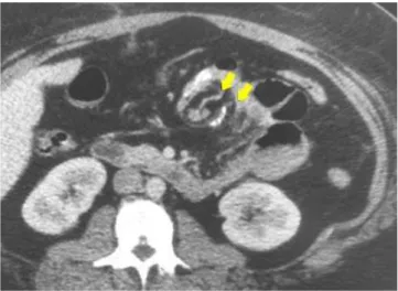
Of the 40 patients, only one (2.5%) had stenosis of the gastric suture (Figure 1).
b) Internal hernia
It was the most frequent complication. Of the 40 patients, five had it (12.5%).
Of the five, three demonstrated mesenteric fat planes densification and the presence of a herniated jejunal segment, located above the gastric level (Figure 2)
The other two patients had rotation of the mesenteric vessels (whirl sign) (Figure 3), mushroom appearance of the distended loops in the left hypochondrium (Figure 4) and also densification of mesenteric fat planes.
c) Anastomotic fistula
Only one patient (2.5%) developed anastomotic leak (Figure 5).
d) Abscess
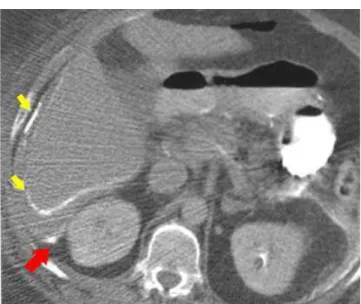
Three patients (7.5%) had abscesses (Figure 6).
DISCUSSION
DISCUSSION
DISCUSSION
DISCUSSION
DISCUSSION
With the increase in obesity, gastroplasty have become increasingly common, as well as digestive and abdominal and complications. Physical examination of these
patients is impaired by obesity itself and so imaging may play a role in clinical research. Often the radiologist is the first to detect the complications of these procedures. For this reason, according to Francis et al. 11, it is of great importance to know the possible complications and how they appear on imaging.
Blachar et al.12 reported that while the potential benefits of laparoscopic Roux-en-Y surgery are evident, little is known about the role of imaging studies in the diagnosis of complications of this procedure. Corroborating this study, it can be seen that many diagnoses were made possible by the CT scan.
Labrunie and Marchiori8, Onopchenko13 and Srikanth et al.14 consider CT imaging the method of choice for investigation of abdominal symptoms, especially
Figure 1 Figure 1 Figure 1 Figure 1
Figure 1 - Axial CT of the abdomen showing dilated proximal intestinal loop (upper arrows) extending to the level of the anastomosis, demarcated by the suture line (right arrow).
Figure 3 Figure 3 Figure 3 Figure 3
-Figure 3 - Axial CT of the abdomen showing rotation (swirl sign) and engorgement of mesenteric vessels (superior arrows) – internal transmesocolic hernia.
Figure 2 Figure 2 Figure 2 Figure 2
of patients undergoing gastric bypass. Patients with nonspecific and vague abdominal symptoms should be promptly submitted to CT with oral and intravenous contrast. In addition to diagnosing obstruction, it can also identify transmesocolic, mesenteric and umbilical hernias and gastric intussusception. It can also assess thickening of the intestine and complications related to pneumoperitoneum, fistulas and collections. Patients included in this study underwent CT examination as routine diagnostic investigation for ab-dominal pain.
Intestinal obstruction is common as a result of adhesion or internal hernia; intussusception has been rarely reported.
Merckle et al. 15 reported that CT can provide detailed view of the anatomy after Roux-en-Y gastric bypass, with all structures clearly demonstrated. For these authors the afferent gastric distention occurring after Roux-en-Y gastric bypass is due to obstruction or swelling of the enteroenterostomy and the imaging modality for this situation is the TC, because all the important structures can best be visualized, such as the excluded stomach, duodenum, low enteroenterostomy and the biliopancreatic loop.
Blachar et al. 12 believe that the combination of clinical and imaging criteria may help distinguish adhesions from bowel obstruction and internal hernias. The ones resulting from internal hernias tend to appear in longer period after the operation and demonstrate the signal of mushroom aspect of the distended loops, accompanied by rotation of the mesenteric vessels. Stenosis of the gastrojejunostomy is probably the result of ischemia and was reported in 3% by these authors, in agreement with this study’s 2.5%.
According to Lockhart et al.10, the laparoscopic Roux-en-Y gastric bypass resulted in reduction of perioperative complications, but the internal hernia remains a problem. The studies of Blachar et al.12, who found 2.8% of internal hernias, and of Higa et al.7, with 2.5%, corroborate this opinion. This study showed a higher percentage (12.5%).
In the paper of Blachar et al.12, patients with fistula presented with fluid collection, with extra-luminal gas seen on CT; this study found two cases of anastomotic leaks, and only one patient displayed a fluid collection. For the same author the most common location of fluid collections was near the anastomosis and in the left upper quadrant, including the peri-splenic space; the same was observed in this study.
According to Labrunie16, imaging studies play a very important role in the diagnosis and evolution of the patient and anastomotic leaks are serious and feared complications in the postoperative period of bariatric surgery because of their high morbidity and mortality. In this sample we obtained the diagnosis of fistula in one patient, whose CT demonstrated extravasation of oral contrast in the left upper quadrant. CT seems to have greater sensitivity in this assessment, associating direct and indirect signals, as well
Figure 6 Figure 6Figure 6 Figure 6
Figure 6 - Axial CT of the abdomen with fluid collection in the periesplenic region and air-fluid level (abscess) (arrow). Figure 5
Figure 5Figure 5 Figure 5
Figure 5 - Axial CT of the abdomen with presence of extra-luminal contrast (fistula) at the lower edge of the liver (upper arrows) and peritoneum (lower arrow). Figure 4
Figure 4Figure 4 Figure 4
as in the search for secondary complications such as collections.
Several signs suggestive of fistula are described in the literature. The visualization of oral contrast extravasation is a direct sign of this complication. Other already described aspects that are considered indirect signs in the literature are mainly collections adjacent to the gastric pouch and free fluid in the abdominal cavity. In this series these factors were also found.
According to Merckle et al.15, abdominal fluid collections, subphrenic abscess and peritonitis are conditions that occur in less than 2% of patients undergoing bariatric operations. These are considered serious complications,
clinical signs may not be enlightening and often the diagnosis is difficult. CT plays a critical role in patients with suspicion of these conditions, should be performed without hesitation and may prevent sepsis, multiple organ failure and death. In this research abscess was observed in 7.5% of patients while Yu et al.17 found abscesses in 15% of patients.
CONCLUSION
CONCLUSION
CONCLUSION
CONCLUSION
CONCLUSION
Total abdominal CT failed to inform the cause of the symptoms in 87.5% of patients seeking medical re-evaluation for symptoms of post-operative bariatric surgery.
R E S U M O R E S U M O R E S U M O R E S U M O R E S U M O
Objetivo Objetivo Objetivo Objetivo
Objetivo: Avaliar por exame de tomografia computadorizada de pacientes submetidos à derivação gastrojejunal em Y-de-Roux, sem anel, para tratamento de obesidade mórbida. MétodosMétodosMétodosMétodos: Estudaram-se 40 pacientes, encaminhados ao serviço de tomografiaMétodos do Hospital Universitário Evangélico de Curitiba para avaliação diagnóstica. Encontravam-se em pós-operatório de cirurgia bariátrica tendo sido operados no mesmo hospital. Foram incluídos pacientes submetidos à operação laparoscópica com sintomas que neces-sitavam de avaliação tomográfica diagnóstica. Excluíram-se pacientes que tinham sido submetidos à cirurgia bariátrica por outras técnicas cirúrgicas; que tinham sido operados por outra equipe; que não concordassem com a administração de contraste iodado por via oral ou endovenosa; e que excediam o limite de peso da mesa de exame. Para análise estatística utilizou-se a média das variáveis. Resultados
Resultados Resultados Resultados
Resultados: Os pacientes apresentaram-se com idade entre 23 a 70 anos e eram 11 homens e 29 mulheres. Não houve alterações extra-abdominais detectáveis pela tomografia de abdômen total; dos 40 pacientes avaliados, 30 apresentavam achados tomográficos dentro do limite da normalidade. A presença de estenose na anastomose gastrojejunal foi encontrada em um paciente; hérnia interna ocorreu em cinco; fístula anastomótica em um e abcesso em três dos pacientes estudados. ConclusãoConclusãoConclusãoConclusãoConclusão: A tomografia de abdome total não conseguiu informar a causa dos sintomas dos pacientes operados em 87,5% dos pacientes que procuraram re-avaliação médica por sintomas pós-operatórios da cirurgia bariátrica.
Descritores: Descritores: Descritores: Descritores:
Descritores: Pacientes. Tomografia. Anastomose em-Y de Roux. Obesidade mórbida. Cirurgia bariátrica.
REFERENCES
REFERENCES
REFERENCES
REFERENCES
REFERENCES
1. Nassif PAN, Malafaia O, Bopp D, Ribas FM. Lima BRD. Correlação entre gastroplastia e prevalência de colelíase no primeiro ano de pós-operatório. Rev med Paraná. 2007;65(2):12-5.
2. Zorrilla PG, Salinas RJ, Salinas-Martinez AM. Vertical banded gastroplasty-gastric bypass with and without the interposition of jejunum: preliminary report. Obes Surg. 1999. 9(1):29-32. 3. Brasil. Ministério da Saúde. Secretaria de Vigilância em Saúde.
Obesidade: SUS realiza três novos tipos de cirurgia para redução do estômago [internet]. Brasília/DF; Ministério da Saúde. 2005. Acesso em: 23/07/2005. Disponível em: http://portal.saude.gov.br/ p o r t a l / a p l i c a c o e s / n o t i c i a s / n o t i c i a s _ d e t a l h e . c f m ? co_seq_noticia=17608.
4. Capella JF, Capella RF. An assessment of vertical banded gastroplasty-Roux-en-Y gastric bypass for the treatment of morbid obesity. Am J Surg. 2002;183(2):117-23.
5. Buchwald H. Bariatric surgery for morbid obesity: health implications for patients, health professionals, and third-party payers. J Am Coll Surg. 2005;200(4):593-604.
6. Garrido Júnior AB. Cirurgia em obesos mórbidos: experiência pes-soal. Arq Bras Endocrinol Metab. 2000;44(1):106-110.
7. Higa KD, Boone KB, Ho T, Davies OG. Laparoscopic Roux-en-Y gastric bypass for morbid obesity: technique and preliminary results of our first 400 patients. Arch Surg. 2000;135(9):1029-33; discussion 1033-4.
8. L a b r u n i e E M , M a r c h i o r i E . O b s t r u ç ã o i n t e s t i n a l p ó s -gastroplastia redutora pela técnica de higa para tratamento da obesidade mórbida: aspectos por imagem. Radiol bras. 2007;40(3):161-5.
9. Blachar A, Federle MP, Dodson SF. Internal hernia: clinical and imaging findings in 17 patients with emphasis on CT criteria. Radiology. 2001;218(1):68-74.
10. Lockhart ME, Tessler FN, Canon CL, Smith JK, Larrison MC, Fineberg NS, et al. Internal hernia after gastric bypass: sensitivity and specificity of seven CT signs with surgical correlation and controls. AJR Am J Roentgenol. 2007;188(3):745-50.
11. Francisco MC, Barella SM, Abud TG, Vilar VS, Reibscheid S, Arasaki CH, et al. Análise radiológica das alterações gastrintestinais após cirurgia de Fobi-Capella. Radiol bras. 2007;40(4):235-8.
13. Parakh S, Soto E, Merola S. Diagnosis and management of internal hernias after laparoscopic gastric bypass. Obes Surg. 2007;17(11):1498-502.
14. Srikanth MS, Keskey T, Fox SR, Oh KH, Fox ER, Fox KM. Computed tomography patterns in small bowel obstruction after open distal gastric bypass. Obes Surg. 2004;14(6):811-22.
15. Merkle EM, Hallowell PT, Crouse C, Nakamoto DA, Stellato TA. Roux-en-Y gastric bypass for clinically severe obesity: normal appearance and spectrum of complications at imaging. Radiology. 2005;234(3):674-83.
16. Labrunie EM, Marchiori E, Tubiana JM. Fístulas de anastomose superior pós-gastroplastia redutora pela técnica de Higa para tra-tamento da obesidade mórbida: aspectos por imagem. Radiol bras. 2008;41(2):75-9.
17. Yu J, Turner MA, Cho SR, Fulcher AS, DeMaria EJ, Kellum JM, et al. Normal anatomy and complications after gastric bypass surgery; helical CT findings. Radiology. 2004;231(3):753-60.
Received on 25/10/2011
Accepted for publication 15/12/2011 Conflict of interest: none
Source of funding: none
How to cite this article: How to cite this article:How to cite this article: How to cite this article:How to cite this article:
Ribas FM, Nassif PAN, Ribas CPM, Dietz UA, Tuon F, Wendler E, Enokawa MS, Ferri KR. Postoperative abdominal ct findings in patients submitted to roux-en-y gastric bypass without ring. Rev Col Bras Cir. [periódico na Internet] 2012; 39(3). Disponível em URL: http:// www.scielo.br/rcbc