Reliability of a rapid hematology stain for sputum cytology*
Confiabilidade da coloração hematológica rápida para citologia de escarroJéssica Gonçalves, Emilio Pizzichini, Marcia Margaret Menezes Pizzichini, Leila John Marques Steidle, Cristiane Cinara Rocha,
Samira Cardoso Ferreira, Célia Tânia Zimmermann
Abstract
Objective: To determine the reliability of a rapid hematology stain for the cytological analysis of induced sputum
samples. Methods: This was a cross-sectional study comparing the standard technique (May-Grünwald-Giemsa
stain) with a rapid hematology stain (Diff-Quik). Of the 50 subjects included in the study, 21 had asthma, 19 had COPD, and 10 were healthy (controls). From the induced sputum samples collected, we prepared four slides: two were stained with May-Grünwald-Giemsa, and two were stained with Diff-Quik. The slides were read independently by two trained researchers blinded to the identification of the slides. The reliability for cell counting using the two techniques was evaluated by determining the intraclass correlation coefficients (ICCs) for intraobserver and interobserver agreement. Agreement in the identification of neutrophilic and eosinophilic sputum between the
observers and between the stains was evaluated with kappa statistics. Results: In our comparison of the two
staining techniques, the ICCs indicated almost perfect interobserver agreement for neutrophil, eosinophil, and macrophage counts (ICC: 0.98-1.00), as well as substantial agreement for lymphocyte counts (ICC: 0.76-0.83). Intraobserver agreement was almost perfect for neutrophil, eosinophil, and macrophage counts (ICC: 0.96-0.99), whereas it was moderate to substantial for lymphocyte counts (ICC = 0.65 and 0.75 for the two observers, respectively). Interobserver agreement for the identification of eosinophilic and neutrophilic sputum using the
two techniques ranged from substantial to almost perfect (kappa range: 0.91-1.00). Conclusions: The use of
Diff-Quik can be considered a reliable alternative for the processing of sputum samples.
Keywords: Sputum\analysis; Sputum\cytology; Azure stains.
Resumo
Objetivo: Determinar a confiabilidade da coloração hematológica rápida para a análise do escarro induzido.
Métodos: Estudo transversal comparando a técnica padrão (coloração May-Grünwald-Giemsa) com a coloração hematológica rápida (panótico rápido). Participaram do estudo 50 indivíduos (21 asmáticos, 19 portadores de DPOC e 10 controles). Após a coleta do escarro induzido, foram preparadas 4 lâminas, sendo 2 coradas por May-Grünwald-Giemsa e 2 por panótico rápido. As lâminas foram lidas de forma independente por dois pesquisadores capacitados para o exame de escarro induzido e cegados para a identificação das lâminas. A confiabilidade para as contagens celulares dos dois métodos foi avaliada pela determinação dos coeficientes de correlação intraclasse (CCI) para as concordâncias intraobservador e interobservador. As concordâncias na identificação de escarro neutrofílico e eosinofílico entre observadores e entre as duas colorações foram calculadas
por estatística kappa. Resultados: Nas duas colorações, os CCI apontaram concordância interobservador quase
perfeita para as contagens de neutrófilos, eosinófilos e macrófagos (variação do CCI: 0,98-1,00) e substancial para as contagens de linfócitos (variação do CCI: 0,76-0,83). Na análise intraobservador, a concordância foi quase perfeita para as contagens de neutrófilos, eosinófilos e macrófagos (variação do CCI: 0,96-0,99) e de moderada a substancial para as contagens de linfócitos (CCI = 0,65 e 0,75 para observadores 1 e 2, respectivamente). A concordância interobservador na identificação de escarro eosinofílico e neutrofílico para os dois métodos de coloração variou entre substancial e quase perfeita (variação kappa: 0,91-1,00). Conclusões: O panótico rápido pode ser considerado uma alternativa confiável para o processamento de amostras de escarro.
Descritores: Escarro\análise; Escarro\citologia; Corantes azur.
*Study carried out at the Federal University of Santa Catarina University Hospital, Florianópolis, Brazil.
Correspondence to: Jéssica Gonçalves. Universidade Federal de Santa Catarina, Hospital Universitário, Núcleo de Pesquisa em Asma e Inflamação das Vias Aéreas, Campus Universitário, Trindade, CEP 88040-970, Florianópolis, SC, Brasil.
Tel. 55 48 3721-8134. E-mail: [email protected] Financial support: None.
shorter; whereas the standard stain requires 34 min, the rapid stain is performed within 2 min.
Shortening the total processing time would be extremely important for improving the viability of the technique. In addition, the cost of Diff-Quik is considerably lower than that of the standard stain. Therefore, the validation of this technique is important, especially for developing countries, such as Brazil.
The objective of the present study was to assess the reliability of Diff-Quik for the cytological analysis of induced sputum samples.
Methods
The study included 50 patients, of whom 21 were adult patients with uncontrolled asthma, characterized by an Asthma Control
Questionnaire(13) score greater than 1.7 in the
previous week and objectively confirmed (in the 3 previous years) by reversible airflow limitation
(a > 12% increase in FEV1 and a > 200 mL
increase in FEV1 after inhalation of a
short-acting bronchodilator) in participants with
airflow limitation (an FEV1/FVC ratio < 0.7);
19 COPD patients aged > 40 years who had a history of respiratory symptoms associated with moderate or severe airflow obstruction (an
FEV1 < 50% of predicted and an FEV1/FVC ratio
< 0.7), were receiving any type of medication for COPD, and were (current or former) heavy smokers with a smoking history of > 20 pack-years; and 10 healthy nonsmokers who had no respiratory symptoms and whose diagnostic status was objectively confirmed by normal spirometry results. The study excluded patients who had respiratory infection in the four previous weeks, those who had severe diseases of other systems, those who had other known pulmonary diseases, and pregnant women.
The study was conducted at the Center for Research on Asthma and Airway Inflammation, located at the Federal University of Santa Catarina University Hospital in the city of Florianópolis, Brazil, and was approved by the local Human Research Ethics Committee (Process no. 2093; FR 437236, issued on November 28, 2011). All participants gave written informed consent after being given a detailed explanation of the study.
Participants underwent pre- and post-bronchodilator spirometry with a computerized spirometer (Koko; PDS Instrumentation, Inc., Louisville, CO, USA), in accordance with the
Introduction
The understanding of the mechanisms of diseases and their correct diagnosis has been made possible by analysis of body fluids in several areas of medicine. In the past, sputum analysis was considered not to be reliable or reproducible enough to assist in the understanding of the
mechanisms of respiratory diseases.(1) More recently,
significant advances related to the processing of sputum samples have allowed the method of sputum examination to become feasible, reproducible, valid, and responsive to interventions. Several researchers have used this method to study the various aspects of airway inflammation in asthma. The use of sputum examination has been further extended to COPD, cystic fibrosis, chronic cough, idiopathic pulmonary fibrosis, and other respiratory diseases.
Inflammation is central in the pathogenesis of airway diseases and is considered responsible for their symptoms, airflow obstruction, exacerbations,
and secondary structural changes.(2) Therefore,
airway inflammation plays an extremely significant role in two major obstructive respiratory tract
diseases: asthma and COPD.(3) Both in asthma and
in COPD, there is great heterogeneity in clinical and inflammatory characteristics, which result
in different clinical phenotypes.(4) Consequently,
there is a need to characterize the phenotype of patients in order to optimize their clinical
management, particularly in more severe cases.(4-6)
Examination of induced sputum is currently considered reliable, reproducible, discriminative of different types of inflammation, and responsive to interventions. Therefore, it has been an important tool in the management of inflammatory diseases
of the airways.(7) In addition, sputum induction is
a safe minimally invasive technique,(8) making it
possible to identify the type of inflammation and
its intensity.(9,10) However, its use in clinical practice
is still very restricted because of the method for sputum induction and for the processing of sputum samples, which is laborious and time-consuming and requires highly trained personnel. Induced sputum slides for differential cell counts, both in research and in clinical practice, have been stained with May-Grünwald-Giemsa or with Wright-Giemsa. These stains are part of
a group called “Romanowsky stains”.(11) Diff-Quik
(a rapid hematology stain), which is also based
on the Romanowsky technique,(12) uses similar
grade methanol, and then they were immersed in a May-Grünwald stain solution and a dilute May-Grünwald solution (1:1). Immediately afterward, the slides were immersed in a freshly prepared Giemsa stain solution (dilution 1:10) and subsequently dried. The whole procedure lasted exactly 34 min, as recommended by the equipment manufacturer.
Diff-Quik was performed manually. The process included initial immersion of the slides in solution no. 1 (0.1% triarylmethane), moving up and down continuously for 5-10 seconds (5-10 one-second immersions). Subsequently, extensions were immersed in solution no. 2 (0.1% xanthene), repeating the same procedure. After draining, the slides were immersed in solution no. 3 (0.1% thiazine), repeating the same procedure. The slides were rinsed with distilled water and allowed to
air-dry.(12) This staining procedure took a maximum
of 2 min to complete. Two researchers, trained in reading induced sputum slides, independently counted 400 non-squamous cells on the slides stained either with May-Grünwald-Giemsa or Diff-Quik. Because the slides were coded, the slide readers were prevented from identifying their respective pairs or previous reading results.
The reliability for cell counting using the two techniques (standard stain vs. tested stain) was evaluated by determining the intraclass correlation coefficients (ICCs) for intraobserver and interobserver agreement, and Bland & Altman
plots were used.(17) The interpretation of ICCs was
based on the classification proposed by Landis
& Koch.(18) Agreement in the identification of
eosinophilic sputum (eosinophils ≥ 3%)(6) and
neutrophilic sputum (neutrophils > 64%)(19)
between the observers and between the stains
was evaluated with kappa statistics.(18) Differences
between the characteristics of the groups studied were examined by ANOVA and the Bonferroni test in the post hoc analysis. The statistical tests were two-tailed, and the level of significance was set at 5%. The Statistical Package for the Social Sciences, version 18.0 (SPSS Inc., Chicago, IL, USA) was used for the analyses.
Results
Sputum induction was performed in 62 individuals, 50 (80.6%) of whom were able to produce an adequate sample, with cell viability greater than 50%. Twelve induced sputum samples were considered inadequate because
American Thoracic Societyguidelines(14) The
reference values used were those of Crapo et al.(15)
Subsequently, sputum induction was performed in accordance with the method described by
Pizzichini et al.(16) The procedure involved
inhalation of an isotonic saline aerosol (0.9%) followed by serial inhalation of increasing concentrations of hypertonic saline aerosol (3%, 4%, and 5%) via a Fisoneb ultrasonic nebulizer (Fisons, Pickering, Ontario, Canada). Aerosol inhalation was continued for 1-2 min, according to the level of bronchoconstriction severity before the procedure, and was followed by measurement
of FEV1. Participants were instructed to rinse their
mouths with water, swallow the water, and blow their noses in order to reduce contamination by saliva or postnasal discharge. They were then asked to cough and expectorate the sputum into a clean container. These procedures were repeated consecutively, with the solution concentration being increased every 7 min for 21 min or until
there was a ≥ 20% decrease in FEV1.
The processing of sputum samples was started within 2 h of collection, which is the longest time
reported in the literature.(2) The thick portions of
the expectorated material were selected with the naked eye or under visualization with an inverted microscope, and the sputum was separated from the saliva. The selected fractions were treated with 0.1% DTT at a ratio of four times the fraction volume. This mixture was homogenized with a Pasteur pipette and agitated on a desktop shaker for 15 min. Dulbecco’s PBS was added thereto in an amount that was four times the initial volume of sputum selected, and the resulting suspension was filtered to remove cell debris and undissolved mucus. Subsequently, total leukocyte counts were performed with a modified Neubauer hemocytometer, excluding squamous cells. Cell viability was determined by the trypan blue exclusion test. The sample was adjusted to
1.0 × 10⁶ cells/mL, and we prepared four slides,
which were coded. In the present study, all of the slides were prepared using the cytocentrifugation method (cytospin). After air-drying, two slides were stained with May-Grünwald-Giemsa, and the other two were stained with Diff-Quik (the technique under study).
May-Grünwald-Giemsa staining was performed
with an automated system (Sysmex sp1000iTM;
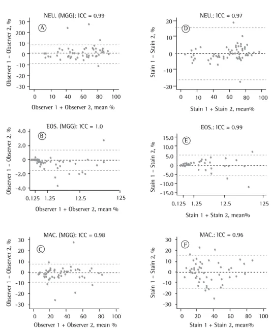
was substantial (ICC = 0.76). For the slides stained by the Diff-Quik technique, the medians and percentiles were also very close between the two observers, and the ICC indicated almost perfect interobserver agreement for eosinophil, neutrophil, macrophage, and lymphocyte counts (ICC = 1.00, 0.99, 0.99, and 0.83, respectively).
Regarding intraobserver agreement, the ICC values for the two observers for the differential cell counts on the pairs of cytospin slides stained by either of the two studied techniques indicated that it was almost perfect for neutrophils (ICC = 0.97 for both), eosinophils (ICC = 0.99 and 0.98), and macrophages (ICC = 0.96 for both). For lymphocyte counts, intraobserver agreement was substantial for observer 1 (ICC = 0.75) and moderate for observer 2 (ICC = 0.65). These
results are shown graphically in Figure 3.(17)
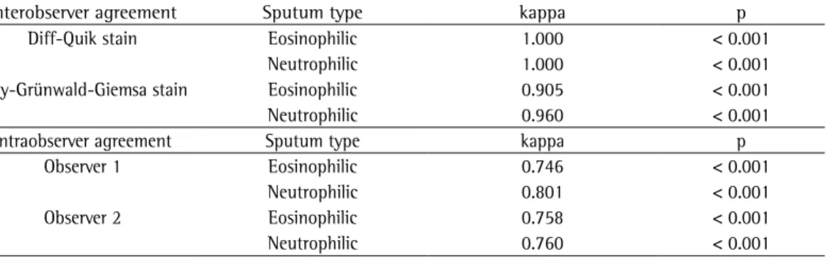
Interobserver agreement for the identification of eosinophilic and neutrophilic sputum using the two techniques ranged from substantial to almost perfect, as shown in Table 2. However, although intraobserver agreement was substantial, it was lower than was interobserver agreement.
Discussion
The results of the present study show that cytospin slides stained either by the May-Grünwald-Giemsa technique or with Diff-Quik yield similar cell counts, with high intraobserver and intraobserver agreement. These results demonstrate the reliability of the Diff-Quik technique for use in the processing of induced sputum samples. This fact is relevant because the Diff-Quik technique is simpler, allows a reduction in sample processing of excessive salivary contamination (> 20% of
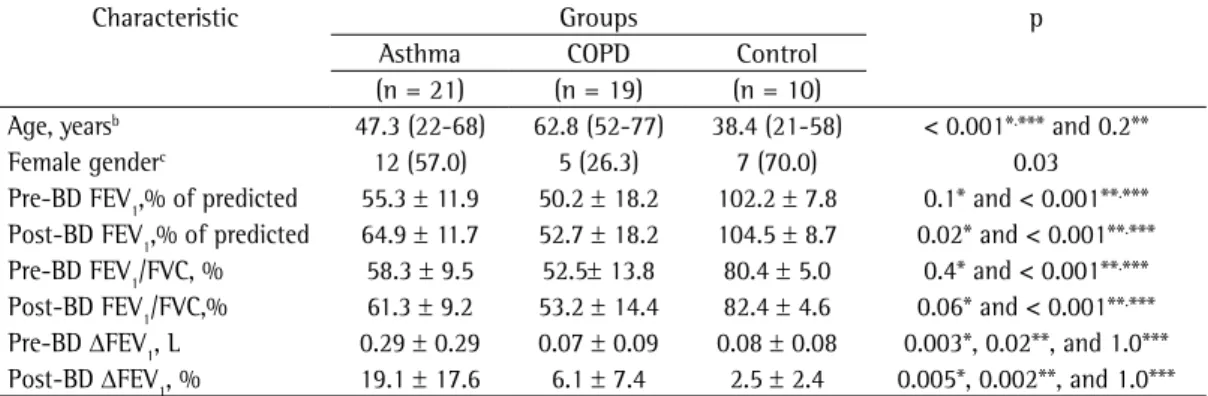
squamous cells), low cell viability (< 50%), or insufficient material to prepare the slides. The demographic, clinical, and functional characteristics of the participants are shown in Table 1. The groups were distinct and well characterized, as demonstrated by their demographic, clinical, and functional characteristics.
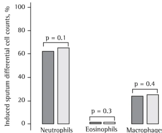
The cellular characteristics of induced sputum were as expected for the different groups studied. The sputum of asthma patients was characterized by a significantly higher proportion of eosinophils than that found in the sputum of COPD patients and healthy controls. In contrast, the sputum of COPD patients showed a significant increase in total cell counts and in the proportion of neutrophils when compared with that of controls. The control group showed a significantly higher proportion of macrophages than did the other two groups. Figure 1 shows the proportions of neutrophils, eosinophils, and macrophages in the different groups studied. In Figure 2, the median proportions of neutrophils, eosinophils, and macrophages, in the study sample as a whole, are separated by type of stain used. No significant differences were found for the cell counts on the slides stained by either of the two techniques used.
The results for interobserver agreement for induced sputum differential cell counts on the cytospin slides stained by the May-Grünwald-Giemsa technique show that the medians and percentiles were similar between the two observers, and the ICCs indicated almost perfect agreement for eosinophil, neutrophil, and macrophage counts (ICC = 1.00, 0.99, and 0.98, respectively). Interobserver agreement for lymphocyte counts
Table 1 - Demographic, clinical, and functional characteristics of the participants.a
Characteristic Groups p
Asthma COPD Control
(n = 21) (n = 19) (n = 10)
Age, yearsb 47.3 (22-68) 62.8 (52-77) 38.4 (21-58) < 0.001*.*** and 0.2**
Female genderc 12 (57.0) 5 (26.3) 7 (70.0) 0.03
Pre-BD FEV1,% of predicted 55.3 ± 11.9 50.2 ± 18.2 102.2 ± 7.8 0.1* and < 0.001**.***
Post-BD FEV1,% of predicted 64.9 ± 11.7 52.7 ± 18.2 104.5 ± 8.7 0.02* and < 0.001**.***
Pre-BD FEV1/FVC, % 58.3 ± 9.5 52.5± 13.8 80.4 ± 5.0 0.4* and < 0.001**.***
Post-BD FEV1/FVC,% 61.3 ± 9.2 53.2 ± 14.4 82.4 ± 4.6 0.06* and < 0.001**.***
Pre-BD ΔFEV1, L 0.29 ± 0.29 0.07 ± 0.09 0.08 ± 0.08 0.003*, 0.02**, and 1.0***
Post-BD ΔFEV1, % 19.1 ± 17.6 6.1 ± 7.4 2.5 ± 2.4 0.005*, 0.002**, and 1.0***
Pre-BD: pre-bronchodilator; and post-BD: post-bronchodilator. aValues expressed as mean ± SD, except where otherwise
indicated. bValue expressed as mean (range). cValue expressed as n (%). *Asthma group vs. COPD group. **Asthma group
staining techniques was found to be substantial. In addition, interobserver agreement for the identification of eosinophilic and neutrophilic sputum was almost perfect. These results again demonstrate the reliability of Diff-Quik, because they confirm its accuracy for identifying the different inflammatory phenotypes. However, the results also showed that interobserver agreement was higher than intraobserver agreement for the identification of the phenotypes. This difference could be due to variability in cell content on the slides stained by either of the two techniques. Although intraobserver agreement was substantial, this particular result suggests caution and a need for further studies to identify the reason for this variability.
Interobserver reproducibility for differential leukocyte counts in induced sputum samples has
been previously reported.(20-22) In 1997, one group of
authors reported high interobserver reproducibility for all cell types studied. Those authors also found lower agreement for lymphocytes than for the other cell types. The lower agreement for the proportions of lymphocytes was considered to be due to the very small amount of this cell type in the induced sputum samples. In the present study, we also found lower agreement for lymphocyte counts. However, this was not emphasized because it is a known fact that these variations are not clinically relevant.
In one study,(21) there was good interobserver
agreement for neutrophil, eosinophil, and macrophage counts, and, again, this agreement was lower for lymphocyte counts; the justification for the lower repeatability rate was related not only to the scarcity of lymphocytes in the samples time of up to 32 min without impairing sample
quality, and is considerably cheaper.
To our knowledge, this was the first study to evaluate the reliability of the Diff-Quik technique for use in induced sputum cytology, by comparing it with a standard staining technique, i.e., the May-Grünwald-Giemsa technique. It is important to evaluate the reliability and reproducibility of the results in order to confirm the accuracy of the results obtained by using Diff-Quik. In the present study, the reliability of Diff-Quik was tested by two distinct strategies. The first strategy was to calculate the ICCs for the cell counts performed by two independent observers, blinded to the identification of the slides. Although the stains used in the present study could be identified by the appearance of the slides, differing codes were used to prevent the identification of the respective
pairs of slides. Previous studies(20-22) have shown
that intraobserver and interobserver agreement for cell counts on cytospin slides stained with Wright-Giemsa and May-Grünwald-Giemsa could be considered perfect, but that it depended on the
degree of salivary contamination on the slides.(21)
The results obtained with Diff-Quik in the present study are in line with those of the aforementioned studies. The second strategy was to examine the reliability of Diff-Quik for identifying eosinophilic sputum and neutrophilic sputum. This is relevant because, in clinical practice, sputum examination is used to identify phenotypes of severe asthma, predict response to treatment, and decrease the number of asthma exacerbations by control of eosinophilic inflammation.
Intraobserver agreement for the identification of eosinophilic and neutrophilic sputum by the two
In
du
ce
d
spu
tu
m
di
ffe
re
n
ti
al
c
el
l
cou
n
ts
,
%
100
80
60
40
20
0
Neutrophils Eosinophils Macrophages Control Asthma COPD
100
80
60
40
20
0
In
du
ce
d
spu
tu
m
di
ffe
re
n
ti
al
c
el
l
cou
n
ts
,
%
Neutrophils Eosinophils Macrophages p = 0.1
p = 0.4
p = 0.3
Figure 1 - Induced sputum differential cell counts in the three groups studied.
Methods for refinement of sputum examination have greatly contributed to its accuracy and
reproducibility.(23) However, previous studies have
primarily focused on the steps preceding the preparation of cytospin slides and the liquid
phase of sputum.(24-27)
In 2003, a study comparing the results and costs of three techniques for analysis of induced but also to the difficulty in identifying these cells.
One group of authors(21) confirmed the results
of a previous study,(20) demonstrating that the
reproducibility of induced sputum differential cell counts is affected by salivary contamination and low cell viability. In the present study, samples with > 20% salivary contamination or < 50% viability were considered inadequate for analysis.
30 20 10 0 -10 -20 -30
0 20 40 60 80 100 Stain 1 + Stain 2, mean% Stain 1 + Stain 2, mean%
Stain 1 + Stain 2, mean%
MAC.: ICC = 0.96
Sta in 1 – Sta in 2, % Sta in 1 – Sta in 2, % Sta in 1 – Sta in 2, % F E
EOS.: ICC = 0.99
15.0 10.0 5.0 0 -5.0 -10.0 -15.0
0.125 1.25 12.5 125 D
NEU.: ICC = 0.97 20
10
0
-10
-20
0 10 40 60 80 100
30 20 10 0 -10 -20 -30
0 20 40 60 80 100 MAC. (MGG): ICC = 0.98
C O bs erv er 1 – O bs erv er 2, % O bs erv er 1 – O bs erv er 2, % O bs erv er 1 – O bs erv er 2, %
Observer 1 + Observer 2, mean % Observer 1 + Observer 2, mean % Observer 1 + Observer 2, mean %
A
NEU. (MGG): ICC = 0.99 30 200 10 0 -10 -20 -30
0 10 40 60 80 100
B
EOS. (MGG): ICC = 1.0
0.125 1.25 12.5 125 4.0
2.0
0
-2.0
-4.0
In summary, the high degree of agreement for the cell counts on the cytospin slides stained either by the May-Grünwald-Giemsa technique or with Diff-Quik attests to the reliability of the latter stain, which justifies the recommendation that it be used when the objective is to reduce sample processing time and induced sputum costs.
References
1. Djukanovic R, Sterk PJ, editors. An atlas of induced sputum--an aid for research and diagnosis. New York: Parthenon Publishing Group; 2004.
2. Pizzichini E, Efthimiadis A, Pizzichini MM, Hargreave FE. Sputum Examination for indices of airway inflammation: laboratory procedures. Lund, Sweden: Astra Draco AB; 1997.
3. Silva LC, Hetzel JL. Pneumonias obstrutivas. In: Condutas em Pneumologia. Silva LC, editor. Rio de Janeiro: Revinter; 2001. p. 261-352.
4. Pavord ID, Sterk PJ, Hargreave FE, Kips JC, Inman MD, Louis R, et al. Clinical applications of assessment of airway inflammation using induced sputum. Eur Respir J Suppl. 2002;37:40s-43s. PMid:12361362
5. Pavord ID, Brightling CE, Woltmann G, Wardlaw AJ. Non-eosinophilic corticosteroid unresponsive asthma. Lancet. 1999;353(9171):2213-4. http://dx.doi.org/10.1016/ S0140-6736(99)01813-9
6. Jayaram L, Pizzichini MM, Cook RJ, Boulet LP, Lemière C, Pizzichini E, et al. Determining asthma treatment by monitoring sputum cell counts: effect on exacerbations. Eur Respir J. 2006;27(3):483-94. http://dx.doi.org/10. 1183/09031936.06.00137704
7. Polombini BC, Moreira JS, Miorim MC, Villanova CA. Exame físico-valorização do exame de escarro. In: Silva LC, editor. Condutas em Pneumologia. Rio de Janeiro: Revinter; 2001. p. 67-78.
8. Pizzichini E, Pizzichini MM, Leigh R, Djukanovic R, Sterk PJ. Safety of sputum induction. Eur Respir J Suppl. 2002;37:9s-18s. PMid:12361366
9. Pavord ID, Pizzichini MM, Pizzichini E, Hargreave FE. The use of induced sputum to investigate airway inflammation. Thorax. 1997;52(6):498-501. http:// dx.doi.org/10.1136/thx.52.6.498
10. Pin I, Gibson PG, Kolendowicz R, Girgis-Gabardo A, Denburg JA, Hargreave FE, et al. Use of induced sputum cell counts to investigate airway inflammation in asthma.
sputum samples was published.(28) The techniques
consisted of one of the following: preparation of smears from sputum not treated with DTT; preparation of smears from sputum treated with DTT; or preparation of cytospin slides from sputum treated with DTT. Although the first two techniques reduced the time and cost of sputum sample analysis, Spearman’s correlation coefficient, which ranged from 0.57 to 0.64 for eosinophil counts and from 0.51 to 0.57 for neutrophil counts ((p < 0.01 for both), can be considered inadequate for these types of techniques. The authors concluded that the technique that uses cytospin slides prepared from samples treated with DTT is more suitable for research purposes
and for use in specialized centers.(28)
Some of the aspects that hinder the widespread use of induced sputum are its complexity and the sample processing time. Two previous studies have attempted to reduce the complexity of
sample processing,(29,30) both using a sputum
filtration device (Accufilter; Cellometrics, Hamilton, Ontario, Canada), which would be an alternative for standardizing the processing of induced sputum samples. The device consists of a kit with a tube used for weighing and treating sputum selected from saliva, connected to a filter and to a reception tube containing DTT, saline solution, and trypan
blue. One of those studies(30) sought to determine
the validity of using this device in the analysis of sputum samples, by comparing it with the standard method. The study reported ICCs that indicate good reproducibility for eosinophil and neutrophil counts between the methods; however, the ICCs indicated a reduction in cell viability and total cell counts, as well as an increase in the proportion of squamous epithelial cells, with the use of that device.
Table 2 - Interobserver and intraobserver agreement for the identification of eosinophilic and neutrophilic sputum in the evaluation of slides stained by either of the two studied techniques.
Interobserver agreement Sputum type kappa p
Diff-Quik stain Eosinophilic 1.000 < 0.001
Neutrophilic 1.000 < 0.001
May-Grünwald-Giemsa stain Eosinophilic 0.905 < 0.001
Neutrophilic 0.960 < 0.001
Intraobserver agreement Sputum type kappa p
Observer 1 Eosinophilic 0.746 < 0.001
Neutrophilic 0.801 < 0.001
Observer 2 Eosinophilic 0.758 < 0.001
Clin Exp Allergy. 1999;29(2):248-52. http://dx.doi. org/10.1046/j.1365-2222.1999.00483.x
22. Spanevello A, Migliori GB, Sharara A, Ballardini L, Bridge P, Pisati P, et al. Induced sputum to assess airway inflammation: a study of reproducibility. Clin Exp Allergy. 1997;27(10):1138-44. http://dx.doi. org/10.1111/j.1365-2222.1997.tb01150.x
23. Efthimiadis A, Spanevello A, Hamid Q, Kelly MM, Linden M, Louis R, et al. Methods of sputum processing for cell counts, immunocytochemistry and in situ hybridisation. Eur Respir J Suppl. 2002;37:19s-23s. PMid:12361358 24. Pizzichini E, Pizzichini MM, Efthimiadis A, Evans S, Morris
MM, Squillace D, et al. Indices of airway inflammation in induced sputum: reproducibility and validity of cell and fluid-phase measurements. Am J Respir Crit Care Med. 1996;154(2 Pt 1):308-17. http://dx.doi.org/10.1164/ ajrccm.154.2.8756799
25. Kelly MM, Leigh R, Horsewood P, Gleich GJ, Cox G, Hargreave FE. Induced sputum: validity of fluid-phase IL-5 measurement. J Allergy Clin Immunol. 2000;105(6 Pt 1):1162-8. http://dx.doi.org/10.1067/mai.2000.106375 26. Popov TA, Pizzichini MM, Pizzichini E, Kolendowicz R,
Punthakee Z, Dolovich J, et al. Some technical factors influencing the induction of sputum for cell analysis. Eur Respir J. 1995;8(4):559-65. PMid:7664854 27. Efthimiadis A, Pizzichini MM, Pizzichini E, Dolovich
J, Hargreave FE. Induced sputum cell and fluid-phase indices of inflammation: comparison of treatment with dithiothreitol vs phosphate-buffered saline. Eur Respir J. 1997;10(6):1336-40. http://dx.doi.org/10.1183/090 31936.97.10061336
28. Saraiva-Romanholo BM, Barnabé V, Carvalho AL, Martins MA, Saldiva PH, Nunes Mdo P. Comparison of three methods for differential cell count in induced sputum. Chest. 2003;124(3):1060-6. http://dx.doi.org/10.1378/ chest.124.3.1060
29. Pizzichini E, Pizzichini MM, Hargreave FE. Induced sputum (IS) cell counts: a new device to standardize processing in clinical trials and practice. In: International Conference of American Thoracic Society, 2004, Orlando. Am J Respir Crit Care Med. 2004;169:A817.
30. Chaboillez S, Dasgupta A, Prince P, Boulet LP, Lemiere C. A kit to facilitate and standardize the processing of sputum for measurement of airway inflammation. Can Respir J. 2013;20(4):248-52. PMid:23717819 Thorax. 1992;47(1):25-9. http://dx.doi.org/10.1136/
thx.47.1.25
11. Horobin RW. How Romanowsky stains work and why they remain valuable - including a proposed universal Romanowsky staining mechanism and a rational troubleshooting scheme. Biotech Histochem. 2011;86(1):36-51. http://dx.doi.org/10.3109/10520295.2010.515491 12. Laborclin Produtos para Laboratório Ltda [homepage on the
Internet]. Pinhais: Laborclin [cited 2013 Jan 1]. Instruções de uso - panótico rápido. [Adobe Acrobat document, 2p.]. Available from: http://www.interlabdist.com.br/dados/ produtos/bula/doc/13764277764820b3e23b11e.pdf 13. Juniper EF, O’Byrne PM, Guyatt GH, Ferrie PJ, King
DR. Development and validation of a questionnaire to measure asthma control. Eur Respir J. 1999;14(4):902-7. http://dx.doi.org/10.1034/j.1399-3003.1999.14d29.x 14. Standardization of Spirometry, 1994 update.
American Thoracic Society. Am J Respir Crit Care Med. 1995;152(3):1107-36. http://dx.doi.org/10.1164/ ajrccm.152.3.7663792
15. Crapo RO, Morris AH, Gardner RM. Reference spirometric values using techniques and equipment that meet ATS recommendations. Am Rev Respir Dis. 1981;123(6):659-64. PMid:7271065
16. Pizzichini E, Pizzichini MM, Efthimiadis A, Hargreave FE, Dolovich J. Measurement of inflammatory indices in induced sputum: effects of selection of sputum to minimize salivary contamination. Eur Respir J. 1996;9(6):1174-80. http://dx.doi.org/10.1183/09031936.96.09061174 17. Bland JM, Altman DG. Comparing methods of
measurement: why plotting difference against standard method is misleading. Lancet. 1995;346(8982):1085-7. http://dx.doi.org/10.1016/S0140-6736(95)91748-9 18. Landis JR, Koch GG. The measurement of observer
agreement for categorical data. Biometrics. 1977;33(1):159-74. http://dx.doi.org/10.2307/2529310
19. Belda J, Leigh R, Parameswaran K, O’Byrne PM, Sears MR, Hargreave FE. Induced sputum cell counts in healthy adults. Am J Respir Crit Care Med. 2000;161(2 Pt 1):475-8. http://dx.doi.org/10.1164/ajrccm.161.2.9903097 20. Efthimiadis A, Pizzichini MM, Pizzichini E. The influence
of cell viability and squamous epithelial cell contamination on the repeatability of sputum differential cell counts (abstract). Am J Respir Crit Care Med. 1995;151:A384. 21. Ward R, Woltmann G, Wardlaw AJ, Pavord ID. Between-observer repeatability of sputum differential cell counts. Influence of cell viability and squamous cell contamination.
About the authors
Jéssica Gonçalves
Pharmacist. Clinical Analysis Department, Federal University of Santa Catarina University Hospital, Florianópolis, Brazil.
Emilio Pizzichini
Adjunct Professor. Federal University of Santa Catarina University Hospital, Florianópolis, Brazil.
Marcia Margaret Menezes Pizzichini
Professor. Graduate Program in Medical Sciences, Federal University of Santa Catarina University Hospital, Florianópolis, Brazil.
Leila John Marques Steidle
Cristiane Cinara Rocha
Coordinating Nurse. Center for Research on Asthma and Airway Inflammation, Federal University of Santa Catarina University Hospital, Florianópolis, Brazil.
Samira Cardoso Ferreira
Pharmacist. Clinical Analysis Department, Federal University of Santa Catarina University Hospital, Florianópolis, Brazil.
Célia Tânia Zimmermann