Stimulus electrodiagnosis and motor and functional
evaluations during ulnar nerve recovery
Luciane F. R. M. Fernandes1, Nuno M. L. Oliveira1, Danyelle C. S. Pelet2, Agnes F. S. Cunha2, Marco A. S. Grecco3, Luciane A. P. S. Souza1
ABSTRACT | Background: Distal ulnar nerve injury leads to impairment of hand function due to motor and sensorial changes. Stimulus electrodiagnosis (SE) is a method of assessing and monitoring the development of this type of injury.
Objective: To identify the most sensitive electrodiagnostic parameters to evaluate ulnar nerve recovery and to correlate these parameters (Rheobase, Chronaxie, and Accommodation) with motor function evaluations. Method: A prospective cohort study of ten patients submitted to ulnar neurorrhaphy and evaluated using electrodiagnosis and motor assessment at two moments of neural recovery. A functional evaluation using the DASH questionnaire (Disability of the Arm, Shoulder, and Hand) was conducted at the end to establish the functional status of the upper limb. Results: There was
signiicant reduction only in the Chronaxie values in relation to time of injury and side (with and without lesion), as well as signiicant correlation of Chronaxie with the motor domain score. Conclusion: Chronaxie was the most sensitive SE parameter for detecting differences in neuromuscular responses during the ulnar nerve recovery process and it was the only parameter correlated with the motor assessment.
Keywords: chronaxie; ulnar nerve; evaluation studies; disability evaluation; rehabilitation; movement.
BULLET POINTS
• Stimulus electrodiagnosis is a reliable, noninvasive method of identifying neural regeneration.
• Chronaxie was the most sensitive parameter for assessing ulnar regeneration.
• Chronaxie and motor evaluation should be used to monitor neural regeneration.
HOW TO CITE THIS ARTICLE
Fernandes LFRM, Oliveira NML, Pelet DCS, Cunha AFS, Grecco MAS, Souza LAPS. Stimulus electrodiagnosis
and motor and functional evaluations during ulnar nerve recovery. Braz J Phys Ther. 2016 Mar-Apr; 20(2):126-132 .
http://dx.doi.org/10.1590/bjpt-rbf.2014.0138
1 Departamento de Fisioterapia Aplicada, Universidade Federal do Triângulo Mineiro (UFTM), Uberaba, MG, Brazil 2 Curso de Fisioterapia, Universidade Federal do Triângulo Mineiro (UFTM), Uberaba, MG, Brazil
3 Departamento de Cirurgia, Universidade Federal do Triângulo Mineiro (UFTM), Uberaba, MG, Brazil
Received: Dec. 18, 2014 Revised: June 30, 2015 Accepted: Oct. 06, 2015
Introduction
Injury to the ulnar nerve is one of the most common upper limb peripheral nerve lesions. Eser et al.1 conducted a retrospective study and found that most cases involved lesions of the ulnar nerve, with 337 cases (27%), followed by lesions of the median nerve, with 273 cases (22%). A complete and detailed evaluation of the hand is essential in order to identify a suitable treatment and achieve the best response to therapy. Rosén and Lundborg2 developed a model for the speciic evaluation of median and ulnar nerve
lesions, considering three domains: motor, sensory, and pain/discomfort. In the motor domain, evaluation of the median and ulnar nerves involves testing the strength of key hand muscles, along with dynamometry measurements of handgrip strength.
Regarding functional evaluation, the DASH (Disability of the Arm, Shoulder, and Hand) questionnaire3, which consists of three modules, employs a series of questions related to different tasks involving the upper limbs. This instrument was developed to measure dysfunction and physical symptoms in the upper limbs and to evaluate progress over time3.
electrostimulation, ensuring use of the most suitable
electrical pulse for treatment of a speciic lesion4,5. SE is one of the most objective means of evaluating and monitoring the evolution of a peripheral nerve lesion4,6,7, and of guiding the use of therapeutic electrostimulation5. In the present study, we attempted to test the eficiency and the importance of SE as a
‘tool-of-the-trade’ for physical therapists, particularly in ulnar nerve recovery.
There are no reports in literature concerning the use of SE in upper limb peripheral nerve lesions.
No protocols have been deined for electrostimulation,
and no studies have investigated the relationship between SE parameters and the results of physical therapeutic evaluation. If a good relationship is found, it could emphasize the importance of SE in the process of nerve lesion recovery and rehabilitation. Thus, the hypothesis of this study was that the SE parameters correlate with motor scores during peripheral ulnar lesion recovery. The objective of this study, therefore, was to identify the most sensitive parameters to use for the evaluation of ulnar nerve recovery and to correlate the SE parameter values with motor performance. The neuromuscular responses, which were obtained using electrodiagnosis during the recovery process after neurorrhaphy of the ulnar nerve, were evaluated, and the electrodiagnosis parameters were correlated with the results of motor assessments. Our objective was to determine whether the motor gains are linked to neural recovery, showing a possible new use for SE.
Method
Subjects
An observational, prospective cohort study was carried out to investigate the recovery of the ulnar nerve after neurorrhaphy. All subjects received information about the objectives and procedures of the study and signed an informed consent form, in accordance with regulation 466/12 of the Brazilian National Health Council. The study was approved by the ethics committee of Universidade Federal do Triângulo Mineiro (UFTM), Uberaba, MG, Brazil (protocol number 1663). The inclusion criteria selected patients with ulnar nerve lesions in the region of the wrist and distal forearm and who had been submitted to neurorrhaphy and then underwent physical
therapy during the irst three months after surgery.
The exclusion criteria were refusal to participate in the study, postoperative complications (infection), failure
to attend the evaluations, and absence of muscular response during the electrodiagnosis examination.
A sample size of nine patients was determined, based on the standard deviation values of 3.03 obtained for
Chronaxie in a pilot project involving ive patients
with ulnar nerve lesions. Sample size estimation was calculated using Power and Sample v.3.0.4 software
with power of 80% and α=0.05. The Chronaxie
variable was selected due to its recognized importance in electrodiagnostic examinations4,8,9.
The study was conducted with ten patients, including six men and four women with a mean age
of 42 (SD=15) years. In the sample, only one subject
was left-handed, and the right-hand side was more severely affected by lesions acquired in the workplace. All of the patients underwent neurorrhaphy, carried out by the same medical team, and were referred to the same physical therapy service at the UFTM, which followed the same protocol developed for the
study. Nine patients underwent surgery in the irst
three weeks following injury.
Two evaluations of the injured limb were performed:
initial (EV1) and inal (EV2). EV1 was conducted
during the initial phase (between 4 and 6 months post-surgery) and EV2 was performed at a later stage (between 10 and 15 months post-surgery). Evaluations (denoted EVWL) were also made on the contralateral limb (i.e. without lesion). All evaluations were conducted by the same examiner and under the same conditions.
Equipment and functional evaluation
The equipment used for the SE tests included: a) a universal pulse generator (Model Nemesys 941, Quark, Brazil); b) aluminum electrodes (10×5 cm); c) natural plant sponges (10×5 cm); d) an electrodiagnosis pen; and e) evaluation sheets (available from the instrument manual). For the motor assessment, a hydraulic dynamometer (Jamar) was used, and the functional evaluation was conducted using the DASH questionnaire that had been translated and validated for use in Portuguese10,11.
Electrodiagnostic testing
corresponds to the shortest time necessary to produce a muscular contraction, also using a rectangular pulse with an interval of 2.0 s and an amplitude equal to two times the Rheobase obtained previously8,12,14. Accommodation is deined similarly to Rheobase, but
the measurement is performed using an exponential pulse with a period of 1.0 s and an interval between the pulses of 2.0 s8,14. Both Rheobase and Accommodation are measures of intensity and are given in units of milliamps (mA), while Chronaxie is a measure of the duration or width of the pulse and is therefore given in milliseconds (ms)8.
The subjects were placed in the seated position with the upper limb supported and maintaining the
shoulder adduced, the elbow lexed at 90°, the forearm
supine, and the wrist in a neutral position. First, the skin was cleansed with 70% alcohol in order to reduce its impedance. The muscle evaluated was the abductor
of the ifth inger. An SMS (strong muscle stimulation)
current was used to locate the motor point, employing a monopolar technique with two electrodes. One was
a pen-type (active) electrode with an area suficiently
small to be able to stimulate the abductor muscle of
the ifth inger. Dampened gauze was used to cover
the metal tip in order to avoid direct contact with the skin. The other (passive) dispersive electrode had a greater area (in order to diminish the concentration of the electric charge on the skin) and was attached to the contralateral upper limb with an elastic band. The interface between this electrode and the skin was
illed with a dampened sponge on the contractile part of
the brachial biceps in order to close the circuit, following the recommendations provided in the manufacturer’s manual. The stimulation electrode was positioned perpendicularly to the muscle under evaluation, and the pressure and angle of the pen were set after the motor point had been located. The intensity used
was suficient to induce a visible contraction. The SE
was then initiated and the Rheobase, Chronaxie, and Accommodation values were recorded. The test was performed bilaterally, with the contralateral side used as the control.
Evaluation of motor performance
Hand muscle strength and grip strength were determined according to the standardized Rosén and Lundborg motor score procedure2. The muscles used to evaluate hand strength were the abductor of
the ifth inger, the fourth palmar, and the irst dorsal
interosseous. The results were graded from zero to
ive, according to the Highet scale15, and the values obtained for the three muscles were added and divided by 15 (the value for a normal individual).
The position adopted for measurements of grip strength was that recommended by the American Society of Hand Therapists (ASHT)16. Three measurements were performed and the arithmetic mean was calculated and divided by the mean for the healthy side.
Functional evaluation
In this study, only the module of the DASH questionnaire that evaluates functional ability was used. The score obtained varies from 0 to 100%, and the higher the score is, the greater the functional limitation3,10,11. The DASH test was only used in the inal evaluation, in order to measure and describe
functional status.
Data analysis
The normality of the SE data (Rheobase, Chronaxie, and Accommodation) was assessed using the Shapiro-Wilks test, and only the Chronaxie values were shown not to have normal distribution. Mean, standard deviation (SD), median, and maximum and minimum values were obtained through descriptive analysis. For the Chronaxie inferential analysis, the Wilcoxon matched pair (time) and Mann-Whitney U test for independent samples (side) were used. For Rheobase and Accommodation inferential analysis, the Student t test for dependent samples (time) and the Student t test for independent samples (side) were used. Finally, we calculated the correlation between the SE data and the Rosén and Lundborg2 motor domain scores using the Spearman rank test. For all the tests,
the signiicance level was set at 5%. The software
Statistica 7 was used for all analyses.
Results
The SE values obtained were compared considering
the initial (EV1) and inal (EV2) evaluations and
evaluations of the sides with and without lesion (EVWL) (Figures 1-3). Chronaxie was the parameter that best represented recovery of the ulnar nerve (Figure 2).
The Chronaxie values obtained for the sides without lesion were very close to zero, and similar results were
obtained in the inal evaluation. The minimum, mean,
standard deviation, median, and maximum values for Rheobase, Chronaxie, and Accommodation obtained
in the initial (EV1), inal (EV2), and side without
According to statistical analysis, Rheobase did
not present signiicant differences between the times of lesion EV1 and EV2 (p=0.56) or the side evaluated in EV1 (p=0.53) and in EV2 (p=0.88).
Furthermore, the Accommodation values did not
show any signiicant differences between the times
of lesion (p=0.61) or between the sides (EV1: p=0.18 and EV2: p=0.56). On the other hand, the Chronaxie values were signiicantly different between EV1 and EV2 (p=0.01), as well as between the sides tested (EV1: p=0.00 and EV2: p=0.00).
The mean (SD) DASH values of the inal evaluation were 33.1% (SD=21.3%) with minimum of 3.3% and
maximum of 59.2%.
The relationship between the SE values and the Rosén and Lundborg2 motor domain scores was investigated by calculating the Spearman correlation
coeficient (rs) (Table 2).
The Chronaxie parameter was the only parameter
that showed a signiicant negative correlation with the
Rosén and Lundborg2 motor domain score in both the initial and inal evaluations.
Discussion
This study contributes to the literature concerning quantitative evaluation of recovery of the ulnar nerve, using a test that has been largely ignored in clinical
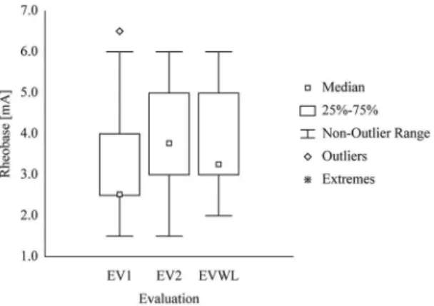
Figure 1. Boxplot of values of the initial (EV1), inal (EV2), and
side without lesion (EVWL) evaluations for Rheobase.
Figure 2. Boxplot of values of the initial (EV1), inal (EV2), and
side without lesion (EVWL) evaluations for Chronaxie.
Figure 3. Boxplot of values of the initial (EV1), inal (EV2), and
side without lesion (EVWL) evaluations for Accommodation.
Table 1. Minimum, Mean, Median, Standard Deviation (SD), and Maximum values of Rheobase, Chronaxie, and Accommodation in
the initial (EV1), inal (EV2), and side without lesion (EVWL) evaluations.
Evaluation Minimum Mean SD Median Maximum
Rheobase [mA]
EV1 1.50 3.40 1.69 2.5 6.50
EV2 1.50 3.75 1.37 3.75 6.00
EVWL 2.00 3.85 1.43 3.25 6.00
Chronaxie [ms]
EV1 0.20 3.93 4.56 2.40 14.00
EV2 0.20 1.51 2.28 0.30 6.00
EVWL 0.10 0.18 0.07 0.20 0.30
Accommodation [mA] EV1 2.00 5.3 3.16 4.25 10.00
EV2 2.50 8.7 5.94 7.50 23.00
practice in recent years. SE is a resource that can be used to aid diagnosis, evaluate the stage of a lesion
and nerve recovery, and deine the parameters used
in electrostimulation5. In addition to SE, the Rosén and Lundborg2 motor domain score and functional evaluation with the DASH questionnaire3,10,11 were used.
The proile of the assessed patients with nerve
lesions was as follows: mostly males (60%); average age of 42 years; right-hand limb most commonly affected (60% of cases), and most common cause of lesion was workplace accident (50%). A similar patient
proile was reported by Eser et al.1, who conducted a study using the data for 938 patients evaluated using electromyography and diagnosed with peripheral nerve lesions located in the upper limbs and the lower limbs. In that study, 71% of patients were male, the average age was 38 years, and the right-hand side was most frequently affected (55%). Most of the lesions (77%) were located in the upper limbs, and the main cause was car accidents (26.9%).
All patients were submitted to the same surgical procedure, which was performed by the same medical team. However, there were differences in clinical scenarios, including scarring processes and recovery times. Furthermore, the individuals showed differences in terms of both regeneration and sensitivity thresholds. Nonetheless, even considering these
factors, Chronaxie proved to be suficiently sensitive
for detecting differences between the lesion phases. It is important to emphasize that, although the SE method was described many years ago, it is rarely used in clinical practice despite the advantages
described above and still requires further scientiic
investigation. It is likely that, in addition to a lack of information in the literature, its poor use could be
related to dificulties encountered during application
of the procedure. The test is detailed and requires an experienced physical therapist for its application
and interpretation of the results. Another dificulty is
related to the equipment required, because there are currently few options commercially available.
However, according to the initial hypothesis, the Chronaxie parameter correlates with motor scores during the recuperation of a peripheral ulnar lesion. Rheobase and Accommodation did not demonstrate any correlation. Chronaxie was the only parameter
that showed signiicant differences between times
of lesion and side with and without lesion. If only the Chronaxie test (based on Rheobase studies) was performed, which is relatively easy, it would be possible to understand the lesion and predict the motor behavior.
Chronaxie and Rheobase was irst deined more
than one hundred years12 ago, and since that time, various researchers have studied these parameters in cases of peripheral nerve lesion4,8,9,17,18. They found that Chronaxie, which provides a measure of the neuromuscular electrical excitation threshold, was the most sensitive parameter for use in detection of nerve lesions. In this study, Chronaxie was also found to be the most sensitive parameter for use in lesion diagnosis and assessment of recovery of the ulnar nerve. The behavior of this parameter during the recovery/regeneration process was similar for all the patients, with high values during the initial phase and low values during the recovery phase. In the latter case, the values were very close to those obtained for the side without lesion.
There have been few reports of Chronaxie values for patients with peripheral nerve lesion. Licht et al.19 associated Chronaxie values with the type and severity
of lesion. The lesions were classiied using six levels
of severity, and the Chronaxie values were: 30-60 ms (neurotmesis); 20-30 ms (total axon degeneration); ~20 ms (partial axon degeneration); 10-20 ms (neuropraxy); 1-10 ms (moderate neuropraxy); and <1 ms (mild neuropraxy). The authors did not provide any information concerning the sample population. In this study, the Chronaxie values obtained in the initial evaluation correspond to normal physiology
and moderate denervation. In the inal evaluation, the
values correspond to light denervation19.
Ervilha and Araújo4 conducted a study of Chronaxie using healthy individuals and individuals who had shown peripheral nerve lesions for more than eight months and less than two years. Seven muscles of the upper limb were evaluated and three Chronaxie
value intervals were deined, depending on the severity of the lesion: the irst (0.13 ms, SD=0.80 ms) was
Table 2. Correlations between the values of the electrodiagnosis parameters and motor domain scores).
rs P-value
EV1 Motor domain X Rheobase 0.313 0.38
Motor domain X Chronaxie –0.757 0.01*
Motor domain X Accommodation 0.396 0.26
EV2 Motor domain X Rheobase 0.355 0.31
Motor domain X Chronaxie –0.794 0.01*
Motor domain X Accommodation 0.247 0.49
representative of normal individuals, the second
(1.5-20 ms) relected moderate peripheral lesion,
and the third (>30 ms) indicated severe lesion and a poor prognosis. Although the duration of the lesion was considered and different muscles were evaluated with the aim of classifying all of the nerves of the upper limb, there were gaps remaining between the established intervals where Chronaxie values were not associated with lesion severity.
Therefore, Chronaxie was found to be a sensitive and useful parameter that could be used to evaluate the process of recovery/regeneration following ulnar nerve lesion. A reduction in Chronaxie towards normal values is indicative of reinnervation or the avoidance
of further degeneration of the muscle iber8. Here, the Chronaxie values obtained in EV2 were very close to the values obtained for the side without lesion, for all but two of the patients.
The Rheobase and Chronaxie parameters were studied by Lee et al.20 in patients suffering from encephalopathy after cerebrovascular accident. The results obtained for the paretic and non-paretic sides were compared, showing that the Rheobase
and Chronaxie values were signiicantly higher for
the paretic side. It could be inferred that reduction in muscular activity in cases of paresis or peripheral nerve lesion contributed to the need for greater stimulation in terms of both intensity and duration.
In the present study, the Rheobase values showed no similarity between patients or during the phases of lesion. High Rheobase values were found for both
the side without lesion and in the inal evaluation of
some of the patients. The Accommodation parameter
has not been the target of scientiic studies in patients
with peripheral nerve lesion, although studies have been conducted with animals8,9,21. Comparisons with the present study were, therefore, not possible.
Comparisons between SE parameters and clinical data for peripheral nerve lesions could not be found in literature. In the present study, Chronaxie showed a
signiicant negative correlation with the values obtained
for the Rosén and Lundborg2 motor domain score. The correlation was negative because, in the process of neural regeneration, the Chronaxie values tended to diminish towards 0.2 ms while the motor domain score increased towards 1.0. The other parameters did not show any correlation with the motor domain score.
In terms of clinical applications, the results of this study reinforce the need for detailed, quantitative and carefully directed evaluation of patients with
peripheral nerve lesions. The indings also indicate the
desirability to reinstate electrodiagnostic evaluation in clinical practice. Chronaxie, especially, is a valuable parameter that can be used in assessments of the recovery/regeneration process. The Chronaxie value is extremely useful for determination of the duration of the electrical impulse used for muscle stimulation and helps in the application of stimulations that are more comfortable22. The optimum duration of an impulse is equal to the Chronaxie of the muscle that it aims to stimulate23. Because the Chronaxie test requires the Rheobase value, we also have an indication of a possible stimulation intensity value.
A limitation of this study was that we did not construct the quadratic and triangular pulse graphics, which might have given us a better idea of the recuperation process. The construction of such graphics should be included in future studies. The use of DASH at baseline could also provide information about functional gain during nerve recuperation.
In conclusion, stimulus electrodiagnosis is a quantitative, noninvasive technique for neuromuscular evaluation and can be used to accompany recovery following neurorrhaphy of the ulnar nerve. The Chronaxie parameter proved to be most sensitive for identifying
differences between initial and inal evaluations of the
limb on the lesion side, as well as between the sides with and without lesion. This parameter also presented correlation with the results of clinical motor domain assessment. The renewed use of electrodiagnosis should therefore be encouraged, and the technique should be included in both clinical practice and academic courses.
Acknowledgements
Financial disclosure statements have been obtained,
and no conlicts of interest have been reported by the
authors or by any individuals in control of the content of this article. The authors would like to thank the patients who participated in this study. The project
was inanced by FAPEMIG and FUNEPU.
References
1. Eser F, Aktekin LA, Bodur H, Atan C. Etiological factors of traumatic peripheral nerve injuries. Neurol India. 2009;57(4):434-7. http://dx.doi.org/10.4103/0028-3886.55614. PMid:19770544.
2. Rosén B, Lundborg G. A new model instrument for outcome after nerve repair. Hand Clin. 2003;19(3):463-70. http:// dx.doi.org/10.1016/S0749-0712(03)00003-9. PMid:12945644. 3. Hudak PL, Amadio PC, Bombardier C, Beaton D, Cole D,
measure: the DASH (disabilities of the arm, shoulder and hand). Am J Ind Med. 1996;29(6):602-8. http://dx.doi. org/10.1002/(SICI)1097-0274(199606)29:6<602::AID-AJIM4>3.0.CO;2-L. PMid:8773720.
4. Ervilha UF, Araujo RC. Estudo sobre a frequência de distribuição da cronaxia e a sua correlação com distintos graus de lesões nervosas periféricas. Rev Bras Fisioter. 1997;1(2):45-50.
5. Coutinho E L, Brasileiro OSM, Parizotto NA, Carmo JM, Carrinho PM. Fisioterapia-abordagem clínica e terapêutica das lesões nervosas periféricas. In: Tatagiba M, Mazzer N, Aguiar PHP, Pereira CU. Nervos periféricos: diagnóstico e tratamento clínico e cirúrgico. Rio de Janeiro: Revinter; 2003. p. 210-25.
6. Parry GJ. Electrodiagnostic studies in the evaluation of peripheral nerve and brachial plexus injuries. Neurol Clin. 1992;10(4):921-34. PMid:1331738.
7. Koka R, Hadlock TA. Quantification of functional recovery following rat sciatic nerve transection. Exp Neurol. 2001;168(1):192-5. http://dx.doi.org/10.1006/exnr.2000.7600. PMid:11170734.
8. Russo TL, França C, Castro C, Salvini TF. Alterations of chronaxie, rheobase and accommodation in denervated skeletal muscle submitted to electrical stimulation. Rev Bras Fisioter. 2004;8(2):169-75.
9. Russo TL, Peviani SM, Freria CM, Gigo-Benato D, Geuna S, Salvini TF. Electrical stimulation based on chronaxie reduces atrogin-1 and myoD gene expressions in denervated rat muscle. Muscle Nerve. 2007;35(1):87-97. http://dx.doi. org/10.1002/mus.20668. PMid:17034040.
10. Orfale AG, Araújo PMP, Ferraz MB, Natour J. Translation into Brazilian Portuguese, cultural adaptation and evaluation of the reliability of the disabilities of the arm, shoulder and hand questionnaire. Braz J Med Biol Res. 2005;38(2):293-302. http://dx.doi.org/10.1590/S0100-879X2005000200018. PMid:15785841.
11. Cheng HMS, Sampaio RF, Mancini MC, Fonseca ST, Cotta RMM. Disabilities of the arm, shoulder and hand (DASH): factor analysis of the version adapted to Portuguese/ Brazil. Disabil Rehabil. 2008;30(25):1901-9. http://dx.doi. org/10.1080/09638280701749342. PMid:19061116. 12. Irnich W. The terms “chronaxie” and “rheobase” are 100
years old. Pacing Clin Electrophysiol. 2010;33(4):491-6. http://dx.doi.org/10.1111/j.1540-8159.2009.02662010;33(4):491-6.x. PMid:20132498.
13. Ashley Z, Sutherland H, Lanmuller H, Unger E, Li F, Mayr W, et al. Determination of the chronaxie and rheobase of denervated limb muscles in conscious rabbits. Artif
Organs. 2005;29(3):212-5. http://dx.doi.org/10.1111/j.1525-1594.2005.29037.x. PMid:15725219.
14. Paternostro-Sluga T, Schuhfried O, Vacariu G, Lang T, Fialka-Moser V. Chronaxie and accommodation index in the diagnosis of muscle denervation. Am J Phys Med Rehabil. 2002;81(4):253-60. http://dx.doi.org/10.1097/00002060-200204000-00003. PMid:11953542.
15. Seftchick JL, Detullio LM, Fedorczyk JM, Aulicino PL. Clinical examination of the hand. In: Skirven TM, Osterman AL, Fedorczyk JM. Rehabilitation of the hand and upper extremity. 6th ed. St Louis: Mosby; 2011.
16. Fess EE. The need for a consistent international language in hand rehabilitation. J Hand Ther. 1993;6(4):329. http:// dx.doi.org/10.1016/S0894-1130(12)80337-1. PMid:8124449. 17. Fernández AM. Electrodiagnóstico y electroestimulación
de músculos denervados. Fisioterapia. 2001;23:23-35. http:// dx.doi.org/10.1016/S0211-5638(01)72970-7.
18. Robinson AJ, Snyder-Mackler L. Eletrofisiologia clínica: eletroterapia e teste eletrofisiológico. São Paulo: Art Med; 2002.
19. Licht S, Jornet A, Garcia-Alsina J. Electrodiagnóstico y electromiografía. Barcelona: Jims; 1970.
20. Lee W-D, Kim J-H, Lee J-U, Kim M-Y, Lee L-K, Yang S-M, et al. Differences in rheobase and chronaxie between the paretic and non-paretic sides of hemiplegic stroke patients: a pilot study. J Phys Ther Sci. 2013;25(6):717-9. http://dx.doi.org/10.1589/jpts.25.717. PMid:24259837. 21. Alves JSM, Leal-Cardoso JH, Santos-Junior FFU, Carlos
PS, Silva RC, Lucci CM, et al. Limb immobilization alters functional electrophysiological parameters of sciatic nerve. Braz J Med Biol Res. 2013;46(8):715-21. http://dx.doi. org/10.1590/1414-431X20132626. PMid:23969978. 22. Seitz O, Gillert O. Klinischer Beitrag zur modernen
Reizstromdiagnostik. Biomed Tech Eng. 1957;2(6):182-8. 23. Spielhoz N. Estimulação elétrica do músculo desnervado. In:
Nelson RM, Hayes KW, Currier DP, editors. Eletroterapia clínica. Barueri: Manole; 2002. p. 411-46.
Correspondence
Luciane Fernanda Rodrigues Martinho Fernandes
Universidade Federal do Triângulo Minerio Departamento de Fisioterapia Aplicada Av. Frei Paulino, 30, Bairro Abadia CEP 38025-180, Uberaba, MG, Brasil