Biological Comparison between Three Clones of
Trypanosoma cruzi
and the Strain of Origin (Bolivia) with
Reference to Clonal Evolution Studies
P Penin
+, C Gamallo*, JA de Diego/
+Unidad de Parasitología y Medicina Tropical, Departamento de Medicina Preventiva y Salud Pública, Universidad Autónoma de Madrid, Arzobispo Morcillo 4, 28029 Madrid, España *Departamento de Anatomía
Patológica, Hospital La Paz, Madrid, España
After isolating three clones of Trypanasoma cruzi (Bolivia), we first characterized them accord-ing to parasitaemia, pleomorphism and virulence, and then histopathologically. The study’s interest lies on the hypothesis that clonal evolution of T. cruzi has a major impact on biologically relevant proper-ties of this parasite. Data obtained from the studies of parasitaemia, pleomorphism and virulence showed no differences between the groups studied. As a final point, the histopathological study shows us a muscular tissue tropism both in clones and in their mother strain (Bolivia). In this paper, we conclude that Bolivia strain and clones isolated from it, pertaining to the same major cloneshare similar biologi-cal properties.
Key words: Trypanosoma cruzi histopathological study clonal variability -biological characterization
+Corresponding author. Fax: 34-1-397.5353
Received 3 March 1995 Accepted 23 January 1996
The high biochemical heterogeneity of the causal agent of Chagas’ disease, Trypanasoma cruzi , has been known for some time, it manifests a great diversity of medical and biological proper-ties which in turn could be the origin of such clini-cal variability in the disease (Morel et al. 1980, Dvorak 1984, Tibayrenc et al. 1986, Tibayrenc & Ayala 1991).
Extensive genetic studies of populations show the multiclonal structure of natural populations of T. cruzi, (Tibayrenc et al. 1986, Tibayrenc & Ayala 1988), which might be the reason for such varied behaviour. The distinctive characteristics of clonal lines are potentially important to the knowledge of human disease, particularly when groups of clones are genetically heterogenous, as happens in the case of T. cruzi. Having obtained a serie of clones and compared them with the mother strain of T. cruzi (Bolivia) for parasitaemia, morphology, virulence and particularly histopathology (tissular tropism), we have tried to verify the hypothesis that the clones behave as independent genetic entities.
MATERIALS AND METHODS
Strain of T. cruzi - Bolivia strain was isolated from Triatoma vitticeps captured in Vittichi (Bo-livia) in 1971. This strain has been maintained fro-zen in our laboratory. The strain belongs to
“ma-jor clone” 20, according to Tibayrenc and Ayala’s classification (1988), where natural populations of T. cruzi are subdivided into clones, of which a few are able to spread unchanged over large geographi-cal areas and long periods of time, these are the “major clones”, the authors express the hypothesis that the major clones play an important role in Chagas’ diseases epidemiology and pathogenicity. Cloning procedure - A drop of blood from a mouse’s tail was put into 1ml of sterile saline solu-tion, a microdrop of the homogenized solution was placed on a cover slip which was then inverted over a concave slide and examined under the micro-scope. The drop filled a microscopic field with objective of 40X.
When a single tripanosoma was seen, it was washed with a saline solution, inoculated in the medium of N.N.N. culture, and incubated at 26oC. Observation began at tenth day postinoculation.
Experimental animals - Male mice Mus musculus of the Swiss strain Ico (OF1: IOPS Caw), maintained under the following conditions: pho-toperiod: 12 hr light/12 hr darkness, temperature; 21oC ± 1oC, humidity: 55 ± 10%, air renewal: 10 to 15 changes/hr.
With the data obtained parasitaemia curves were done using arithmetic mean and standard devia-tion.
Virulence study - The virulence of each clone and of the original Bolivia strain was evaluated by determining the mortality of inoculated mice, fol-lowing the experiments of Phillips (1960) and Andrade and Andrade (1968).
Histopatological study - Three sets of mice were sacrificed for each of the clones and the Bo-livia strain at intervals of seven days during the acute infection phase starting at the 7th day of in-fection.
The following organs were extracted from each mouse: heart, brain, skeletal muscle (quadriceps), spleen, liver and colon. All the organs were placed in jars of saline formol 10%. The organs were ob-served macroscopically before being cut up and placed in paraffin. Subsequently, cuts of 5 µm were made in each organ and dyed with Hematoxilin-Eosin.
Once dyed, the cuts in each organ were exam-ined microscopically for inflammatory lesions and/ or pseudocysts, and graded on a 3-point scale: # less than 25% of affected tissue: +
# between 25% and 50% of affected tissue: ++ # more than 50% of affected tissue: +++
In the heart specimens a more detailed study was carried out with the different cardiac chambers being examined for inflammatory lesions and/or pseudocysts.
RESULTS
Blood study: examination of the parasitemia -In the Bolivia strain this becames measurable after the second week, reaching its maximum peak at 28 days with more than 100 parasites in 100 micro-scopic fields with 100X objective. This parasitaemia fell sharply becoming negative on day 56.
The three clones became positive at the 7th day and reached their maximum peak at day 21, Clone II being the one that reached the greatest parasitaemia with approximately 450 parasites in 100 microscopic fields. In Clone II parasitaemia fell sharply at day 35 becoming negative at day 42. With Clone III the same thing happened, be-coming negative at day 49. Clone I fell more gradu-ally, becoming undetectable at day 63 (Fig. 1).
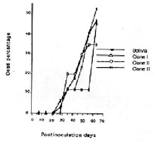
Virulence study - Mortality commenced be-tween 21 and 28 days postinoculation, and reached 40%-60% in every case at day 63 (Fig. 2).
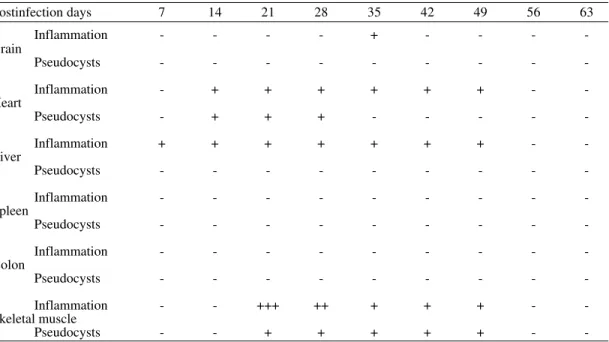
Histopathological study - In the anatomopathol-ogical study of sections of the heart, brain, liver, spleen, skeletal muscle and colon of mice infected with T. cruzi (Bolivia) and clones I, II and III iso-lated from it, notable similarity in tissular tropism could be seen.
Fig. 1: parasitaemia evolution among Trypanosoma cruzi strain Bolivia, Clone I, Clone II and Clone III.
Fig. 2: percentage comparative of mortality among Trypanosoma cruzi strain Bolivia, Clone I, Clone II and Clone III.
TABLE I
Histopathological study of Trypanosoma cruzi (Bolivia strain)
Postinfection days 7 14 21 28 35 42 49 56 63
Inflammation - - - +
Brain
Pseudocysts - - -
-Inflammation - - ++ ++ ++ ++ +++ ++ + Heart
Pseudocysts - - ++ ++ +++ +++ ++ ++ +
Inflammation + + + + + + + + +
Liver
Pseudocysts - - -
-Inflammation - - -
-Spleen
Pseudocysts - - -
-Inflammation - - -
-Colon
Pseudocysts - + - - -
-Inflammation - - + + + + + + +++
Skeletal muscle
Pseudocysts - + + + + + + ++ ++
+ < 25%; ++ between 25% and 50%; +++ > 50% of the surface affected
Fig. 3: amastigotes of Trypanosoma cruzi into myocardic fiber of mice infected with Bolivia strain. H/E X 400.
of the acute phase, more than 50%.
In Clone I (Table II), however, the inflamma-tory effect on the heart is very slight, and altogether absent in the liver; the predominant effect is muscu-lar, with inflammatory infiltration in the brain and
TABLE II
Histopathological study of Trypanosoma cruzi (Clone I)
Postinfection days 7 14 21 28 35 42 49 56 63
Inflammation - - - - + + + + +
Brain
Pseudocysts - - -
-Inflammation - - + + + ++ ++ + +
Heart
Pseudocysts - - - + +
-Inflammation - - -
-Liver
Pseudocysts - - -
-Inflammation - - -
-Spleen
Pseudocysts - - -
-Inflammation - - - + + + - -
-Colon
Pseudocysts - - - + ++ + - -
-Inflammation - ++ + + +++ ++ ++ + + Skeletal muscle
Pseudocysts - - + + ++ ++ - -
-+ < 25%; -+-+ between 25% and 50%; -+-+-+ > 50% of the surface affected
Clone II (Table III) shows little alteration in the heart and slightly more in the muscle (Fig. 4), though never more than 25% of the affected area. Pieces infected with this Clone II showed also iso-lated hyaline necrosis of non-parasitized
myocar-dial fibers. This clone had as a peculiarity an he-patic inflammation throughout the study.
Clone III (Table IV) shows cardiac and muscle alterations, in addition to cerebral inflammation which was evident in 78.6% of the mice studied.
TABLE III
Histopathological study of Trypanosoma cruzi (Clone II)
Postinfection days 7 14 21 28 35 42 49 56 63
Inflammation - - - - + - - -
-Brain
Pseudocysts - - -
-Inflammation - + + + + + + -
-Heart
Pseudocysts - + + + - - - -
-Inflammation + + + + + + + -
-Liver
Pseudocysts - - -
-Inflammation - - -
-Spleen
Pseudocysts - - -
-Inflammation - - -
-Colon
Pseudocysts - - -
-Inflammation - - +++ ++ + + + -
-Skeletal muscle
Pseudocysts - - + + + + + -
Fig. 4: inflammatiory infiltration and pseudocyst in skeletal mucle of mice infected with Clone II of Trypanosoma cruzi. H/E X 400.
TABLE IV
Histopathological study of Trypanosoma cruzi (Clone III)
Postinfection days 7 14 21 28 35 42 49 56 63
Inflammation - + + + + + + +
-Brain
Pseudocysts - - -
-Inflammation - - + ++ ++ + + +
-Heart
Pseudocysts - - + + + - - -
-Inflammation - + - - -
-Liver
Pseudocysts - - -
-Inflammation - - -
-Spleen
Pseudocysts - - -
-Inflammation - - -
-Colon
Pseudocysts - - -
-Inflammation - + ++ +++ ++ ++ ++ ++ -Skeletal muscle
Pseudocysts - + ++ ++ + + - -
-+ < 25%; -+-+ between 25% and 50%; -+-+-+ > 50% of the surface affected
Due to the possible existence of non-specific lymphocytary proliferation we have to be very cau-tious with the interpretation of the inflammatory data (De Diego et al. 1991). Hearts and skeletal muscles disclose diffuse and focal inflammatory
infiltration with macrophages, eosinophils, lym-phocytes and plasma cells, never occupying areas bigger than 50% of the total tissue.
and varied in intensity and extension from case to case. Infiltration with polymorphonuclear eosino-phils was identified in several cases.
DISCUSSION
Trypanosoma cruzi, the etiological agent of Chagas’ disease, shows great biochemical variabil-ity which enables a large variety of medical and biological properties to develop, depending on its geographic distribution. Many strains also develop variations over time (Morel et al. 1980, Dvorak 1984, Tibayrenc et al. 1986, Tibayrenc & Ayala 1991).
The general objective of this work has been to verify the hypothetical correlation between natu-ral clones and their genetic variability.
Specifically, a comparative study has been done between three clones from Bolivia strain isolated from their natural vector; these clones have been compared among themselves and each of them with the mother strain, observing if they behave as in-dependent genetic entities.
The high biochemical and medical heteroge-neity of T. cruzi has already been shown (Dvorak 1984, Brenière et al. 1984, Tibayrenc et al. 1986, Tibayrenc & Ayala 1988, Tibayrenc et al. 1990), the principal question being whether the high clonal diversity might be responsible for all or part of this variability (Tibayrenc & Ayala 1987).
This genetic heterogeneity can be expressed phenotypically in different ways, such as in their different degrees of virulence. Differences in phenotypical variation can be detected in the anti-genic composition of various strains of T. cruzi. When the clones are isolated from various strains and compared, they differ according to their anti-genic composition, sequences of nucleotides of k-DNA, electrophoresis of isoenzymes, virulence, histotropism, and pathology.
In this work, a blood study was done, and in all cases the pattern of evolution of parasitaemia was similar.
No important variations were observed in viru-lence of any of the clones, or in the mother strain Bolivia, although recent studies in the cloning of T. cruzi show differences related mainly with the degree of virulence between those isolated from the mother strain (Andrade et al. 1985, Postan et al. 1987).
Blood pleomorphism of Bolivia strain has an absolute predominance of stout forms during the acute phase, which conforms to the characteristics of Andrade’s group II (1974). Previous classifica-tions by Brener and Chiari (1963) referring only to blood pleomorphism do not find these charac-teristics. However, research by Ribeiro et al. (1988) corroborates the predominance of stout forms in
the Bolivia strain in infections in Swiss mice. In our study, in Bolivia strain as in their iso-lated clones, as infection time passes, stout forms become a majority; this fact would make them fit within the second group of Brener and Chiari (1963), which gathers the strains with a predomi-nance of stout forms and agrees with that described by Andrade (1974) for her group II.
In Clone III the last day the number of stout forms fall, with the slender forms increasing si-multaneously, as described by Dvorak (1976) who finds that the morphogenesis of blood forms be-gins with a predominance of broad-stout, giving way to slender as happens in our study.
We can thus summarise that both Bolivia strain and clones show relatively slow multiplication with irregular peaks of parasitaemia, virulence with mean mortality into the acute phase and predomi-nance of stout forms that fit within the framework of Andrade’s group II (1974).
The histopathological study has demonstrated a clear tendency towards muscular tissue both in clones as in the Bolivia strain. However, we find some specific differences as follows: (a) clones II and III have a skeletal muscular tropism, (b) Bo-livia strain is plainty cardiac, (c) clone I displays both muscular and cardiac alterations, with an im-portant effect in the intestine. These preferences have been established as a function of the inten-sity of the presence of T. cruzi in each organ. De-spite the strains’ predilection for certain types of tissue, they can invade other tissues (Koberle 1968, Hoare 1972, Braun & Titto 1985).
Both the mother strain and the clones that fall within the framework of Andrade’s Group II (1974) in their blood pleomorphism, exhibit the pattern of typically cardiac histopathological alterations, notwithstanding inflammatory lesions and pseudocysts that develop in other organs.
As a result we find that there are differences between the mother strain T. cruzi (Bolivia) and clones I, II, and III; this could be explained by con-sidering that: (1) strain may have pluripotencial pathogenic effects; (2) changes in histopathogical patterns are considered to depend as much on the characteristics of the host as on the parasite (Postam et al. 1987); as tropism results from interaction be-tween parasite and host membranes, an alteration of either of these characteristics could change the tropism.
be the result of a natural selection of its clones. In addition, Bolivia strain was checked by enzime pro-files and these were similar to other included in one of the “major clones” studied by Tibayrenc and Ayala (1988), and very different from those of other major clones (unpublished data), being the underlining data that all stocks pertaining to a given major clone, independly of their geographical and host origin, could share common biological prop-erties that could be radically different from the properties of other clones.
REFERENCES
Andrade SG 1974. Caracterização de cepas do
Trypanosoma cruzi isoladas no Recôncavo Baiano.
Rev Patol Trop 3: 65-121.
Andrade SG, Andrade Z 1968. Patologia da doença de Chagas experimental de longa duração. Rev Inst Med Trop São Paulo 10: 180-187.
Andrade V, Andrade SG, Barral-Netto M, Pontes AL, Castro R 1985. Avaliação do comportamento de diferentes cepas do Trypanosoma cruzi na infecção de seis linhagens isogênicas de camundongos. Rev Soc Bras Med Trop 18: 143-154.
Braun M, Titto E 1985. Immune response to
Trypanosoma cruzi. An approach to the pathogen-esis of Chagas disease. Acta Physiol Pharmacol Lat Am 35: 1-48.
Brener Z, Chiari E 1963. Variações morfológicas observadas em diferentes amostras de Trypanosoma cruzi. Rev Soc Bras Med Trop 5: 220-224. Brénière SF, Poch O, Seales H, Tibayrenc M, Lemesre
JL, Antezana G 1984. Specific humoral depression in chronic patients infected by Trypanosoma cruzi.
Rev Soc Bras Med Trop 26: 254-258.
De Diego JA, Penin P, del Rey J, Mayer R, Gamallo C 1991. A comparative pathological study of three strains of Ttrypanosoma cruzi in an experimental model. Histol Histopath 6: 199-206.
Dvorak J A 1976. New in vitro approach to quantitation of Trypanosoma cruzi vertebrate infections, p. 109-120. In American Trypanosomiasis Research. WHO Scientific Publication No. 318, Washington. Dvorak JA 1984. The natural heterogeneity of
Trypanosoma cruzi. Biological and medical impli-cations. J Cell Biol 24: 357-360.
Hoare CA 1972. Trypanosoma (Schizotrypanum) cruzi,
p. 332-380. In CA Hoare The Trypanosomes of mam-mals. Blackwell Scientific Publication, Oxford. Köberle F 1968. Chagas’ disease and Chagas’ syndrome:
the pathology of American Trypanosomiasis, p. 63-116. In F Köberle Advances in Parasitology. Aca-demic Press, New York.
Lumsden WHR, Herbert WJ, Mc Neillage GJC 1973.
Techniques with Trypanosomes. Churchill Livingstone. Edimburg, London, 183 pp.
Morel C, Chiari E, Plessmann E, Mattei DM, Romanha AJ, Simpson L 1980. Strain and clones of
Trypanosoma cruzi can be characterized by pattern of restriction endonuclease products of kinetoplast DNA. Proc Natl Acad Sci USA 77: 6810-6814. Phillips NR 1960. Experimental studies on the
quantita-tive transmission of Trypanosoma cruzi: consider-ations regarding standartization of material. Am Trop Med Parasitol 54: 60-70.
Postan MC, Daniel JP, Dvorak JA 1987. Comparative studies of the infection of Lewis rats with four
Trypanosoma cruzi clones. Trans R Soc Trop Med Hyg 81: 415-419
Ribeiro RD, Lopes RA, García TAR, Campos A 1988. Histopathological study of the mammary gland in
Trypanosoma cruzi infected mice. Parasitol Res 74: 290-292.
Tibayrenc M, Ayala FJ 1987. Trypanosoma cruzi popu-lations; more clonal than sexual. Parasitol Today 7: 228-232.
Tibayrenc M, Ayala FJ 1988. Isoenzyme variability in
Trypanosoma cruzi, the agent of Chagas’ disease. Genetical, taxonomical and epidemiological signifi-cance. Evolution 42: 277-292.
Tibayrenc M, Ayala FJ 1991. Towards a population genetic of microorganisms: the clonal theory of para-sitic protozoa. Parasitol Today 7: 228-232. Tibayrenc M, Kjellerg F, Ayala FJ 1990. A clonal theory
of parasitic protozoa: the population structures of
Entamoeba, Giardia, Leishmania, Naegleria, Plas-modium, Trichomonas and Trypanosoma, and their medical and taxonomical consequences. Proc Natl Acad Sci USA 87: 2414-2418.