Spermatogenesis and Nucleolar Activity in
Triatoma klugi
(Triatomine, Heteroptera)
Laiana Cristina da Costa, Maria Tercília Vilela de Azeredo-Oliveira and Ester Tartarotti
Departamento de Biologia, Instituto de Biociências, Letras e Ciências Exatas,
Universidade Estadual Paulista “Julio de Mesquita Filho”, São José do Rio Preto, SP, Brazil.
Abstract
Triatoma klugi is a Chagas disease vector in the Rio Grande do Sul State. Triatominae chromosomes are holocentric and sex chromosomes segregation is post-reductional. In this paper we describe the karyotype of maleT. klugi and a meiotic analysis including the nucleolar behavior during spermatogenesis. Testis cells were analyzed after lacto-acetic orcein and silver nitrate staining. Two autosomes and the heterochromosomes presented nucleolar activity (Ag-NORs) during diplotene-diakinesis. The analysis of metaphase I and II revealed a karyotype with 2n = 20+XY. In metaphase I a prominent nucleolar mass was observed in the cell periphery and small silver grains were detected in metaphase II. During anaphase, the chromosomes segregated in parallel and a typical holocentric late migration be-havior was observed. The restoration of the nucleolus was an important feature in this phase. During telophase nu-cleolar masses persisted and in early spermiogenesis the spermatids presented a small peripheral mass until elongation. The present study is a contribution to the study of chromatin behavior and nucleolar persistence in meio-sis.
Key words: Triatoma klugi, Triatomine, cytogenetics, nucleolus, karyotype, meiosis.
Received: August 27, 2007; Accepted: January 16, 2008.
Introduction
The hematophagous insects belonging to the Tria-tominae subfamily are best known as triatomines. These ar-thropods are vectors of the Chagas disease etiological agent, the protozoanTrypanosoma cruzi (T. cruzi). The dis-ease is endemic in South America with pronounced rele-vance in heart illnesses. It is considered thatT. cruziinfects about 11 million people in endemic regions (Diaset al., 2002). Chagas disease represents the third most common parasitic affection, after malaria and schistosomiasis (Guzmán-Bracho, 2001).
The Triatominae subfamily includes 137 insect spe-cies (Galvão et al., 2003) divided into six tribes:
Alberproseniini, Bolboderini, Cavernicolini,
Linshcosteini, Rhodniini, and Triatomini. Eighteen triato-mine genera have been described: Alberprosenia, Belminus, Bolbodera, Microtriatoma, Parabelminus, Cavernicolai, Linshcosteus, Psammolestes, Rhodnius, Dipetalogaster, Eratyrus, Hermanlentia, Meccus, Mepraia, Nesotriatoma, Paratriatoma, Panstrongylus,and
Triatoma(Galvãoet al., 2003).
Triatomines are homogeneous in relation to their hematophagous behavior and all species are potential vec-tors of Chagas disease (Tartarottiet al., 2004).Triatoma klugi(T. klugi) is an important vector in sylvan environ-ments and is distributed throughout the state of Rio Grande do Sul. This species is part of the “oliverai complex”, which also includesT. matogrossensis,T. williami,T. guazuand
T. jurbergi(Carcavalloet al., 2001).Triatoma klugi speci-mens susceptible to different lineages of Trypanosoma cruzishowed an infection similar to that observed in Triat-oma infestans (T. infestans)(Joukoskiet al., 2000). Under laboratory conditions this species presented an infection rate for theT. cruzilineages 60% higher than previously ob-served (Emmanuelle-Machadoet al., 2002).
Cytogenetic studies revealed that the typical number of chromosomes in Triatominae is 2n = 22, with 20 auto-somes and two sex chromoauto-somes (XX, XY) (Ueshima, 1979). Most species have 20 autosomes, except for Triat-oma rubrofasciata(22A),Panstrongylus megistus andT. nitida(18A) (Dujardinet al., 2000). Fifty species have had their karyotypes described and 30 of them were shown to possess 2n = 20A+XY (Panzeraet al., 1998; Morielle and Azeredo-Oliveira, 2004). The variation in the number of chromosomes was mainly due to the presence of different sex determining systems. The subfamily displayed a frag-Send correspondence to Ester Tartarotti. Departamento de
Biolo-gia, Instituto de Biociências, Letras e Ciências Exatas, Univer-sidade Estadual Paulista “Julio de Mesquita Filho”, Rua Cristóvão Colombo 2265, Jardim Nazareth, 15054-000 São José do Rio Preto, SP, Brazil. E-mail: [email protected].
mentation of the males X chromosome and the following systems have been observed: XY, X1X2Y and X1X2X3Y. These three systems have been observed in the genus Triat-oma (Panzera et al., 1998). A fourth sex determination mechanism, X1X2Y1Y2, has been recently described in
Mepraia spinolai(Frías and Atria, 1998).
Triatomines have holokinetic chromosomes, which are characterized by the absence of centromere in the pri-mary chromosome constriction (Pérezet al., 2000). These insects possess a peculiar behavior during meiosis, in which the first meiotic division is reductional for autoso-mes and equational for the sex chromosoautoso-mes (González-Garcíaet al., 1996).
Several authors have studied the nucleolus organizer regions (NORs) in Heteroptera (Camacho et al., 1985; Fossey and Liebenberg, 1995; Cattani and Papeschi, 2004). In the subfamily Triatominae the nucleolus persists during meiosis (Tartarotti and Azeredo-Oliveira, 1999b; Morielle and Azeredo-Oliveira, 2004). Some studies performed in insects indicated that silver nitrate stained structures could represent kinetochore or centromere material (Sujaet al., 1991; Rufas and Gosálvez, 1982). Ultrastructural analysis of Tricholepidion (Insecta) spermiogenesis evidenced a lump of diffuse granular material, the related to the centrio-le in early spermatids (Dallaiet al., 2001). This structure re-sembles the nucleolar bodies present in triatomine spermatids.Triatoma klugihas been recently described by Carcavalloet al.(2001) and it is morphologically related to
T. oliveirai, justifying its inclusion in the oliveirai complex (Emmanuelle-Machadoet al., 2002). The purpose of this paper was to describe the karyotype of maleTriatoma klugi
and to analyze the chromosomes meiotic behavior and nu-cleolar cycle during spermatogenesis.
Materials and Methods
Specimens of Triatoma klugi (subfamily Triato-minae, family Reduviidae, order Heteroptera; Carcavalloet al., 2001) were supplied by the Araraquara Special Health Service (SESA), part of the Epidemiology Department of the Public Health Faculty of University of São Paulo (USP).
Triatoma klugiis found in gaps between rocks and is distributed throughout the district of Nova Petrópolis, in the state of Rio Grande do Sul. These insects are predominantly black; with a central yellow spot on each connexivum seg-ment (Carcavalloet al., 2001).
We analyzed testes from 20 young adults. After the seminiferous tubules were squashed, part of the material was stained with lacto-acetic orcein (De Vaioet al.1985) and part was submitted to silver nitrate staining (Howell and Black, 1980). Measurements of the sex chromosomes were performed with the Image Tool version 3 for Win-dows (UTHSCSA, 1995/2002) in ten metaphase I cells. The photomicrographs were taken in Zeiss-Jenaval photo-microscope.
Results
Karyotype and meiosis ofTriatoma klugi
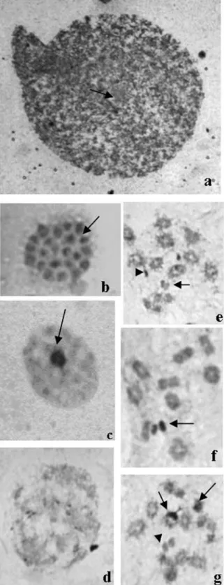
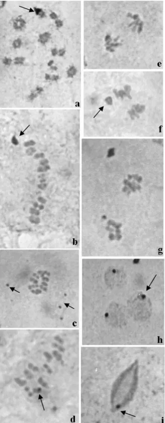
Triatoma klugimales (Figure 1a) presented a karyo-type with 2n = 22, consisting of 20 autosomes and a XY pair (Figure 1b). The Y chromosome (2,21±0,12µm) was larger than the X (1,85±0,16µm). The polyploid nuclei of nutritious cells from the seminiferous tubules walls pre-sented on average five positive heteropycnotic corpuscles (Figure 2a). In spermatogonial interphase cells, six positive heteropycnotic corpuscles were observed (Figure 2b). In the diffuse stage of prophase I, the nuclei had one or two positive heteropycnotic corpuscles, probably formed by sex chromosomes (Figure 2c-d), and were more compacted during the diffuse phase (Figure 2 e-f). During diplotene-diakinesis autosome bivalents presented chiasmata and the sex chromosomes were highly condensed (Figure 2g). At metaphase I the ten autosome bivalents and the sex chro-mosomes were arranged on the metaphase plate (Figure 3a) and usually disposed in a ring configuration (Figure 3b). In metaphase II, ten autosomes and one sex chromosome were present (Figure 3c-d). During anaphase II, late migrating chromosomes were seen (Figure 3e-f), which were some-times also observed later at telophase II (Figure 3g). Posi-tive heteropycnotic corpuscles were observed in early spermatids, but not in (Figure 3h) cells at more advanced differentiation stages (Figure 3i).
Silver nitrate staining
After silver nitrate staining the polyploid nuclei of nutritious cells from the seminiferous tubules walls pre-sented a central nucleolus (Figure 4a). Chromosomes joined by chromatin filaments were observed at sperma-togonial metaphases (Figure 4b), while a central/lateral nu-cleolus and small nucleolar bodies could be seen in sperma-tocytes (Figure 4c). Dispersed nucleolar masses were present at the beginning of the diffuse stage (Figure 4d) and during diplotene-diakinesis the sex chromosomes some-times presented an active NOR (Figures 4e-g). At these same stages NORs were detected on two autosome biva-lents (Figure 4g) or, alternatively, a prominent NOR was visible in one autosome bivalent (Figure 5a); in both
tions the sex chromosomes presented inactive NORs (Fig-ure 4g and 5a). A large peripheral nucleolar mass was ob-served during metaphase I (Figure 5b) and small silver stained corpuscles were common during metaphase II (Fig-ure 5c). In this phase, at least one autosome presented an ac-tive NOR (Figure 5d). At anaphase, the chromosomes seg-regated to the poles in a parallel configuration and were late migrating, as it is typical for holokinetic chromosomes (Figure 5e). In this stage a prominent nucleolar mass reap-peared (Figure 5f) and at telophase nucleolar remnants could be detected (Figure 5g). Early spermatids also had a small peripheral nucleolar mass (Figure 5h) and during the
Figure 2- Testicular tubules ofTriatoma klugiafter lacto-acetic orcein staining. (a) Polyploid nuclei with on average five heteropycnotic corpus-cles, arrows indicate corpuscles; (b) Spermatogonial interphase cells with six pycnotic corpuscles, the arrow indicates the corpuscles; (c-d) Nuclei at the beginning of prophase I (diffuse stage) presenting one or two con-densed corpuscles formed by the sex chromosomes, arrows indicate cor-puscles; (e-f) Nuclei in early prophase I, sexual corpuscle is indicated by arrow; (g) Nucleus in diplotene-diakinesis, arrow indicates bivalent auto-somes and the arrowhead points to the pseudobivalent sex chromoauto-somes.
elongation stage, spermatids still presented a small silver stained mass (Figure 5i). The masses seen in Figures 5h and 5i probably represent the centriole adjunt already observed in ultrastructural studies in insects (Dallaiet al.2001).
Discussion
Heteroptera have been very important for cytogenetic studies because they possess holocentric chromosomes, late chromosome migration in anaphase and a diffuse stage that lasts from prophase I until early diplotene (Ueshima, 1979; Hughes-Schrader and Schrader, 1961). The typical autosome number (A) in triatomines is 20 (Panzeraet al., 1996) and maleTriatoma klugihad 20 autosomes and two sex chromosomes (20A+XY). This diploid number is the same found inT. brasiliensis, T. infestans, T. guasayana, T. lecticularia, T. matogrossensis, T. pseudomaculata, T. rubrovaria, T. sordida, T. circummaculata, T. delpontei, T. dimidiata, T. maculata, T. melanosoma, T. pallidipennis, T. patagonicaandT.platensis(Panzeraet al.1996; Panzeraet al., 1998). Additionally, in the genusTriatomathe Y chro-mosome is larger than the X (Pérezet al.1997; Pérezet al.
2000; Pérezet al., 2005), as seen inT.klugi. This karyo-typical similarity indicates evolutionary conservation in
Triatoma.
Meiosis in triatomines characteristically present posi-tive heteropycnotic blocks at prophase I (Panzera et al.
1995). InT. klugi,the X and Y chromosomes were associ-ated appearing as a positive heteropycnotic pseudobivalent at this stage. These heteropycnotic elements have already been observed in other triatomine species, such as: T. brasiliensis, T. lecticularis, T. rubrovaria, T. sordida (Ta-vares and Azeredo-Oliveira, 1997), Panstrongylus megistus, P. herreri (Tartarotti and Azeredo-Oliveira, 1999a) andT. vitticeps(Severi-Aguiaret al.2006). How-ever, this phenomenon is not typical of Heteroptera, in whichBelostoma dentatumshowed bivalent autosomes in continuous condensation while both X chromosomes were decondensed and negatively heteropycnotic during diaki-nesis (Papeschi and Bidau, 1985).
InT. klugithe X and Y chromosomes were laterally paired (pseudobivalency) at diplotene-diakinesis and the sex pseudobivalent showed a terminal association during metaphases I and II (Figure 3b). This is possibly due to the presence of available extremities at both ends which are free to interact with spindle fibers. Nevertheless, a lateral association has been observed during metaphase II inT. infestans(Pérezet al.2000).
The sex chromosomes presented two kinds of config-uration during metaphases I and II inT. klugi: they could be positioned at the center of a ring formed by autosomes or, alternatively, could be found at the periphery of the equato-rial plate, which was usually observed in linear metaphases. Such configurations are typical of triatomines and the first type predominated in T. platensis, T. infestans and T. delpontei(Panzeraet al., 1995). Both configurations were
observed inRhodnius domesticus(Morielle and Azeredo-Oliveira, 2004), Panstrongylus megistus and P. herreri
(Tartarotti and Azeredo-Oliveira, 1999a).
Late chromosome migration was observed during meiotic anaphase inT. klugi, a phenomenon also described inPanstrongylus herreri(Tartarotti and Azeredo-Oliveira, 1999a), T. brasiliensis, T. delpontei, T. lecticularia, T. rubrovariaandT. sordida(Tavares and Azeredo-Oliveira, 1997). Late migrating chromosomes can appear either spontaneously or as a consequence of radiation in somatic or germinative cells (Hughes-Schrader and Schrader, 1961).
InT. klugian interaction between microtubules and chromosome ends was specifically seen at anaphase I (Fig-ure 1f-g). The kinetic activity was located at one extremity of the chromatids because the migration is parallel to the spindle axis. This phenomenon was also detected in T. infestans suggesting that the interaction of microtubules during meiosis possesses at least two steps: first, an initial holocentric interaction which determines the stabilization of chromosomes; and second, a restricted interaction to-wards the end that involves segregation (Pérezet al., 2000).
The restriction of kinetic activity to chromosome ends is a general characteristic of holocentric chromo-somes. It has been suggested that these characteristics can facilitate the meiotic processes, such as crossing over, chiasmata terminalization and bivalent orientation on the metaphase plate (Pimpinelli and Goday, 1989).
InT. klugithe chromosomes behave as bivalents in metaphase I even after chiasmata terminalization. In Hete-roptera bivalents usually display a single chiasma in any chromosome region and its presence persists beyond metaphase I (Pérezet al., 1997). The sex chromosomes of
T. klugiwere univalent and pseudobivalent at metaphase I and were predominantly univalent at metaphase II. The sex chromosomes appeared as univalents at metaphase I of the
Antiteuchusspecies of heteropterans, while in metaphase II they were pseudobivalents (Lanzone and Souza, 2006). Nevertheless, a pseudobivalent XY was observed in both the first and second meiotic divisions in the Heteroptera
Graphosoma italicum(González-Garcíaet al.1996). Silver staining revealed impregnated bodies in the polyploid nuclei ofT. klugi. Nucleolar bodies have also been observed in testis polyploid nuclei ofPanstrongylus megistusandP. Herreri(Tartarotti and Azeredo-Oliveira, 1999b). The increase in nucleolar bodies in polyploid nu-clei may be related to increased rRNA synthesis (Tavares and Azeredo-Oliveira, 1997).
During meiotic prophase and metaphases I and II of
T. klugi, an alternation of ribosomal genes activity between autosomes and sex chromosomes was observed. The same phenomenon has already been describedP. megistusandP. herreri(Tartarotti and Azeredo-Oliveira, 1999b) and indi-cates that not all ribosomal genes are necessarily active. The presence of NORs in heterochromosomes is a common
phenomenon in insects and severalDrosophilaspecies, for instance, have ribosomal genes located on their sex chro-mosomes (Bicudo, 1984).
Dispersed nucleolar bodies were observed at meta-phase II inT.klugiand a large nucleolar mass was also ob-served during metaphase I, anaphase I and telophase I. This confirms the occurrence of “nucleolar material persistence” as suggested by Tartarotti and Azeredo-Oliveira (1999b) and provides evidence that the nucleolus does not com-pletely disappear during meiosis in triatomines, but is reor-ganized from pre-existing nucleolar elements.
Nevertheless, some authors suggest that silver nitrate impregnation may also reveal other chromosome struc-tures. Silver staining ofSchistocerva gregaria(Orthoptera) early spermatids apparently revealed kinetochore or centro-mere material, while silver stained structures in elongated spermatids were attributed to a particular kind of con-densed chromatin. Another study performed during meio-sis in Orthopera led to the conclusion that silver staining reveals three distinct chromosome structures: (i) active nu-cleolar organizer regions (ii) kinetochores and (iii) cores that run along each chromatid except at their distal ends (Sujaet al., 1991).
In T. klugi, silver staining observed in early sper-matids disappears during elongation. Post-meiotic NORs reactivation has already been observed in mammalian cells submitted to silver staining (Hofgartneret al., 1979). It is possible that the conservation of post-meiotic NORs reacti-vation in vertebrates and invertebrates indicates the need for post-meiotic RNA synthesis related to differentiation. Alternatively, the silver stained structures seen in sper-matids may represent he centriole adjunt observed in ultra-structural studies of spermiogenesis. An electron micro-scope analysis of Tricholepidion (insecta) showed that, after meiosis is completed, the spermatid has only a single centriole located close to the nucleus. Also connected to the centriole is a lump of diffuse granular material, named the centriole adjunt. The terms “centriole adjunt” or “post-nuclear body” have been given to aggregates of granular masses, which initially accumulate around the centriole and that are believed to differentiate later into various sperm components (Dallaiet al., 2001).
This study presents the description of the karyotype of maleTriatoma klugiand contributes to a better under-standing of chromatin and nucleolar behavior during meio-sis.
Acknowledgments
The authors are grateful to Dr. José M. Soares Barata, Director of the Insectarium (Araraquara, SP), Epidemiol-ogy Department, Public Health Faculty (São Paulo, SP), and to the technicians of the Insectarium Mr. João Luis Molina Gil and Mr. João Mauricio Nóbrega da Silva Filho for providing the insects. We also thank Elza Mitiko Sato for the literature review, Hederson Vinicius de Souza for
help in chromosome measurements, to David Roy Michael Mercer for the English revision, and to FAPESP for finan-cial support.
References
Bicudo HEMC (1984) Variabilidade das regiões organizadoras de nucléolos nos eucariotos. Cienc Cult 440-447 (Abstract in English).
Camacho JPM, Belda J and Cabrero J (1985) Meiotic behaviour of the holocentric chromosomes ofNezara viridula(Insecta, Heteroptera) analysed by C-banding and silver impregna-tion. Can J Genet Cytol 27:490-497.
Carcavallo RU, Jurberg J, Lent H, Galvão C, Steindel M and Pinto CJC (2001) Nova espécie do complexo oliveirai (Nova denominação para o complexo matogrossensis) (Hemiptera, Reduviidae, Triatominae) do estado do Rio Grande do Sul, Brasil. Mem Inst Oswaldo Cruz 96:71-79.
Cattani MV and Papeschi AG (2004) Nucleolus organizing re-gions and semi-persistent nucleolus during meiosis in Spartocera fusca(Thunberg) (Coreidae, Heteropeta). Here-ditas 140:105-111.
Dallai R, Lupetti P, Frati F, Nardi F and Afzelius BA (2001) Sperm ultrastructure and spermiogenesis in the relic species Tricholepidion gertschi Wygodzinsky (Insecta, Zygento-ma). Tissue Cell 33:596-605.
De Vaio ES, Grucci B and Castagnino A (1985) Meiotic differ-ences between three triatomine species (Hemiptera, Reduviidae). Genetica 67:185-191.
Dias JCP, Silveira AC and Schofield CJ (2002) The impact of Chagas control in Latin America: A review. Mem Inst Os-waldo Cruz 97:603-612.
Dujardin JP, Schofield CJ and Panzera F (2000) Les vecteurs de la maladie de Chagas. Recherches taxonomiques, biologiques et genetiques. Acad R Sci dóutre-Mer Bruxelles 24:1-162. Emmanuelle-Machado P, Koerich LB, Joukoski DDB,
Carva-lho-Pinto CJ, Grisard EC and Steindel M (2002) Biology of Triatoma klugiCarcavallo, Jurberg, Lent and Galvão 2001 (Heteroptera, Reduviidae) under laboratory conditions: Ef-fects of distinct blood sources and susceptibility to Trypanosoma cruziandTrypanosoma rangeli, Brazil. Mem Inst Oswaldo Cruz 97:583-587.
Frias D and Atria J (1998) Chromosomal variation, macro-evolution and possible parapatric specition in Mepria spinolai(Porter) (Hemiptera, Reduviidae). Genet Mol Biol 21:179-184.
Fossey A and Liebenberg H (1995) Meiosis and nucleolar struc-tures in the stink bug Carlisis wahlbergi Stal (Coreidae, Heteroptera). Cytobios 81:7-15.
Galvão C, Carcavallo RU, Rocha DS and Jurberg J (2003) A checklist of the current valid species of the subfamily Triato-minae Jeannel, 1919 (Hemiptera, Reduviidae) and their geo-graphical distribution, with nomenclatural and taxonomic notes. Zootaxa 202:1-36.
González-García JM, Antonio C, Suja JA and Rufas JS (1996) Meiosis in holocentric chromosomes: Kinetic activity is ran-domly restricted to the chromatid ends of sex univalents in Graphosoma italicum (Heteroptera). Chromosome Res 4:124-132.
Hofgartner FJ, Schmid M, Krone W, Zenzes MT and Engel W (1979) Pattern of activity of nucleolus organizers during spermatogenesis in mammals as analyzed by silver-staining. Chromosoma 71:197-216.
Howell WM and Black DA (1980) Controlled silver staining of nucleolus organizer regions with protective colloidal devel-oper: I-step method. Experientia 36:1014-1015.
Hughes-Schrader S and Schrader F (1961) The kinetochore of the Hemiptera. Chromosoma 12:327-350.
Joukoski DDB, Grisard EC and Steindel M (2000) Evaluation of the vectorial capacity ofTriatoma klugi, a new triatomine species. Mem Inst Oswaldo Cruz 95(Suppl.II):333. Lanzone C and Souza MJ (2006) Chromosome complement and
meiosis in three species of the Neotropical bug genus Antiteuchus(Heteroptera, Pentatomidae, Discocephalinae). Genet Mol Biol 29:49-55.
Morielle A and Azeredo-Oliveira MTV (2004) Description of the nucleolar activity and karyotype in germinative cell lines of Rhodnius domesticus(Triatominae, Heteroptera). Caryolo-gia 57:31-37.
Panzera F, Perez R, Panzera Y, Alvarez F, Scvortzoff E and Salvatella R (1995) Karyotype evolution in holocentric chromosomes of three related species of triatomines (Hemíptera-Reduviidae). Chromosome Res 3:143-150.
Panzera F, Pérez R, Hornos S, Panzera Y, Cestau R, Delgado V and Nicolini P (1996) Chromosome numbers in Triatominae (Hemíptera-Reduviidae): A review. Mem Inst Oswaldo Cruz 91:515-518.
Panzera F, Scvortzoff E, Pérez R, Panzera Y, Hornos S, Cestau R, Nicolini P, Delegado V, Alvarez F, Mazzella MC, et al. (1998) Cytogenetics of triatomines. In: Carcavallo RU, Galíndez IG, Jurberg J and Lent H (eds) Atlas dos Vetores da Doença de Chagas nas Américas. Editora Fiocruz, Rio de Ja-neiro, pp 621-664.
Papeschi AG and Bidau CJ (1985) Chromosome complement and male meiosis in four species ofBelostomaLatreille (Hete-roptera-Belostomatidae). Rev Bras Genet 8:249-261. Perez R, Panzera F, Page J, Suja JA and Rufas JS (1997) Meiotic
behaviour of holocentric chromosomes: Orientation and se-gregation of autosomes inTriatoma infestans(Heteroptera). Chromosome Res 5:47-56.
Pérez R, Rufas J, Suja J and Panzera F (2000) Meiosis in holo-centric chromosomes: Orientation and segregation of in
autosome and sex chromosomes in Triatoma infestans
(Heteroptera). Chromosome Res 8:17-25.
Pérez R, Rufas J, Suja J and Panzera F (2005) Cytogenetic analy-sis of experimental hybrids in species of Triatominae (Hemiptera-Reduviidae). Genetica 125:261-270.
Pimpinelli S and Goday C (1989) Centromere organization in meiotic chromosomes of Paracaris univalents. Chromo-soma 98:160-166.
Rufas JS and Gosálvez F (1982) Development of silver stained structures during spermatogenesis ofSchistocerca gregaria (FORSK) (Orthoptera, Acrididae). Caryologia 35:261-267. Severi-Aguiar GDC, Lourenço LB, Bicudo HEMC and
Azeredo-Oliveira MTV (2006) Meiosis aspects and nucleolar activity inTriatoma vitticeps(Triatominae, Heteroptera). Genetica 126:141-151.
Suja JA, De La Torre J, Gimenéz-Abián JF, Garcia de la Vega C and Rufas JS (1991) Meiotic chromosome structure. Kine-tochores and chromatid cores in standard and B chromo-somes of Arcyptera fusca(Orthoptera) revealed by silver staining. Genome 34:19-27.
Tartarotti E and Azeredo-Oliveira MTV (1999a) Meiosis patterns
of holocentric chromosomes in Triatomines genus
Panstrongylus. Cytologia 64:235-240.
Tartarotti E and Azeredo-Oliveira MTV (1999b) Patterns of nu-cleolar activity during spermatogenesis of two triatomines, Panstrongylus megistusandP. herreri. Caryologia 52:177-184.
Tartarotti E, Azeredo-Oliveira MTV and Ceron CR (2004) Pro-blemática vetorial da doença de Chagas. Arq Cienc Saude 11:44-47 (Abstract in English).
Tavares MG and Azeredo-Oliveira MTV (1997) Cytogenetic studies on holocentric chromosomes of five species of tria-tomines (Heteroptera, Reduviidae). Cytobios 89:51-61. Ueshima N (1979) Hemiptera II: Heteroptera. In: John, B. Animal
Cytogenetics, v. 3: insecta 6. Gebrüder Borntraeger, Berlim, pp 1-117.
Internet Resources
UTHSCSA image tool for windows. Version 3. San Antonio: the University of Texas Health Science in San Antonio, c 1995/2002, http://www.uthscsa.edu.
Associate Editor: Yatiyo Yonenaga-Yassuda