Surgical repair of pseudoaneurysms and complex arteriovenous
fistula between popliteal vessels
Correção cirúrgica de pseudoaneurismas e fístula arteriovenosa
complexa entre vasos poplíteos
Adenauer Marinho de Oliveira Góes Junior1,2
*
, Carolina Pinheiro de Oliveira1, Camilla Castilho Maia1, Bruno Campos Xavier1, Silvia Karinny Brito Calandrini de Azevedo1
Abstract
An arteriovenous fistula (AVF) is an abnormal and permanent communication between an artery and a vein caused by penetrating traumas or iatrogenic injuries. A penetrating trauma to the endothelial wall can lead to formation of pseudoaneurysms (PSA) and to formation of an AVF. Here, the authors present the case of a patient with a complex AVF of popliteal vessels, associated with popliteal artery pseudoaneurysm, suggested by clinical features and imaging exams, and treated with conventional surgery due to unavailability of a stent graft with appropriate diameter and because endovascular surgery isn’t provided at the service where this patient was operated.
Keywords: arteriovenous fistula; wounds and injuries; popliteal artery; popliteal vein; aneurysm, false; surgery.
Resumo
A fístula arteriovenosa (FAV) é uma comunicação anormal e permanente entre uma artéria e uma veia devido a traumas penetrantes e lesões iatrogênicas. O trauma penetrante na parede arterial pode levar à formação de pseudoaneurismas (PSA) e, se houver lesão venosa concomitante, à formação de uma FAV. Os autores apresentam o caso de um paciente portador de FAV complexa de vasos poplíteos associada a pseudoaneurisma de artéria poplítea, sugeridos a partir de exames clínicos e exames de imagem, e tratados por cirurgia convencional devido à indisponibilidade de um stent
graft com diâmetro apropriado, além de a cirurgia endovascular não estar disponível no serviço em que o paciente foi operado.
Palavras-chave: fístula arteriovenosa; ferimentos e lesões; artéria poplítea; veia poplítea; falso aneurisma; cirurgia.
1 Centro Universitário do Estado do Pará (CESUPA), Belém, PA, Brasil.
2 Hospital Metropolitano de Urgência e Emergência (HMUE), Ananindeua, PA, Brasil.
Financial support: None.
Conflicts of interest: No conflicts of interest declared concerning the publication of this article. Submitted: January 20, 2018. Accepted: May 29, 2018.
INTRODUCTION
An arteriovenous fistula (AVF) is an abnormal and permanent communication between an artery and a
vein,1,2 which is generally associated with penetrating
traumas and iatrogenic injuries.1,3 The duration
of clinical presentation and the time that elapses between trauma and diagnosis vary and may even run to decades.4 Penetrating traumas to the artery
wall can cause formation of pseudoaneurysms and, if there is also venous damage, to development of AVF.1 However, simultaneous occurrence of both
clinical conditions is a rare complication that has been described little.5
The superficial femorals are the vessels most often involved (22%), followed by the popliteal vessels (16%).6 Trauma to popliteal vessels involves significant risk of amputation.7 An AVF is primarily diagnosed
on the basis of clinical status, with localized murmurs and thrills, edema, and venous ulcers. Diagnostic investigation tends to be pursued using imaging exams, such as Doppler echography, angiotomography, and sometimes angiography.1
Part I – clinical situation
The patient was a 53-year-old male who had been wounded in the left thigh by a cartridge belt 2 years and 9 months previously.
The patient complained of pain in the left lower limb, which had developed edema, varicose veins, ochrodermatitis and ulceration of the anterior surface of the leg. The limb involved had no palpable distal pulses and thrill and murmur were detectable from the groin to the proximal third of the leg (with greatest intensity in the popliteal fossa). Pulsation was also noted along the entire length of the patient’s thigh.
Angiotomography revealed AVF of the left popliteal vessels, a left popliteal artery pseudoaneurysm with a 2.6 cm diameter and an aneurysm of the popliteal vein with a 5 cm diameter (Figure 1).
Part II – what was done
The decision was taken to perform open surgery to repair the vessels involved. The procedure was conducted under general anesthesia.
With the patient in ventral decubitus, with a pneumatic cuff already in place at the base of the left thigh (in case of need for urgent hemostasis), an italic-S incision was made in the left popliteal region, revealing large dilations of the popliteal vessels. Dissection of these vessels was complicated by fibrosis and diffuse bleeding caused by venous hypertension of the limb, but it was not necessary to inflate the pneumatic cuff. The infragenicular segment of the popliteal artery was dissected and repaired, but because of the large volume of the dilatations of
Figure 1. (A) and (B) Angiotomography reconstructions from different angles, showing simultaneous impregnation of the arterial
the vessels involved, the surgical field was not large enough to obtain proximal control safely.
The patient was repositioned in dorsal decubitus, to enable a longitudinal incision to be opened along the medial surfaces of the left thigh and leg and proximal and distal control of the vessels involved was achieved. When the artery was clamped, stopping flow through the fistula, arterial hypotension set in and vasoactive drugs were needed.
Arterial and venous ligatures were performed proximal and distal of the AVF and then a graft was constructed from the supragenicular to infragenicular
segments of the popliteal artery using a length of the contralateral great saphenous vein reversed (with proximal end-to-side and distal end-to-end anastomoses). Since the patient was hemodynamically unstable, the operation was concluded without reconstruction of the deep vein system (Figure 2).
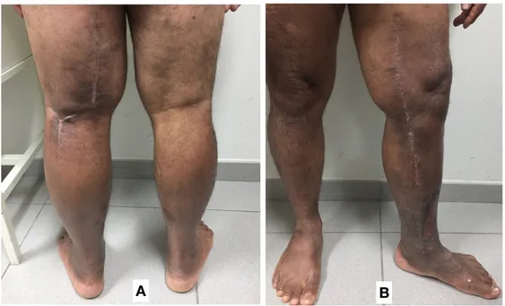
The patient has been in outpatients follow-up for 6 months. The surgical wounds and the venous ulcer have healed and there has been no further increase in edema of the limb compared to the preoperative baseline and the limb has exhibited satisfactory functional recovery (Figure 3).
Figure 2. (A) Posterior italic-S surgical access; n: retractors applied to the sciatic nerve; av: aneurysm of the popliteal vein; v: popliteal
vein; (B) Medial surgical access; the arrow indicates the proximal anastomosis of the graft to the supragenicular popliteal artery; the arrowhead indicates the distal anastomosis of the graft to the infragenicular popliteal artery.
Figure 3. Photographs taken 6 months after the operation, showing the healed surgical scars. (A) Posterior italic-S surgical access;
DISCUSSION
In high output FAVs diagnosed late, the low resistance flow causes arteriomegaly proximal of the AVF.8 There are two theories to explain this dilatation.
According to the first theory, shear forces resulting from the increased flow rate at the site cause increased production of endothelium-derived relaxing factor, which in turn provokes dilatation of arterial smooth muscle. The second theory states that late increase of blood flow in the artery proximal to the AVF causes destruction of the elastic fibers in this segment, with progression to arteriomegaly.4
Many different procedures for repair of pseudoaneurysms and AVFs have been described: aneurysmorrhaphy, resection of the aneurysmal segment and interposition of prosthetic or venous grafts, placement of stent-grafts, and combination procedures.9
Endovascular surgery offers the advantages of reduced morbidity and mortality, shorter length of hospital stay, and preservation of the great saphenous vein,7
although there is still a lack of long-term follow-up studies.10 However, for treatment of true aneurysms
of the popliteal artery, it is known that conventional surgery offers superior long-term patency, particularly in younger patients and when arterial reconstruction is performed using a venous graft.3
In the case described here, conventional surgery was chosen. One of the factors that influenced the decision to employ open surgery was that no covered stent with an appropriate diameter was available,9 because the arteriomegaly proximal to the AVF resulted in a difference in caliber between the artery proximal and distal of the AVF and there is no conical covered stent that could fit the disproportion between these diameters. Possibly, the lack of a suitable conical stent could have been dealt with by releasing multiple covered stents with gradually increasing calibers overlapping each other, using tapered stent-grafts.11-16 However, the long-term efficacy of this endovascular option is lacking evidence from studies, especially in the topography of the knee joint. Furthermore, endovascular surgery is not available at the service where this patient was treated.
In this case, surgical treatment was initiated with a posterior access to the popliteal vessels. Posterior access should be used when the objective is to expose only the segments of the popliteal vessels posterior to the joint.7 However, the large volume
of the venous aneurysm interfered with the arterial dissection procedure and the posterior approach did not provide sufficient access to safely control the proximal artery. The patient was therefore turned over to dorsal decubitus and a medial access was
performed, providing good exposure and enabling the initial incision to be extended.7
When we analyzed the angiotomography images retrospectively, we concluded that the posterior access should not have been attempted. We recommend that in future cases the level of the lesion in relation to the patella should be used as a reference for planning surgery and that medial access is preferable when the lesion is higher than the upper margin of the patella.
In cases such as the one reported here, the priority is arterial reconstruction, which was achieved with a graft constructed using the contralateral great saphenous vein, preserving the saphenous vein in the operated limb for venous return, because of the possible need to ligate the deep vein system. Whenever possible, the deep vein system should also be reconstructed by venorrhaphy, resection, and anastomosis or grafting. Venous ligature should be avoided because of the possibility of chronic venous hypertension of the limb and its medium and long-term clinical repercussions.1
In the case reported here, popliteal vein ligature was performed because of the risk of thrombosis of the large venous aneurysm and of emboli after the AVF was closed. Additionally, reconstruction with a venous graft, which had been planned initially, was not performed because of hemodynamic instability.
REFERENCES
1. Santos EP Jr, Batista RRA, Felici FM, Correia VE, Oliveira MB, Alves RF. Correção endovascular de fístula arteriovenosa traumática em ilíaca interna com stent revestido. J Vasc Bras. 2014;13(1):48-52. http://dx.doi.org/10.1590/jvb.2014.010.
2. Rogel-Rodríguez JF, Zaragoza-Salas T, Díaz-Castillo L, Noriega-Salas L, Rogel-Rodríguez J, Rodríguez-Martínez JC. Fístula arteriovenosa femoral postraumática, tratamiento endovascular. Cir Cir. 2017;85(2):158-63. http://dx.doi.org/10.1016/j.circir.2015.10.010. PMid:26763666.
3. McVeigh PZ, Kayssi A, Lo A, Rajan DK, Oreopoulos GD, Roche-Nagle G. Repair of popliteal aneurysm and spontaneous arteriovenous fistula in a patient with Marfan syndrome. J Vasc Surg Cases. 2016;2:137-40.
4. Góes AMO Jr, Jeha SAH, Franco RSM. Tratamento híbrido para fístula arteriovenosa entre vasos poplíteos. J Vasc Bras. 2014;13(4):325-9. http://dx.doi.org/10.1590/1677-5449.0024.
5. Azevedo ACA, Taveira TS, Cristino MAB, Barros MVL. Pseudoaneurisma de artéria femoral associado a fístula arteriovenosa iatrogênica. Arq Bras Cardiol. 2015;28(4):231-5.
6. Espinosa SDT, Carrillo LRV, Hernández FG. Tratamiento quirúrgico y endovascular de las fístulas arteriovenosas secundarias a trauma vascular. Orthotips. 2013;9(2):99-103.
7. Zizi O, Naouli H, Jiber H, Bouarhroum A. Fistule artérioveineuse poplitée post-traumatique associée à un faux anévrysme. JMV-Journal de Médecine Vasculaire. 2017;42(1):46-9. http://dx.doi. org/10.1016/j.jdmv.2017.01.006.
graves: desafio terapêutico. J Vasc Bras. 2014;13(1):34-8. http:// dx.doi.org/10.1590/jvb.2014.007.
9. Moreira RWC, Carrilho DDR, Costa KMAH, Pinheiro RBB. Correção cirúrgica de aneurismas saculares de fístula arteriovenosa para hemodiálise utilizando a técnica de aneurismorrafia. J Vasc Bras. 2011;10(2):165-7. http://dx.doi.org/10.1590/S1677-54492011000200012.
10. Oliveira FM, Macedo AAR, Rodrigues APM, et al. Trauma inguinal penetrante com formação de fístula arteriovenosa e pseudoaneurisma: relato de caso. J Vasc Bras. 2015;14(4):364-7. http://dx.doi.org/10.1590/1677-5449.003315.
11. Góes AMO Jr, Jeha SAH. Stent Graft-in-Stent Graft as a Rescue Technique for Endovascular Treatment of Giant Extracranial Internal Carotid Aneurysm. Case Rep Surg. 2016;2016:2656421. PMid:27752387.
12. Nigro G, Gatta E, Pagliariccio G, Grilli C, Carbonari L. Use of the Gore hybrid vascular graft in a challenging high-lying extracranial carotid artery aneurysm. J Vasc Surg. 2014;59(3):817-20. http:// dx.doi.org/10.1016/j.jvs.2013.04.044. PMid:23777810.
13. Janjua N, Alkawi A, Georgiadis AL, Kirmani JF, Qureshi AL. Covered stent graft for treatment of a pseudoaneurysm and carotid blowout syndrome. J Vasc Interv Neurol. 2008;1(1):5-8. PMid:22518207.
14. Lesley WS, Weigele JB, Chaloupka JC. Outcomes for overlapping stents in the extracranial carotid artery. Catheter Cardiovasc Interv. 2004;62(3):375-9. http://dx.doi.org/10.1002/ccd.20090. PMid:15224307.
15. Amistà P, Barbisan D, Beghetto M, Cavasin N, Zucchetta P, Frego M. Three-stent placement for treatment of carotid artery pseudoaneurysm. A case report. Interv Neuroradiol. 2006;12(4):339-43. http://dx.doi.org/10.1177/159101990601200408. PMid:20569592.
16. Stager V, Gandhi R, Stroman D, Timaran C, Broker H. Traumatic internal carotid artery injury treated with overlapping bare metal
stents under intravascular ultrasound guidance. J Vasc Surg. 2011;53(2):483-6. http://dx.doi.org/10.1016/j.jvs.2010.08.032. PMid:20875711.
*
Correspondence:
Adenauer Marinho de Oliveira Góes Junior Rua Domingos Marreiros, 307/802 - Umarizal CEP 66055-210 - Belém (PA), Brasil Tel.: +55 (91) 981279656 E-mail: adenauerjunior@gmail.com
Author information
AMOGJ - Full member, Sociedade Brasileira de Angiologia e Cirurgia Vascular (SBACV); Vascular surgeon, Hospital Metropolitano de Urgência e Emergência (HMUE); Coordinator, Residência Médica de Cirurgia do Trauma (HMUE); Professor of Vascular Surgery, Centro Universitário do Estado do Pará (CESUPA). CPO, CCM, BCX, SKBCA - Medical students, Centro Universitário do Estado do Pará (CESUPA).
Author contributions
Conception and design: AMOGJ Analysis and interpretation: AMOGJ Data collection: AMOGJ, CPO, CCM, BCX, SKBCA Writing the article: AMOGJ, CPO, CCM, BCX, SKBCA Critical revision of the article: AMOGJ Final approval of the article*: AMOGJ, CPO, CCM, BCX, SKBCA Statistical analysis: N/A Overall responsibility: AMOGJ