Validation of a Rapid and Sensitive LC-MS/
MS Method for Determination of
Exemestane and Its Metabolites, 17
β
-Hydroxyexemestane and 17
β
-Hydroxyexemestane-17-O-
β
-D-Glucuronide:
Application to Human Pharmacokinetics
Study
Ling-Zhi Wang1,2*, Sok-Hwei Goh3, Andrea Li-Ann Wong1,4, Win-Lwin Thuya1, Jie-Ying Amelia Lau1, Seow-Ching Wan1, Soo-Chin Lee1,4, Paul C. Ho3, Boon-Cher Goh1,2,4*
1Cancer Science Institute of Singapore, National University of Singapore, 14 Medical Drive, Singapore 117599, Singapore,2Department of Pharmacology, Yong Loo Lin School of Medicine, National University Health System, Singapore 117599, Singapore,3Department of Pharmacy, Faculty of Science, National University of Singapore, 18 Science Drive 4, Singapore 117543, Singapore,4Department of Haematology & Oncology, National University Health System, Singapore 119074, Singapore
*[email protected](LZW); [email protected](BCG)
Abstract
A novel, rapid and sensitive liquid chromatography-tandem mass spectrometric (LC-MS/ MS) method was developed and validated for the evaluation of exemestane pharmacokinet-ics and its metabolites, 17β-dihydroexemestane (active metabolite) and 17β -dihydroexe-mestane-17-O-β-D-glucuronide (inactive metabolite) in human plasma. Their respective D3 isotopes were used as internal standards. Chromatographic separation of analytes was achieved using Thermo Fisher BDS Hypersil C18 analytic HPLC column (100 × 2.1 mm, 5μm). The mobile phase was delivered at a rate of 0.5 mL/min by gradient elution with 0.1 % aqueous formic acid and acetonitrile. The column effluents were detected by API 4000 tri-ple quadrupole mass spectrometer using electrospray ionisation (ESI) and monitored by multiple reaction monitoring (MRM) in positive mode. Mass transitions 297>121m/z, 300
>121m/z, 299>135m/z, 302>135m/z, 475>281m/z, and 478>284m/zwere moni-tored for exemestane, exemestane-d3, 17β-dihydroexemestane, 17β -dihydroexemestane-d3, 17β-dihydroexemestane-17-O-β-D-glucuronide, and 17β-dihydroexemestane-17-O-β -D-glucuronide-d3 respectively. The assay demonstrated linear ranges of 0.4–40.0 ng/mL,
for exemestane; and 0.2–15.0 ng/mL, for 17β-dihydroexemestane and 17β
-dihydroexe-mestane-17-O-β-D-glucuronide, with coefficient of determination (r2) of>0.998. The preci-sion (coefficient of variation) were10.7%, 7.7% and 9.5% and the accuracies ranged from 88.8 to 103.1% for exemestane, 98.5 to 106.1% for 17β-dihydroexemestane and 92.0 to 103.2% for 17β-dihydroexemestane-17-O-β-D-glucuronide. The method was successfully a11111
OPEN ACCESS
Citation:Wang L-Z, Goh S-H, Wong AL-A, Thuya W-L, Lau J-YA, Wan S-C, et al. (2015) Validation of a Rapid and Sensitive LC-MS/MS Method for Determination of Exemestane and Its Metabolites, 17β-Hydroxyexemestane and 17β
-Hydroxyexemestane-17-O-β-D-Glucuronide: Application to Human Pharmacokinetics Study. PLoS ONE 10(3): e0118553. doi:10.1371/journal. pone.0118553
Academic Editor:Junxuan Lu, Texas Tech Univ School of Pharmacy, UNITED STATES
Received:October 30, 2014
Accepted:January 20, 2015
Published:March 20, 2015
Copyright:© 2015 Wang et al. This is an open access article distributed under the terms of the Creative Commons Attribution License, which permits unrestricted use, distribution, and reproduction in any medium, provided the original author and source are credited.
Data Availability Statement:All relevant data are within the paper.
applied to a pharmacokinetics/dynamics study in breast cancer patients receiving exemes-tane 25mg daily orally. For a representative patient, 20.7% of exemesexemes-tane in plasma was converted into 17β-dihydroexemestane and 29.0% of 17β-dihydroexemestane was inacti-vated as 17β-dihydroexemestane-17-O-β-D-glucuronide 24 hours after ingestion of exe-mestane, suggesting that altered 17-dihydroexemestane glucuronidation may play an important role in determining effect of exemestane against breast cancer cells.
Introduction
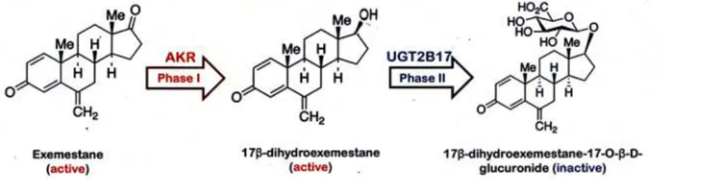
Breast cancer has remained the most common cancer among females in Singapore over the last forty years.[1] Currently, the role of exemstane (Exe) in breast cancer treatment is evolving; traditionally used as an adjuvant medication for hormone receptor positive breast cancer, new trials have investigated its role as adjuvant medication in metastatic cancer and preventive medication in high-risk women.[2–6] In the adjuvant setting, Exe, similar to the other third-generation compounds, revealed improved relapse-free survival compared to tamoxifen mono-therapy when administered as sequential mono-therapy.[7] Exe is the first steroidal aromatase inhibi-tor (AI) which inhibitsin vivoformation of oestrogens (estrone and estradiol), thereby reducing stimulation for breast cancer cell proliferation.[8] Variability in response and side ef-fect profile has been observed in patients in many AI clinical trials and the underlying mecha-nism remains undefined.[9–13] Recently, the major metabolic pathway of Exe has been delineated; as a reduction of double bond in 17 keto group via aldo-keto reductase (AKR) to form 17β-dihydroexemestane (17DhExe) is a major pathway for exemestane phase I metabo-lism. [14] In addition, 17DhExe has been reported to be an active metabolite which is subse-quently inactivated by glucuronidation to 17β-dihydroexemestane-17-O-β-D-glucuronide (Exe17Oglu). [15,16] Inactivation of 17DhExe is catalysed by the enzyme UDP-gluconoryl-transferase 2B17 (UGT2B17). Exemestane’s major metabolism pathway is summarized in Fig. 1. It has also been identified that 60–70% of Asians suffer from homozygous gene deletion of UGT2B17, which can result in reduced glucuronidation of 17DhExe and increased exposure to this active metabolite. [16] Therefore, simultaneous quantification of Exe, 17DhExe, and Exe17Oglu can aid in determination of the impact of homozygous UGT2B17 gene deletion on thein vivometabolic profile of Exe. [17]
To date, two reported analytical methods have been developed and validated for determina-tion of Exe and 17DhExe; one is a HPLC-UV assay with low limit of quantificadetermina-tion (LLOQ) of 10 ng/mL for both parental drug and 17DhExe [18] and the other is a LC-MS/MS method with
Fig 1. Exemestane major metabolism pathway.
doi:10.1371/journal.pone.0118553.g001 and analysis, decision to publish, or preparation of
the manuscript.
LLOQ of 0.2 ng/ml for Exe and 0.1 ng/ml for 17DhExe.[19] Compared to HPLC-UV method, the LC-MSMS method demonstrated superior sensitivity and specificity which are critical to accurately quantify analytes of interest. Nevertheless, no analytical method has been estab-lished to concurrently quantify Exe and its two key metabolites (17DhExe and Exe17Oglu) within one chromatographic run. This is a major research gap to determine the influence of UGT2B17 variants on the glucuronidation of 17DhExe in human plasma samples. In order to support a clinical trial of Exe in breast cancer patients in Singapore National University Hospi-tal (NUH), a simple, novel and sensitive LC-MS/MS method was developed for determination of plasma concentrations of these three compounds simultaneously. The well-validated meth-od has been successfully applied to determine the plasma concentrations of Exe and two im-portant metabolites in clinical plasma samples of breast cancer patients.
Experimental
Chemicals and Reagents
Exemestane, 17β-hydroxyexemestane, and 17β-hydroxyexemestane-17-O-β-D-glucuronide, the reference standards, were purchased from Toronto Research Chemicals Inc. (North York, Ontario, Canada). Exemestane-d3, 17β-hydroxyexemestane-d3 and 17β -hydroxyexemestane-d3–17-O-β-D-glucuronide, the stable isotope-labelled internal standards (IS), were also pur-chased from Toronto Research Chemicals Inc. (North York, Ontario, Canada).
Methanol, acetonitrile and formic acid (100%, v/v) were purchased from Merck (Darmstadt, Germany). Milli-Q water from Milli-Q Plus system (Millipore, Milford, MA, USA) was used throughout the study.
Drug-free blank human plasma was obtained from healthy donors from the National Uni-versity Hospital (NUH), Singapore.
Chromatographic and Mass Spectrometric Conditions
LC-MS/MS analysis was performed using an API 4000 triple quadrupole mass spectrometer (Applied Biosystems, MDS SCIEX, Ontario, Canada). The HPLC system was composed of Agi-lent 1100 series gradient pump, degasser, autosampler and column oven (AgiAgi-lent Technologies, Germany). The data was acquired and processed using Analyst software (Version 1.4.2, Ap-plied Biosystems, MDS SCIEX, Ontario, Canada).
The chromatographic separation of analytes from endogenous compounds was performed on a BDS Hypersil C18 column (100 × 2.1 mm, 5μm, Thermo Fisher Scientific, Waltham,
Massachusetts, United States), which was preceded by a SecurityGuard cartridge (4.0 × 3.0 mm, Phenomenex, CA, United States). The column and the autosampler were both maintained at 24°C.
The mass spectrometer was equipped with an electrospray ionisation source and it was op-erated in the positive ion detection mode in the multiple reaction monitoring (MRM).
Preparation of Stock Solutions, Calibration and Quality Control
Standards
Exe17Oglu-d3. Three quality control (QC) working solutions of Exe were prepared by serial di-lution with methanol to achieve concentrations of 30, 75, and 250 ng/mL. Similarly, three QC working solutions of 17DhExe and Exe17Oglu were prepared separately by serial dilution with methanol to achieve concentrations of 15, 40, and 100 ng/mL. All stock and working solutions were stored at 4°C.
Calibrator and Quality Control Sample Preparation
FiveμL of each standard working solution and 10μL of the IS working solution were placed
into 1.5 mL polypropylene (PP) centrifuge tube. An aliquot of human plasma (50μL) was
added, and the PP tube was vortexed for 30 s. Proteins were precipitated by addition of 150 uL of acetonitrile subsequently and the PP tube was vortexed for another 30 s. Next, the PP tube was centrifuged at 35,000 × g for 10 min at 4°C. Thereafter, 150μL of the supernatant was
transferred to a second 1.5 mL PP tube and the supernatant dried by concentrator plus (Eppen-dorf) for 60 min at 45°C. The dried residue was reconstituted with 50μL of acetonitrile—0.1%
formic acid mixture (25:75, v/v) and vortexed for 30 s. After that, the tubes were centrifuged at 35,000 × g for 10 min at 4°C. Forty microliters of the resulting supernatant was transferred to a 250μL glass insert in an autosampler vial. A volume of 30μL was injected per run for
quantita-tive analysis by LC-MS/MS.
Construction of Standard Calibration Curve
The standard calibration curves were constructed using six concentrations. The calibrators were prepared at the following concentrations: 0.4, 1.0, 2.0, 5.0, 10.0, 20.0, and 40.0 ng/mL for Exe; and 0.2, 0.5, 1.0, 2.0, 5.0, 7.5, and 15.0 ng/mL, for 17DhExe and Exe17Oglu. The standard calibration curves of Exe, 17DhExe, and Exe17Oglu were generated using the peak area ratios of Exe to Exe-d3, 17DhExe to 17DhExe-d3, and Exe17Oglu to Exe17Oglu-d3 respectively.
Bioanalytical Method Validation
A full method validation was performed according to guidelines for bioanalytical method vali-dation by the United States Food and Drug Administration. [20]
Selectivity, Carry-over and Linearity
Six different sources of blank human plasma were tested for interference in the selectivity test. Blank wash samples were analysed right after the highest concentration calibration stan-dards, for a total of six times, in the carry-over test.
The coefficients of determination (r2) of the standard calibration curves were used in the linearity test.
Accuracy and Precision
Matrix Effect
The matrix effect was validated by analysing the ratios of analytes and IS peak areas in the matrix-based tubes to those in the reference tubes. The validation was carried out on QC samples in quadruplicate at each concentration.
For the matrix-based tube, 50μL of blank human plasma was placed in a 1.5 mL PP tube.
150μL of acetonitrile was added subsequently and the PP tube was vortexed for another 30 s.
The PP tube was centrifuged at 35,000 × g for 10 min at 4°C. Thereafter, 150μL of the
super-natant was transferred to a second 1.5 mL PP tube. Five microliters of each QC working solu-tion and 10μL of the IS working solution were added, and the PP tube was vortexed for 30 s.
The mixture was subsequently dried. The dried mixture was reconstituted with 50μL of
ace-tonitrile—0.1% formic acid mixture (25:75, v/v) and vortexed for 30 s. The reconstituted mix-ture was centrifuged at 35,000 × g for 10 min at 4°C. 40μL of the reconstituted mixture was
transferred to a 250μL glass insert in an autosampler vial for analysis.
For the reference tube, the procedure was repeated with 50μL of milliQ water replacing the
blank human plasma.
Recovery
The recovery was investigated by analysing the ratios of analytes and IS peak areas in the tube spiked before extraction to those in the tube spiked after extraction. The validation was carried out on QC samples in quadruplicate at each concentration.
For the tube spiked before extraction, the steps were carried out as in Section of Calibrator and Quality Control Sample Preparation.
For the tube spiked after extraction, the steps were carried out as in Section of Matrix Effect for the matrix-based tube.
Stability
Stability of the analytes in human plasma was determined using QC samples in triplicates at each concentration.
(a) Short-term Temperature Stability
Intervals of 4, 8, and 24 hours were selected for stability testing. Nine aliquots of each QC concentration were prepared in human plasma and kept on the bench-top. Three aliquots of each QC concentration were taken at each time interval after 4, 8, and 24 hours. Sample prepa-ration, as stipulated in Section 2.4 with the exception of spiking with the standard working so-lutions, was then carried out to analyse the samples.
(b) Freeze-thaw Stability
Three, six, and nine freeze-thaw cycles were selected for stability testing. For each set (i.e. three, six, and nine freeze-thaw cycles), three aliquots of each QC concentration were prepared in human plasma, stored at -80°C until completely frozen, and thawed unassisted at room tem-perature. Upon completion of thawing, the samples were refrozen at -80°C. The freeze-thaw cycle was then repeated for a total of two, five, and eight times respectively to execute three, six, and nine freeze-thaw cycles. Sample preparation, as stipulated in Section of Calibrator and Quality Control Sample Preparation with the exception of spiking with the standard working solutions, was then carried out to analyse the samples.
Dilution factor
by a factor of three before sample preparation. Actual concentrations of the patient samples were then back-calculated by multiplication of the quantified concentrations by three. Valida-tion of the diluValida-tion procedure was carried out using spiked plasma samples of Exe at 50.0, 60.0, and 70.0 ng/mL in quadruplicates. No extrapolation of the calibration was performed to quan-tify Exe at concentrations above 40.0 ng/mL.
Application in human plasma
Human plasma samples were obtained from breast cancer patients with post-menopausal, hor-mone receptor positive advanced breast cancer enrolled in a pharmacokinetics/dynamics study at National University Hospital, Singapore. This clinical trial has been approved by the Nation-al HeNation-althcare Group Domain Specific Review Board (DSRB), the InstitutionNation-al Review Board (IRB) of National University Hospital. Written informed consent was obtained. It was docu-mented in a DSRB-approved written informed consent form and signed by the subject or sub-ject’s legally authorized representative. An oral dose of exemestane (25 mg) was administered daily, starting from day 1. Plasma samples were collected on day 29 before dosing, and 0.5, 1, 2, 4, 6, 8 and 24 hours after exemestane ingestion.
The samples were stored frozen at -80°C and thawed unassisted at room temperature prior to analysis. Sample preparative procedure was carried out as stipulated in Calibrator and Quali-ty Control Sample Preparation, with the exception of spiking of the standard working solu-tions. Quantification of patient samples was derived and calculated using interpolation within the standard calibration curve. The area under the curve (AUC) in a plot of concentrations of Exe and its metabolites were calculated through non-compartmental analysis using pharmaco-kinetic software (WinNonlin 5.3.).
Results and Discussion
LC-MS/MS optimization
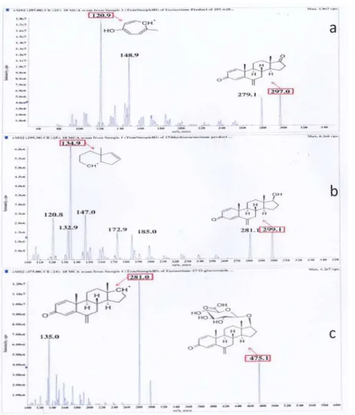
Mass spectrometric parameters were optimised and the product ion mass spectra of each ana-lyte under optimised conditions were as follows. Based onFig. 2, mass transitions 297>
121m/zand 300>121m/zwere monitored for Exe (C20H24O2, molecular weight: 296.40) and Exe-d3 (C20H21O2D3, molecular weight: 299.43) respectively. Mass transitions 299>135m/z
and 302>135m/zwere monitored for 17DhExe (C20H26O2, molecular weight: 298.43) and 17DhExe-d3 (C20H23O2D3, molecular weight: 301.44) respectively. Mass transitions 475>281
m/zand 478>284m/zwere monitored for Exe17Oglu (C26H34O8, molecular weight: 474.54) and Exe17Oglu-d3 (C26H31O8D3, molecular weight: 477.56) respectively. The desolvation
tem-perature was set at 500°C, the electrospray ionisation source 5500 V, and the optimised en-trance potential 10 V. The optimised declustering potentials were set at 61 V for Exe, Exe-d3, 17DhExe, and 17DhExe-d3; and 81 V, for Exe17Oglu and Exe17Oglu-d3. The optimised colli-sion energies were set at 33 V for Exe and Exe-d3; 29 V for 17DhExe and 17DhExe-d3; 23 V for Exe17Oglu and Exe17Oglu-d3. The optimised collision cell exit potentials were set at 8 V for Exe, Exe-d3, 17DhExe, and 17DhExe-d3; 14 V for Exe17Oglu and
Exe17Oglu-d3 respectively.
A total of three reversed-phase HPLC columns were investigated for chromatographic sepa-ration of analytes from endogeneous interferences and among analytes. BDS Hypersil C18 col-umn was chosen as the final chromatographic colcol-umn due to the successful baseline separation of analytes, reduced run-time and symmetrical chromatographic peaks achieved.
due to the reduced run-time, baseline separation among the peaks of analytes and from the endogenous substances.
Formic acid 0.1% and acetic acid 0.1% were investigated as aqueous solvents for HPLC mo-bile phase. Formic acid 0.1% was chosen as the final aqueous solvent due to the increased sensi-tivity and better peak shape achieved.
Isocratic elution and gradient elution were investigated as programmes for chromatographic separation. Gradient elution was chosen as the final chromatographic programme due to the successful baseline separation of analytes, reduced endogeneous interference, and sharpened analyte peaks. In addition, Exe and 17DhExe are much more hydrophobic than Exe17Oglu. Hence, only the gradient elution can analyze Exe and its metabolites simultaneously within a short run time. The mobile phase was composed of 0.1% aqueous formic acid (Phase A) and acetonitrile (Phase B). The following gradient programme was used: 0.00–0.10 min: 25% B, 0.10–0.20 min: 25!62% B (linear), 0.20–1.60 min: 62% B, 1.60–1.65 min: 62!25% B
Fig 2. Product ion mass spectra of (a) Exe (b) 17DhExe and (c) Exe17Oglu.
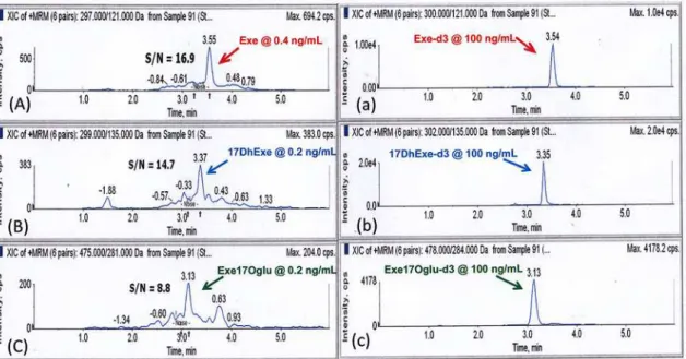
(linear), 1.65–6.00 min: 25% B. The flow rate was set at 0.5 mL/min. The resulting chromato-grams of 3 analytes at their low limit of quantitation are shown inFig. 3.
Extraction Protocol Optimisation
Extraction and pre-concentration methods were optimised to improve the recovery and in-crease the sensitivity of the assay.
Solid-phase extraction, liquid-liquid extraction and direct protein precipitation were carried out to investigate the optimal method for sample extraction.
HLB, WCX, WAX and LC-CN solid phases were investigated for solid-phase extraction. All four solid phases were found to be unable to retain Exe17Oglu and thus unsuitable for
sample extraction.
Ethanol—ethyl acetate mixture (4:1, v/v) and methyl-tert-butyl-ether were investigated as organic solvents for liquid-liquid extraction. Both solvent systems were found to be unable to extract Exe17Oglu and thus unsuitable for sample extraction as well.
Due to the different polarities of the analytes of interest, with Exe and 17DhExe being non-polar compounds, and Exe17Oglu being a highly non-polar substance, solid-phase extraction and liquid-liquid extraction methods were unable to extract all three analytes to a reasonable recov-ery. This may be due to the intrinsic nature of these extraction methods, which take advantage of the different polarities between compounds and interfering substances for separation from endogeneous interferences.
Acetonitrile in two volumes and three volumes were investigated as organic solvent for di-rect protein precipitation. Acetonitrile in three volumes was chosen as the final sample prepa-ration method due to the sufficient sample clean-up and less matrix effect. Furthermore, no interfering peaks were observed in the chromatogram, which validated the use of acetonitrile in three volumes for protein precipitation.
Fig 3. Representative chromatograms of (a) Exe (b) 17DhExe and (c) Exe17Oglu at LLOQ and (a) Exe-d3, (b) DhExe-d3 as well as Exe17Oglu at 100 ng/mL as internal standards.
Method validation
Selectivity, Carry-over and Linearity. Selectivity for the analytes was shown by peak iden-tification of analytes without interferences at their respective lower limits of quaniden-tification (LLOQ, 0.4 ng/mL, for Exe; and 0.2 ng/mL, both for 17DhExe and 17DhExe-17Oglu). A chro-matogram of the standard plasma spiked with three analytes at the concentrations of LLOQ is shown inFig. 3.
No carry-over effect was shown as the injection of wash after that of plasma with highest spiked concentration showed no peaks.
Linearity of the calibration curves were shown by the high r2of>0.998, which implied a
strong correlation between the peak area ratio and concentration of each analyte in their linear ranges of 0.4–40.0 ng/mL, for exemestane; and 0.2–15.0 ng/mL, for 17β-dihydroexemestane and 17β-dihydroexemestane-17-O-β-D-glucuronide.
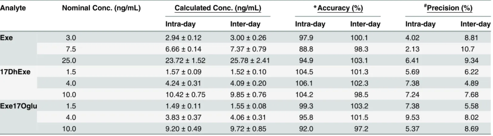
Accuracy and Precision. Results of intra-day and inter-day tests are shown inTable 1. The precision (coefficient of variation) were10.7%, 7.7% and 9.5% and the accuracies ranged from 88.8 to 103.1% for exemestane, 98.5 to 106.1% for 17β-dihydroexemestane and 92.0 to 103.2 for 17β-dihydroexemestane-17-O-β-D-glucuronide. Taken together, both intra-day and inter-day accuracy and precision were within 15%, as stipulated by the FDA guidelines.
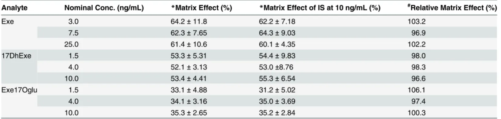
Matrix Effect. Matrix effect was demonstrated to be significant for Exe, 17DhExe, and Exe17Oglu. Ion suppression was significant, especially for Exe17Oglu. However, with the use of stable isotope-labelled IS for all three analytes, the relative matrix effect was demonstrated to be close to 100%, permitting correction of the matrix effect. Results of matrix effect tests are shown inTable 2.
Recovery. The relative recoveries of Exe, Exe-d3, 17DhExe, and 17DhExe-d3 were demon-strated to be greater than 98%. This showed that the extraction protocol was efficient and com-plete for these four compounds. Even though the relative recoveries of Exe17Oglu and
Exe17Oglu-d3 were found to be about 85%, the recoveries of Exe17Oglu and its IS were consis-tent and similar, allowing adjustment of the low recovery and fulfilling the FDA validation guidelines. Results of recovery tests are shown inTable 3.
Table 1. Intra-run and inter-run concentrations, accuracy and precision of QC samples for Exe, 17DhExe and Exe17Oglu.
Analyte Nominal Conc. (ng/mL) Calculated Conc. (ng/mL) *Accuracy (%) #Precision (%)
Intra-day Inter-day Intra-day Inter-day Intra-day Inter-day
Exe 3.0 2.94±0.12 3.00±0.26 97.9 100.1 4.02 8.81
7.5 6.66±0.14 7.37±0.79 88.8 98.3 2.13 10.7
25.0 23.72±1.52 25.78±2.41 94.9 103.1 6.41 9.34
17DhExe 1.5 1.57±0.09 1.52±0.10 104.5 101.3 5.69 6.22
4.0 4.24±0.31 4.09±0.20 106.1 102.3 7.38 4.89
10.0 10.42±0.75 9.85±0.76 104.2 98.5 7.24 7.68
Exe17Oglu 1.5 1.49±0.11 1.55±0.08 99.3 103.2 7.38 5.58
4.0 3.83±0.37 4.06±0.31 95.8 101.5 9.53 8.02
10.0 9.20±0.49 9.72±0.85 92.0 97.2 5.37 8.69
*Expressed as percentage of the mean value (n = 4) measured over the nominal value
#
Expressed as percentage of the standard deviation divided by the mean
Table 2. Matrix Effect of QC Samples for Exe, 17DhExe and Exe17Oglu and their internal standards.
Analyte Nominal Conc. (ng/mL) *Matrix Effect (%) *Matrix Effect of IS at 10 ng/mL (%) #Relative Matrix Effect (%)
Exe 3.0 64.2±11.8 62.2±7.18 103.2
7.5 62.3±7.65 64.3±9.03 96.9
25.0 61.4±10.6 60.1±4.35 102.2
17DhExe 1.5 53.3±5.31 54.4±9.83 98.0
4.0 52.1±3.13 53.0±8.76 98.3
10.0 53.4±4.41 55.3±6.54 96.6
Exe17Oglu 1.5 33.1±4.88 31.2±5.02 106.1
4.0 34.1±3.16 35.0±3.69 97.4
10.0 35.3±2.65 35.2±2.84 100.3
*Expressed as average percentage (n = 4) of peak area of analyte in matrix-based tube with that in reference tube
#
Expressed as ratio of matrix effect on compound to that of IS
doi:10.1371/journal.pone.0118553.t002
Table 3. Recovery of QC Samples for Exe, Exe-d3, 17DhExe, 17DhExe-d3, Exe17Oglu, and Exe17Oglu-d3.
Compound Nominal Concentration (ng/mL) *Recovery (%)
Exe 3.0 109.5
7.5 98.3
25.0 109.1
Exe-d3 10.0 107.9
17DhExe 1.5 106.8
4.0 102.8
10.0 108.3
17DhExe-d3 10.0 109.4
Exe17Oglu 1.5 86.4
4.0 85.3
10.0 84.7
Exe17Oglu-d3 10.0 86.0
*Expressed as mean percentage (n = 4) of peak area of analyte in the tube spiked before extraction to that in the tube spiked after extraction doi:10.1371/journal.pone.0118553.t003
Table 4. Short-term and freeze-thaw stability of QC samples of Exe, 17DhExe and Exe17Oglu.
Stability*
Analyte Nominal Conc. Short-term (hours) Freeze-thaw (cycles)
ng/mL 4 8 24 3 6 9
3.0 106.8 108.7 88.2 103.8 113.9 112.7
Exe 7.5 114.3 108.8 105.1 114.0 112.4 110.9
25.0 112.8 109.6 95.1 109.6 112.0 113.1
1.5 111.1 112.0 109.6 106.0 108.0 110.7
17DhExe 4.0 111.8 109.6 111.8 108.7 110.1 112.5
10.0 109.7 112.0 112.7 108.0 109.7 104.7
1.5 100.7 106.0 95.6 94.9 100.2 107.6
Exe17Oglu 4.0 101.7 105.7 103.5 96.6 107.3 110.7
10.0 107.7 109.7 107.3 106.4 101.9 110.0
Stability. Stabilities of Exe, 17DhExe, and Exe17Oglu were demonstrated to be with-in ± 15% of nomwith-inal concentrations. This with-indicated that the analytes of with-interest was stable with-in human plasma during short-term bench-top storage and freeze-thaw cycles. Results of stability tests are shown inTable 4.
Dilution Factor. Due to the presence of patient samples which exceed the maximum cali-brated concentration of Exe (40.0 ng/mL), dilution was carried out with blank human plasma by a factor of three before sample preparation. Accuracy and precision of the dilution proce-dure prior to sample preparation were within 10%, thus ensuring reliability of the proceproce-dure. Results of accuracy and precision of the dilution procedure are shown inTable 5.
Table 5. Accuracy and Precision of Dilution Procedure for Exe.
Nominal Concentration (ng/mL) Dilution Factor Quantified Concentration(Mean±S.D., ng/mL) *Accuracy (%) #Precision (%)
50.0 3 46.90±1.86 93.8 3.97
60.0 3 58.43±1.02 97.4 1.75
70.0 3 73.88±2.28 105.5 3.09
*Expressed as mean percentage (n = 4) of the mean value measured over the nominal value
#Expressed as percentage of the standard deviation divided by the mean
doi:10.1371/journal.pone.0118553.t005
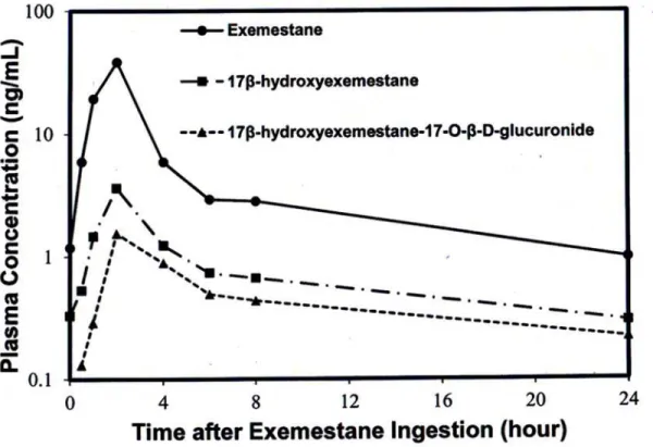
Fig 4. Mean concentrations of a) Exe (b) 17DhExe and (c) Exe17Oglu in plasma samples of one representative breast cancer patient.
Application of Validated LC-MS/MS Method
The developed method for the simultaneous quantification of Exe and its metabolites, 17DhExe, and Exe17Oglu was successfully applied in a pharmacokinetics/dynamics study, in which all the plasma samples taken from one typical patient were quantified for the concentra-tions of Exe, 17DhExe, and Exe17Oglu over 24 hours after exemestane ingestion at dose of 25 mg. Pharmacokinetic profiles were plotted and are shown inFig. 4in which the time-concen-tration profiles of Exe, 17DhExe and Exe17Oglu were well described with 24 hours. The maxi-mum concentrations of Exe, 17DhExe and Exe17Oglu were observed as 38.5, 3.6 and 1.5 ng/mL respectively at 2 hours after exemestane ingestion. The AUC calculated using WinNon-lin software with non-compartmental analysis were 129.6, 24.2 and 15.7 ng/mL for Exe, 17DhExe, and Exe17Oglu respectively.
Conclusions
A simple, novel and specific LC-MS/MS assay for the quantification of Exe and its two main metabolites was developed and fully validated according to the FDA guidelines. This method offers a significant advantage over previously reported methods due to its ability to quantify Exe17Oglu as well. Excellent linearity was demonstrated within the ranges of 1.0–40.0ng/mL for Exe and 0.5–15.0ng/mL for 17DhExe and Exe17Oglu. Accuracy and precision (CV) were well within FDA guidelines of<15%. The method was also successfully utilised for the
quanti-fication of Exe and its two main metabolites in human plasma in a pharmacokinetics/dynamics study. For a representative patient, 20.7% of exemestane in plasma was converted into 17β -dihydroexemestane and 29.0% of 17β-dihydroexemestane was inactivated as 17β -dihydroexe-mestane-17-O-β-D-glucuronide 24 hours after ingestion of exemestane, suggesting that altered 17-dihydroexemestane glucuronidation may play an important role in determining effect of exemestane against breast cancer cells due to genetic difference of UGT2B17 among breast cancer patients.
Acknowledgments
We would like to express our deep appreciation to Ms. Khoo Yok Moi for her meticulous guid-ance on solid-phase extraction optimization, Dr. Fan Lu for her thorough assistguid-ance on the use of WinNonlin software and Ms. Luo Wenxia for her wonderful help through this project.
Author Contributions
Conceived and designed the experiments: WLZ WA GBC PCH LSC. Performed the experi-ments: WLZ GSH TWL LJA WSC. Analyzed the data: WLZ WA GSH PCH. Contributed re-agents/materials/analysis tools: GBC WA LSC. Wrote the paper: WLZ GSH PCH WA.
References
1. Teo MC, Soo KC. Cancer Trends and Incidences in Singapore. Jpn J Clin Oncol., 2013 (43: ):219–24. doi:10.1093/jjco/hys230PMID:23303840
2. Kim SH, Park IH, Lee H, Lee KS, Nam BH, Ro J. Efficacy of exemestane after nonsteroidal aromatase inhibitor use in metastatic breast cancer patients. Asian Pac J Cancer Prev., 2012; 13(3): 979–983. PMID:22631683
3. Zhang Y, Simondsen K, Kolesar JM. Exemestane for primary prevention of breast cancer in postmeno-pausal women. Am J Health Syst Pharm, 2012; 69(16): 1384–1388. doi:10.2146/ajhp110585PMID: 22855103
5. Litton JK, Bevers TB, Arun BK. Exemestane in the prevention setting. Ther Adv Med Oncol., 2012; 4(3): 107–112. doi:10.1177/1758834012438214PMID:22590484
6. Winer EP, Hudis C, Burstein HJ, Chlebowski RT, Ingle JN, Edge SB, et al. American Society of Clinical Oncology Technology Assessment on the Use of Aromatase Inhibitors as Adjuvant Therapy for Women With Hormone Receptor—Positive Breast Cancer: Status Report 2002. Journal of Clinical On-cology, 2002; 20(15): 3317–3327. PMID:12149306
7. Lønning PE, Geisler J. Experience with Exemestane in the Treatment of Early and Advanced Breast Cancer. Expert Opin. Drug Metab. Toxicol., 2008; 4(7):987–997. doi:10.1517/17425255.4.7.987 PMID:18624685
8. Kittaneh M, Glück S. Exemestane in the Adjuvant Treatment of Breast Cancer in Postmenopausal Women. Breast Cancer, Breast Cancer (Auckl), 2011; 5:209–226. doi:10.4137/BCBCR.S6234PMID: 22084574
9. Eisen A, Trudeau M, Shelley W, Messersmith H, Pritchard KI. Aromatase inhibitors in adjuvant therapy for hormone receptor positive breast cancer: A systematic review. Cancer Treatment Reviews, 2008; 34(2):157–174. doi:10.1016/j.ctrv.2007.11.001PMID:18164821
10. Winters L1, Habin K, Gallagher J. Aromatase inhibitors and musculoskeletal pain in patients with breast cancer. Clin J Oncol Nurs. 2007; 11(3):433–9. PMID:17623627
11. Herold CI1, Blackwell KL. Aromatase inhibitors for breast cancer: proven efficacy across the spectrum of disease. Clin Breast Cancer. 2008; 8(1):50–64. doi:10.3816/CBC.2008.n.003PMID:18501059
12. Tomao F1, Spinelli G, Vici P, Pisanelli GC, Cascialli G, Frati L, et al. Current role and safety profile of aromatase inhibitors in early breast cancer. Expert Rev Anticancer Ther. 2011; 11(8):1253–63. doi:10. 1586/era.11.96PMID:21916579
13. Van Asten K1, Neven P, Lintermans A, Wildiers H, Paridaens R. Aromatase inhibitors in the breast can-cer clinic: focus on exemestane. Endocr Relat Cancan-cer. 2014; 21(1):R31–49. doi: 10.1530/ERC-13-0269PMID:24434719
14. Cavalcantia Gde A, Garridoa BC, Leala FD. Detection of new urinary exemestane metabolites by gas chromatography coupled to mass spectrometry. Steroids, 2011; 76:1010–1015. doi:10.1016/j. steroids.2011.04.001PMID:21530565
15. Kamdem LK, Flockhart DA, Desta Z. In Vitro Cytochrome P450-Mediated Metabolism of Exemestane. Drug Metabolism and Disposition, 2011; 39(1):98–105. doi:10.1124/dmd.110.032276PMID: 20876785
16. Sun D, Chen G, Dellinger RW, Sharma AK, Lazarus P. Characterization of 17-dihydroexemestane glu-curonidation: potential role of the UGT2B17 deletion in exemestane pharmacogenetics. Pharmacoge-netics and Genomics, 2010; 20(10):575–585. doi:10.1097/FPC.0b013e32833b04afPMID:20697310
17. Wong NS1, Seah EZh, Wang LZ, Yeo WL, Yap HL, Chuah B, et al. Impact of UDP-gluconoryltransfer-ase 2B17 genotype on vorinostat metabolism and clinical outcomes in Asian women with breast can-cer. Pharmacogenetics and Genomics, 2011; 21(11):760–768. doi:10.1097/FPC.0b013e32834a8639 PMID:21849928
18. Breda M, Pianezzola E, Benedetti MS. Determination of exemestane, a new aromatase inhibitor, in plasma by high-performance liquid chromatography with ultraviolet detection. J Chromatogr., 1993; 620(2):225–31. PMID:8300790
19. Corona G, Elia C, Casetta B, Diana C, Rosalen S, Bari M, et al. A liquid chromatography-tandem mass spectrometry method for the simultaneous determination of exemestane and its metabolite 17-dihydroexemestane in human plasma. Journal of Mass Spectrometry, 2009; 44(6):920–928. doi:10. 1002/jms.1566PMID:19214962