MMP-2, MMP-9, RANKL/RANK, Cathepsin K and
Up-Regulates OPG in an Experimental Periodontitis Model
Aurigena Antunes de Arau´jo1*., Hugo Varela2., Gerly Anne de Castro Brito3 ,
Caroline Addison Carvalho Xavier de Medeiros4, Lorena de Souza Arau´jo5,
Jose´ Heriberto Oliveira do Nascimento6, Raimundo Fernandes de Arau´jo Ju´nior7
1Post Graduation Program Public Health/Post Graduation Program in Pharmaceutical Science/Department of Biophysics and Pharmacology,UFRN, Natal, Rio Grande do Norte, Brazil,2Post Graduation Program Public Health/Department of Dentistry, UFRN, Natal, Rio Grande do Norte, Brazil,3Post Graduation Program in Pharmacology/ Department of Morphology, UFC, Fortaleza, Ceara´, Brazil,4Department of Biophysics and Pharmacology, UFRN, Post Graduation Program in Health and Society, UERN, Natal, Rio Grande do Norte, Brazil,5Department of Dentistry, UFRN, Natal, Rio Grande do Norte, Brazil,6Department of Textile Engineered, UFRN, Natal, Rio Grande do Norte, Brazil,7Post Graduation Program in Functional and Structural Biology/Post Graduation Program Health Science/Department of Morphology, UFRN, Natal, Rio Grande do Norte, Brazil
Abstract
Aims:The aim of this study was to evaluate the effects of azilsartan (AZT) on bone loss, inflammation, and the expression of matrix metallo proteinases (MMPs), receptor activator of nuclear factorkB ligand (RANKL), receptor activator of nuclear factorkB (RANK), osteoprotegerin (OPG), cyclooxygenase-2 (COX-2), and cathepsin K in periodontal tissue in a rat model of ligature-induced periodontitis.
Materials and Methods:Male Wistar albino rats were randomly divided into 5 groups of 10 rats each: (1) nonligated, water; (2) ligated, water; (3) ligated, 1 mg/kg AZT; (4) ligated, 5 mg/kg AZT; and (5) ligated, 10 mg/kg AZT. All groups were treated with saline or AZT for 10 days. Periodontal tissues were analyzed by histopathology and immunohistochemical detection of MMP-2, MMP-9, COX-2, RANKL, RANK, OPG, and cathepsin K. Levels of IL-1b, IL-10, TNF-a, myeloperoxidase (MPO), and glutathione (GSH) were determined by ELISA.
Results:Treatment with 5 mg/kg AZT resulted in reduced MPO (p,0.05) and IL-1b(p,0.05), increased levels of IL-10 (p, 0.05), and reduced expression of MMP-2, MMP-9, COX-2, RANK, RANKL, cathepsin K, and increased expression of OPG.
Conclusions:These findings reveal that AZT increases anti-inflammatory cytokines and GSH and decreases bone loss in ligature-induced periodontitis in rats.
Citation:Arau´jo AAd, Varela H, Brito GAdC, Medeiros CACXd, Arau´jo LdS, et al. (2014) Azilsartan Increases Levels of IL-10, Down-Regulates MMP-2, MMP-9, RANKL/RANK, Cathepsin K and Up-Regulates OPG in an Experimental Periodontitis Model. PLoS ONE 9(5): e96750. doi:10.1371/journal.pone.0096750
Editor:Dominique Heymann, Faculte´ de me´decine de Nantes, France
ReceivedFebruary 24, 2014;AcceptedApril 10, 2014;PublishedMay 12, 2014
Copyright:ß2014 Arau´jo et al. This is an open-access article distributed under the terms of the Creative Commons Attribution License, which permits unrestricted use, distribution, and reproduction in any medium, provided the original author and source are credited.
Funding:The authors have no support or funding to report.
Competing Interests:The authors have declared that no competing interests exist.
* E-mail: [email protected]
.These authors contributed equally to this work.
Introduction
Periodontal disease is a chronic infectious, inflammatory disease of the gums and supporting tissues. Gingival inflammation that accompanies periodontal disease can damage supporting connec-tive tissues and disrupt tooth anchoring to the jawbone. Several modulating agents have been investigated as potential therapies for periodontal disease, including antiproteinases, anti-inflammatory drugs, and bone-sparing drugs [1].
Improved knowledge of the mechanisms involved in the pathogenesis of periodontal disease have led to the use of novel agents to modulate the host response by inhibiting inflammatory mediators. Studies with antihypertensive drugs have shown that they have an anti-inflammatory activity in periodontal disease with reduced bone loss [2–5]. Our group have study the angiotensin II
receptor blocker (ARB). For example, the angiotensin II receptor blocker (ARB) has been implicated as an anti-inflammatory agent that suppresses tumor necrosis factor (TNF)-a-induced activation of nuclear factor (NF)-kB in vascular endothelial cells [6].
Treatment of periodontitis with Telmisartan, angiotensin II receptor blocker (ARB), reduced markers of inflammation, proteases and changed proteins involved in bone remodeling [2]. Similar results were obtained in a study using another ARB, olmesartan [3].
overall incidence of adverse reactions similar to placebo [7]. Our group has been working with antihypertensives, angiotensin II receptor blocker (ARB), and how patients with periodontal disease have systemic diseases such as hypertension, our group aims to investigate whether these antihypertensive drugs interfere with periodontal disease. The aim of present study was to determine the efficacy of Azilsartan in treating periodontal disease.
Materials and Methods
Animals
Experiments were performed on male Wistar rats (180–220 g) housed in standard conditions (12 hour light/dark cycle; 2260,1uC), with ad libitum access to food and water. All animal protocols were approved by the Animal Ethics Committee (No. 28/2012) of the Federal University of Rio Grande do Norte, Brazil. Anesthesia was induced by intraperitoneal injection of 10% ketamine (70 mg/kg, Vetnil, Sa˜o Paulo, Brazil) and 2% xylazine (10 mg/kg, Sa˜o Paulo, Brazil). Experimental Periodontal Disease was induced in rats under anaesthesia induced by ketamine (70 mg/kg administered i.p., 10% Quetamina, VETNIL, Sa˜o Paulo) and xylazine (10 mg/kg administered i.p., 2% Calmium, Sa˜o Paulo) by the placement of a sterile nylon thread ligature (3-0; Polysuture, NP45330, Sa˜o Paulo) around the cervix of the maxillary left second molar [8]. Eleven days after the initial treatment, animals were euthanized with 80 mg/kg thiopental (Crista´lia, Sa˜o Paulo, Brazil).
Drug treatments
AZT (Edarbi, Takeda Pharmaceuticals America, USA) was dissolved in distilled water (vehicle) and administered by oral gavage 1 hour before induction of periodontitis and once daily for 10 days until euthanasia. Animals (n = 10) were assigned randomly to 5 groups: (1) nonligated, water (NL), (2) ligated, water (L), (3) ligated, treated with 1 mg/kg AZT, (4) ligated, treated with 5 mg/ kg AZT, and (5) ligated, treated with 10 mg/kg AZT. Animal drug doses were not based on human dosages, because different genetic features affect pharmacokinetics of drugs. Instead, dosage was based onin vivostudies in rats that examined the effects of AZT on blood pressure [9,10].
Measurement of alveolar bone loss (ABL)
Excised maxillae were fixed in 10% neutral formalin for 24 hours, and maxillary halves were defleshed and stained with 1% aqueous methylene blue to differentiate bone from teeth. Bone loss was measured along the length of each root surface of each molar. ABL was measured in all 5 experimental groups. Three measurements were performed on the first molars (3 roots each) and 2 measurements were performed on the second and third molars (two roots each). Total alveolar bone loss was determined by taking the sum of the measurements from buccal tooth surfaces and subtracting the values of the right maxilla (no ligated control) from those of the left maxilla, in millimeters [11]. Morphometric analyses of alveolar bone were performed using standardized digital photography (Olympus SC30), and the distances (in millimeters) were measured with Imaging Software (analysis getIT 5.1).
Histopathological analyses
Immunohistochemical analyses were performed independently by 2 oral pathologists (R.F.A. Jr. and A.A.A). Sectioning was performed in the morphology and oral pathology laboratory, and slides were analyzed using light microscopy in the Department of Morphology. Five jaws per group were used. Alveolar bone
specimens were harvested, fixed in 10% neutral-buffered formalin, and demineralised in 5% nitric acid. Then, specimens were dehydrated, embedded in paraffin, and sectioned along the molars in the mesiodistal plane for hematoxylin and eosin staining. Sections (4mm) corresponding to the area between the first and second molars where the ligature had been placed were evaluated by light microscopy (640 magnification). Inflammatory cell influx and alveolar bone and cementum integrity were analyzed by a histologist in a single-blind fashion and graded. A score of 0 indicated that inflammatory cell infiltration was absent or sparse and was restricted to the marginal gingival region, and that the alveolar process and cementum were preserved; a score of 1 indicated moderate cellular infiltration throughout the entire gingival insert, minor alveolar resorption, and intact cementum; a score of 2 indicated accentuated cellular infiltration in the gingiva and the periodontal ligament, accentuated degradation of the alveolar process, and partial destruction of the cementum; and 3 indicated accentuated cellular infiltration, complete resorption of the alveolar process, and severe destruction of the cementum [12].
Immunohistochemical analyses
Thin sections of periodontal tissue (4mm) (3 jaws per group) were produced using a microtome and transferred to gelatin-coated slides. Each section was deparaffinised and rehydrated. Gingival and periodontal tissue slices were washed with 0.3% Triton X-100 in phosphate buffer, quenched with endogenous peroxidase (3% hydrogen peroxide), and incubated with the following primary antibodies (Santa Cruz Biotechnology, INTER-PRISE, Brazil) overnight at 4uC: COX-2, 1:400; MMP-2, 1:400; MMP-9, 1:400; RANKL, 1:400; RANK, 1:400; OPG, 1:400 and cathepsin K: 1:400. Slices were washed with phosphate buffer and incubated with streptavidin-HRP-conjugated secondary antibodies (Biocare Medical, Concord, CA, USA) for 30 minutes, and immunoreactivity to MMP-2, MMP-9, RANK, RANK-L, OPG, COX-2, and cathepsin K was visualized using a colorimetric detection kit following the manufacturer’s instructions (TrekAvi-din-HRP Label+Kit, Biocare Medical, Dako, USA).
Myeloperoxidase (MPO) assay
The extent of neutrophil accumulation in gingival samples was measured by assaying MPO activity. Gingival samples (5 per group) were harvested as described above and stored at270uC until use. Samples were homogenized and centrifuged (20006gfor 20 minutes), and MPO activity was determined using a colorimetric method described previously [13]. The results were reported as units of MPO per milligram tissue.
Glutathione (GSH) assay
GSH levels in gingival tissues were measured to approximate antioxidant activity. Gingival samples (5 per group) were stored at 270uC until use. Gingival tissue homogenates (0.25 mL of a 5% tissue solution prepared in 0.02 M EDTA) were added to 320mL of distilled water and 80mL of 50% TCA. Samples were centrifuged at 3000 rpm for 15 minutes at 4uC. The supernatant (400mL) was added to 800mL of 0.4 M Tris buffer pH 8.9 and 20mL of 0.01 M (5,59-dithiobis-(2-nitrobenzoic acid) DTNB. Absorbance was measured at 420 nm, and results were reported as units of MPO per milligram tissue.
IL-1b, IL-10, and TNF-a assay
12.5 ng/mL recombinant mouse IL-1b), IL-10 (detection range: 62.5–4000 pg/mL; lower limit of detection: 12.5 ng/mL recom-binant mouse IL-10), and TNF-a(detection range: 62.5–4000 pg/ mL; lower limit of detection: 50 ng/mL recombinant mouse
TNF-a) were determined using commercial ELISA kits (R&D Systems, Minneapolis, MN, USA), as described previously [15]. All samples were measured at 490 nm.
Briefly, microtitre plates were coated overnight at 4uC with antibodies against mouse TNF-a, IL-1b, and Il-10. After plates were blocked, samples and standards were added at various dilutions in duplicate and incubated at 4uC for 24 hours. Plates were washed 3 times with buffer and antibodies were added to the wells (biotinylated sheep polyclonal anti-TNF-a, anti-IL-1b, or
anti-IL-10 diluted 1:1000 with 1% BSA assay buffer). Plates were incubated at room temperature for 1 hour, washed, and 50mL of avidin-HRP (1:5000) were added. The color reagent o-phenyl-enediamine (50mL) was added 15 minutes later, and the plates were incubated in the dark at 37uC for 15–20 minutes. The enzyme reaction was stopped with H2SO4 and absorbance was measured at 490 nm. Values were expressed in pg/mL.
Statistical analyses
Data are presented as means6standard error, or as medians when appropriate. Analyses of variance and Bonferroni’s tests were used to calculate the means, and the Kruskal-Wallis and Dunn’s tests were used to compare medians (GraphPad Prism 5.0 Software, La Jolla, CA, USA). A p-value of ,0.05 indicated statistical significance.
Results
Effects of AZT treatment on alveolar bone loss in rats with Experimental Periodontal Disease
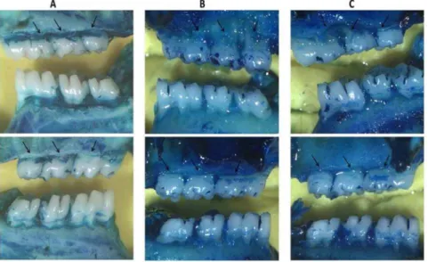
Rats with ligation-induced periodontitis (L) showed significant alveolar bone loss compared with non-ligated animals (NL) (NL = 0.22+0.41 mm; L = 4.661.4 mm; p,0.001). AZT treat-ments (5 mg/kg) reversed alveolar bone loss caused by periodon-titis compared to L animals (AZT 5 mg/kg 2.561.9; L = 4.661.4 mm, p,0.05) (Figure 1). The macroscopic aspects of the NL group with no resorption of the alveolar bone compared to the L group with severe bone resorption and root exposure can be seen in (Figure 2B). Figure 2C shows the macroscopic appearance of the periodontium after ligation and treatment with AZT 5 mg/kg, which led to decreased bone loss compared to ligated animals. Rats treated with 1 mg/kg and 10 mg/kg AZT also showed differences in alveolar bone loss compared to NL animals (AZT 1 mg/kg = 3.260.8 mm; AZT 10 mg/ kg = 4.961.9 mm,p,0.001 andp,0.05, respectively) (Figure 1).
Histological analyses
Figure 3 shows the structures of the periodontium, gingiva, periodontal ligament, alveolar bone, and cementum prior to Figure 1. Effect of AZT treatment on alveolar bone loss
associated with experimental periodontitis (EP) in rats.Values are expressed as means6SEM (Compared to NL ***p,0.001, *p,0.05; Compared to L#p,0.05).
doi:10.1371/journal.pone.0096750.g001
Figure 2. A) Superior: Left/Below: Right, NL group, showing no resorption of the alveolar bone.B) Superior: Left/Below: Right, L group, showing severe bone resorption with root exposure (arrows). C) Superior: Left/Below: Right, L group treated with Azilsartan 5 mg/kg, showing decreased bone resorption (arrows). Images were obtained at an original magnification of 1.76.
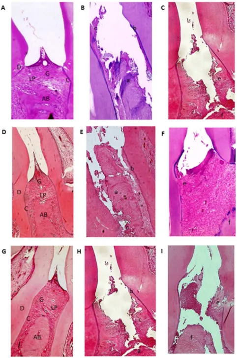
Figure 3. Microscopic analyses.A, D and G: Normal periodontium. B, E, H: Periodontium from a rat with periodontitis (treated with saline) showing alveolar bone and cementum resorption (discontinuous cementum) and inflammatory cell infiltration. C: Treatment with AZT (1 mg/kg) and I: Treatment with AZT (10 mg/kg) showing no reduced inflammation and increased alveolar bone loss. F: Periodontium from a rat with periodontitis (treated with AZT, 5 mg/kg) showing reduced inflammation and decreased alveolar bone loss. Sections were stained with H&E. Original magnification 406. Scale bars = 100mm. G, gingiva; PL, periodontal ligament; D, dentin; AB, alveolar bone; C, cementum; a, bone loss; b, resorption of cementum; c, inflammatory process; e, f, decreased inflammation process and bone loss.
doi:10.1371/journal.pone.0096750.g003
Table 1.Histological analysis of maxillae from rats presenting with periodontal disease, Natal, RN, 2014.
NL L Azt 1 mg/kg Azt 5 mg/kg Azt 10 mg/kg
0 (0-0) 3 (3-3)# 3 (3-3)# 1 (1-2) 3 (2-3)#
#p
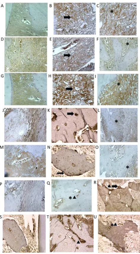
Figure 4. Photomicrographs of periodontal tissue of PD rats treated with AZT, showing immunoreactivity to MMP-2, MMP-9, COX-2, RANK, RANK-L, OPG, and cathapsyn.Rats subjected to saline (A, D, G, J, M, P,S); rats subjected to ligation (B, E, H, K, N, Q,T); rats subjected to ligation and treated with AZT (5 mg/kg) (C, F, I, L, O, R,U). Images are shown at 406magnification. Bar = 100mm. Arrow indicates high or moderate labeling in the periodontal ligament or the alveolar bone. Asterisk indicates mild or moderate labeling in the periodontal ligament or the alveolar bone. Triangle and asterisk indicate mild labeling of OPG in osteoclasts. Triangle and arrow indicate high labeling of osteoclasts. Triangle indicates intense labeling of Cathepsin K on alveolar bone. Asterisk and triangle indicate mild labeling of cathepsin K on alveolar bone.
ligation. Histopathological analyses revealed that alveolar bone loss was reduced in ligated animals treated with 5 mg/kg AZT (p, 0.05) compared to ligated animals treated with 1 mg/kg and 10 mg/kg AZT. Discrete cellular infiltration restricted to the region of the marginal gingiva, preservation of alveolar bone, and intact cementum were observed in animals treated with 5 mg/kg AZT (Figure 3F). In animals subjected to ligation that received no treatment (L), inflammatory cell infiltration and severe destruction of the cementum and alveolar process were observed, animals in this group received median scores of 3 (Figure 3B,E,H; Table 1). Treatment with 5 mg/kg AZT prevented ligation-induced inflam-mation (Figure 3F) and reduced median histopathological scores 1 (1–2) (Table 1).
Immunohistochemical analyses of inflammatory markers
Compared to the NL group, the periodontium of rats in the L group showed marked immunostaining for MMP-2, MMP-9, COX-2, RANK-L, RANK, OPG and Cataphesyn (NL-saline animals, Figure 4A, D, G, J, M, P and S compared to L-saline animals, Figure 4B, E, H, K, N, Q and T). However, treatment with AZT (5 mg/kg) reduced MMP-2, MMP-9, COX-2, RANK-L, RANK, and Cataphesyn, immunostaining in the periodontium of rats subjected to ligation (Figure 4 C, F, I, L, O and U). OPG
immunostaining was mild in the periodontium of the L group, moderate in the NL group, and intense in the group treated with 5 mg/kg AZT (Figure 4R).
Effects of AZT on inflammatory activity and GSH
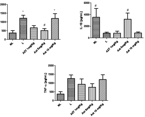
MPO activity was increased in the L group compared to the NL group (p,0.05). The group subjected to ligation and treated with 5 mg/kg AZT showed reduced concentrations of MPO compared to the L group (p,0.05) (Figure 5). Levels of antiinflammatory cytokine IL-1b were decreased in animals treated with 5 mg/kg AZT compared to L group animals (p,0.05). Levels of antiin-flammatory cytokine IL-10 were increased in animals treated with 5 mg/kg AZT compared to L group animals (p,0.05) (Figure 6). The treatment of with AZT no reduced the levels of GSH (p. 0.05) compared with those of the L group (Figure 5).
Discussion
Periodontitis is a chronic inflammatory disease resulting from perturbed homeostasis between the subgingival microbiota and host defenses [16].Given the extensive use of antihypertensive drugs, combined with the fact that periodontal disease affects adults and the elderly, we decided to investigate the effect of antihypertensive drugs on the progression of periodontal disease. In this study, azilsartan treatment produced some results than previous studies by our group using other antihypertensives. Telmisartan and Olmesartan significantly reduced pro-inflamma-tory cytokines such as IL-1b and TNF-a, which resulted in significant preservation of bone in periodontal disease [2,3]. In the periodontium, the activation of IL-1b and TNF-a is known to stimulate the degradation of connective tissue matrix, the activation of osteoclasts, and the resorption of bone [17,18]. IL-1 induces the release of MMPs, whereas TNF-a, which is present in inflamed gingival tissue, is involved in the destruction of tissue [19]. These cytokines play key roles in the breakdown of periodontal tissue through collagenolytic enzymes such as MMPs. The inflamed tissue also expresses COX-2, which may play a role in the formation of arachidonic acid metabolites. In this study, MPO and IL-1b levels were significantly reduced by AZT treatment (5 mg/kg). Reduction in the inflammatory response can be confirmed by observing reduction in tissue COX-2, an enzyme expressed in inflamed tissues, that was markedly reduced in rats treated with Azilsartan (5 mg/kg) compared to levels observed in rats presenting with periodontal disease.
We believe these results may be related to IL-10 increased levels. According to a study by Gaddiset al.(2013), IL-10 plays an important role controlling infection and the progression of periodontal disease [20]. Surface components ofP. gingivalis, such as LPS, lipoproteins, and fimbriae, interact with host-expressed Toll-like receptors (TLRs), key control elements of the innate immune response. TLR activation leads to nuclear translocation of NF-kB and induction of inflammation-related genes, suggesting that exaggerated inflammation mediated by TLRs is a driving factor in periodontal disease [21]. One of the most effective suppressors of TLR-induced inflammatory cytokine production is IL-10, which inhibits innate immune cells by directly inhibiting cytokine transcription [22].
The main problem related to periodontal disease is bone loss, which is caused by activation of lymphocyte pro-inflammatory cytokines, such as IL-1b and TNF-a, and may be mediated by significant increases in IL-10 levels. IL-10 can mediate bone loss through both direct and indirect actions. It inhibits bone resorption indirectly by upregulating osteoprotegerin expression Figure 5. MPO and GSH in NL, L, and groups treated with 1 mg/
and downregulating expression of RANKL [23]. IL-10 mediates bone loss directly by inhibiting osteoclast formation [24].
MMPs are a family of related zinc-containing proteinases that can degrade most of the extracellular matrix. It has been reported that the induction of MMPs (such as MMP-2) in osteoblasts is essential for bone resorption [25]. Excessive production of MMP-2 and MMP-9 can lead to accelerated matrix degradation in pathological conditions such as periodontitis [25]. Tissue destruc-tion caused by MMP-2 and MMP-9 was reduced in animals treated with 5 mg/kg AZT, indicating that IL-10 may be a protective cytokine in periodontal diseases [26]. We also observed reduced RANKL and RANK staining and increased OPG staining. These results are corroborated by studies showing that inhibition of osteoclast formation by IL-10 is mediated through direct actions on osteoclast precursors [24,27], including induced osteoclastogenesis. IL-10 potently reduced the RANKL-induced expression, which are essential for osteoclastogenesis [28]. A study by Liu et al. (2006) suggested that IL-10 inhibits bone resorption by up-regulating OPG expression and down-regulating RANKL expression [23].
The study by Gallet et al. (2006) showed that expression of MMP-1, MMP-2, MMP-9, and RANKL was correlated with expression of IL-1b, TNFa, and interferon-gamma inflammatory reactions and alveolar bone loss. On the other hand, 4 and IL-10 were associated with higher expression of OPG, with lower expression of MMPs and RANKL, and with reduced rates of cellular infiltration in periodontal tissues and alveolar bone loss [29].
The study of Bostanci et al (2011) aimed to monitor quantitative changes in the RANKL–OPG system in gingival crevicular fluid
(GCF) following non-surgical periodontal treatment over a period of 4 months. The data indicated that periodontal treatment does not affect RANKL or OPG levels in chronic periodontitis (CP) patients. Hence, conventional therapy alone cannot modify the overall capacity of the tissue to produce these factors. Levels of RANKL and OPG transiently increased in the GCF 2 months post-treatment, with RANKL changes being statistically significant [30]. Adjunctive therapies for periodontal disease management may modulate RANKL/OPG ratio [31].
Another important marker of bone is Cathepsin K, a cysteine protease that is released from mature osteoclasts to degrade type I collagen, the major organic matrix protein in bone. It is an important mediator of osteoclast activity and is relatively abundant and selective compared to other collagenases in these cells [32]. Therefore, reduction of Cathepsin K in the AZT treated group (5 mg/kg) implies that AZT treatment reduces degradation of the bone matrix.
In conclusion, this study revealed that treatment with 5 mg/kg doses of AZT reduces bone loss, decreased IL-1b, increased IL-10 and decreased MPO levels, and reduced expression of RANKL/ RANK and cathepsin K. These changes increased OPG staining, which controls osteoclastogenesis. Taken together, these data indicate that AZT is an effective treatment to decrease bone loss in ligature-induced periodontitis in rats.
Author Contributions
Conceived and designed the experiments: AAA GACB CACXM RFAJ. Performed the experiments: AAA HV LSA JHON. Analyzed the data: AAA RFAJ. Contributed reagents/materials/analysis tools: GACB CACXM JHON. Wrote the paper: AAA RFAJ HV GACB CACXM.
References
1. Elavarasu S, Sekar S, Murugan T (2012) Host modulation by therapeutic agents. J Pharm Bioallied Sci 4: S256–259.
2. Araujo AA, Souza TO, Moura LM, Brito GA, Aragao KS, et al. (2013) Effect of telmisartan on levels of IL-1, TNF-alpha, down-regulated COX-2, MMP-2, MMP-9 and RANKL/RANK in an experimental periodontitis model. J Clin Periodontol 40: 1104–1111.
3. Araujo AA, Lopes de Souza G, Souza TO, de Castro Brito GA, Saboia Aragao K, et al. (2013) Olmesartan decreases IL-1beta and TNF-alpha levels; downregulates MMP-2, MMP-9, COX-2, and RANKL; and upregulates OPG in experimental periodontitis. Naunyn Schmiedebergs Arch Pharmacol 386: 875–884.
4. de Araujo Junior RF, Souza TO, de Medeiros CA, de Souza LB, Freitas Mde L, et al. (2013) Carvedilol decrease IL-1beta and TNF-alpha, inhibits MMP-2, MMP-9, COX-2, and RANKL expression, and up-regulates OPG in a rat model of periodontitis. Plos One 8: e66391.
5. de Araujo Junior RF, Souza TO, de Moura LM, Torres KP, de Souza LB, et al. (2013) Atorvastatin decreases bone loss, inflammation and oxidative stress in experimental periodontitis. Plos One 8: e75322.
6. Nakano A, Hattori Y, Aoki C, Jojima T, Kasai K (2009) Telmisartan inhibits cytokine-induced nuclear factor-kappaB activation independently of the peroxisome proliferator-activated receptor-gamma. Hypertens Res 32: 765–769. 7. Zaiken K, Cheng JW (2011) Azilsartan medoxomil: a new Angiotensin receptor
blocker. Clinical Therapeutics 33: 1577–1589.
8. Carvalho RS, de Souza CM, Neves JC, Holanda-Pinto SA, Pinto LM, et al. (2010) Effect of venlafaxine on bone loss associated with ligature-induced periodontitis in Wistar rats. J Negat Results Biomed 9: 3.
9. Wienen W, Hauel N, Van Meel JC, Narr B, Ries U, et al. (1993) Pharmacological characterization of the novel nonpeptide angiotensin II receptor antagonist, BIBR 277. Br J Pharmacol 110: 245–252.
10. Wienen W, Richard S, Champeroux P, Audeval-Gerard C (2001) Comparative antihypertensive and renoprotective effects of telmisartan and lisinopril after long-term treatment in hypertensive diabetic rats. Journal of the Renin-Angiotensin-Aldosterone System 2: 31–36.
11. Carvalho Rde S, de Souza CM, Neves JC, Holanda-Pinto SA, Pinto LM, et al. (2013) Vitamin E does not prevent bone loss and induced anxiety in rats with ligature-induced periodontitis. Arch Oral Biol 58: 50–58.
12. Leitao RF, Ribeiro RA, Chaves HV, Rocha FA, Lima V, et al. (2005) Nitric oxide synthase inhibition prevents alveolar bone resorption in experimental periodontitis in rats. J Periodontol 76: 956–963.
13. Souza MH, Troncon LE, Cunha FQ, Oliveira RB (2003) Decreased gastric tone and delayed gastric emptying precede neutrophil infiltration and mucosal lesion formation in indomethacin-induced gastric damage in rats. Brazilian Journal of Medical and Biological Research 36: 1383–1390.
14. Safieh-Garabedian B, Poole S, Allchorne A, Winter J, Woolf CJ (1995) Contribution of interleukin-1 beta to the inflammation-induced increase in nerve growth factor levels and inflammatory hyperalgesia. Br J Pharmacol 115: 1265– 1275.
15. Kendall C, Ionescu-Matiu I, Dreesman GR (1983) Utilization of the biotin/ avidin system to amplify the sensitivity of the enzyme-linked immunosorbent assay (ELISA). J Immunol Methods 56: 329–339.
16. Sanz M, van Winkelhoff AJ (2011) Periodontal infections: understanding the complexity–consensus of the Seventh European Workshop on Periodontology. J Clin Periodontol 38 Suppl 11: 3–6.
17. Graves DT, Li J, Cochran DL (2011) Inflammation and uncoupling as mechanisms of periodontal bone loss. Journal of Dental Research 90: 143–153. 18. Graves D (2008) Cytokines that promote periodontal tissue destruction.
J Periodontol 79: 1585–1591.
19. Yamaguchi M, Kasai K (2005) Inflammation in periodontal tissues in response to mechanical forces. Arch Immunol Ther Exp (Warsz) 53: 388–398. 20. Gaddis DE, Maynard CL, Weaver CT, Michalek SM, Katz J (2013) Role of
TLR2-dependent IL-10 production in the inhibition of the initial IFN-gamma T cell response to Porphyromonas gingivalis. J Leukoc Biol 93: 21–31. 21. Takeda K, Akira S (2004) TLR signaling pathways. Semin Immunol 16: 3–9. 22. Curtale G, Mirolo M, Renzi TA, Rossato M, Bazzoni F, et al. (2013) Negative
regulation of Toll-like receptor 4 signaling by IL-10-dependent microRNA-146b. Proc Natl Acad Sci U S A 110: 11499–11504.
23. Liu DW, Yao SM, Wise GE (2006) Effect of interleukin-10 on gene expression of osteoclastogenic regulatory molecules in the rat dental follicle. Eur J Oral Sci 114: 42–49.
24. Lovibond AC, Haque SJ, Chambers TJ, Fox SW (2003) TGF-beta-induced SOCS3 expression augments TNF-alpha-induced osteoclast formation. Biochem Biophys Res Commun 309: 762–767.
25. Kusano K, Miyaura C, Inada M, Tamura T, Ito A, et al. (1998) Regulation of matrix metalloproteinases (MMP-2, -3, -9, and -13) by interleukin-1 and interleukin-6 in mouse calvaria: association of MMP induction with bone resorption. Endocrinology 139: 1338–1345.
26. Sasaki H, Okamatsu Y, Kawai T, Kent R, Taubman M, et al. (2004) The interleukin-10 knockout mouse is highly susceptible to Porphyromonas gingivalis-induced alveolar bone loss. J Periodontal Res 39: 432–441. 27. Evans KE, Fox SW (2007) Interleukin-10 inhibits osteoclastogenesis by reducing
NFATcI expression and preventing its translocation to the nucleus. BMC Cell Biol 8.
28. Mohamed SGK, Sugiyama E, Shinoda K, Taki H, Hounoki H, et al. (2007) Interleukin-10 inhibits RANKL-mediated expression of NFATc I in part via suppression of c-Fos and c-Jun in RAW264.7 cells and mouse bone marrow cells. Bone 41: 592–602.
29. Garlet GP, Cardoso CR, Silva TA, Ferreira BR, Avila-Campos MJ, et al. (2006) Cytokine pattern determines the progression of experimental periodontal disease induced by Actinobacillus actinomycetemcomitans through the modulation of MMPs, RANKL, and their physiological inhibitors. Oral Microbiol Immunol 21: 12–20.
30. Bostanci N, Saygan B, Emingil G, Atilla G, Belibasakis GN (2011) Effect of periodontal treatment on receptor activator of NF-kappaB ligand and osteoprotegerin levels and relative ratio in gingival crevicular fluid. J Clin Periodontol 38: 428–433.
31. Salvi GE, Lang NP (2005) Host response modulation in the management of periodontal diseases. J Clin Periodontol 32 Suppl 6: 108–129.