Mesenchymal Stem Cell Isolation from the Removed
Medium of Rat’s Bone Marrow Primary Culture and their
Differentiation into Skeletal Cell Lineages
Mohamadreza Baghaban Eslaminejad, Ph.D. , Hamid Nazarian, M.Sc., Leila Taghiyar, M.Sc
Stem Cells Department, Cell Sciences Research Center, Royan Institute, ACECR
Corresponding Address: P.O. Box: 19395-4644, Stem Cell Department, Cell Sciences Research Center, Royan Institute, ACECR, Tehran, Iran
Email: [email protected]
Abstract
Received: 28/Aug/2007, Accepted: 22/Nov/2007
Objective: In all protocols for isolation of mesenchymal stem cells (MSCs), a few days after culture initiation, the medium were discarded along with its contents of non-adherent cells and the adherent cell population kept and expanded as MSCs population. In the present study, attempt was made to expand the cells suspended in removed medium of primary culture and compare them with the adherent cell population.
Materials and Methods: Four days after rat’s bone marrow culture initiation, medium of the culture was collected and its suspended cells were culture-expanded in parallel with adherent cells till passage 3. During the culture period, the cells from either group were statistically compared with respect of the time required for cell confluency (the stage in which cells cover the entire surfaces) as an index of growth rate. At the end, the cells from both cultures were evaluated in terms of their differentiation potential.
Results: The primary culture of the cells from removed medium contained large colonies of spindle-shaped cells that reached into confluency after 5.36±0.5 days, while those from the adherent population possessed small colonies reaching into confluency in 8.09±0.70 days. According to the results, at all studied passages, the cells of removed medium were significantly (p<0.05) achieved confluency in shorter time than the adherent population. Moreover, the cells from either culture could easily differentiate into bone, cartilage and adipose cells.
Conclusions: It seems that some cells from removed medium, usually discarded in medium substitution, are MSCs possessing more growth rate than the primarily adherent cell population.
Keywords: MSCs, Removed Medium, Osteogenic, Chondrogenic and
Adipogenic Differentiation, Adherent Cells
Yakhteh Medical Journal, Vol 10, No 1, Spring 2008, Pages: 65-72
Introduction
Mesenchymal stem cells (MSCs) are defined as non-hematopoietic cells that are able to replicate for a long time while maintaining their multilineage differentiation potential. These cells were first recognized with the capacity to generate three osteoblastic, chondroblastic and adipocytic lineages (1, 2). Many recent research studies have demonstrated that MSCs may possess more extensive differentiation potentials than expected. These cells have been shown that are able to differentiate into many other specialized phenotypes other
than the skeletal lineages including neural cell, pancreatic cell, cardiomyocyte, renal epithelial cell, intestine cell and keratinocyte (3-8).
marrow established a firm attachment on the surfaces and formed aggregates of 2-4 fibroblastic cells. According to Friedenstein and associates’s observations, these aggregates were remained inactive for 2-4 days and then began to multiply. These cells appeared homogenously fibroblastic in appearance after several subcultures. The most important characteristics of MSCs were to produce small colonies resembling small deposits of bone and cartilage. Friedenstein and co-worker’s findings were later extended by several other investigators (9, 10). Subsequent studies have indicated that these cells were occurred in very low frequency in bone marrow samples (11). MSCs were first isolated using their plastic-adherent properties (2) and till now this property was utilized as current method for MSCs isolation from variety of species including human, mouse, rat, cat, canine, rabbit, pig, baboon by many researchers (12-18). Although macrophage, endothelial cells, lymphocyte and smooth muscle cells are kinds of adherent cells that are able to attach and contaminate MSCs culture but they gradually eliminate during the subcultures leaving the MSCs purified. The routine method for MSCs isolation from bone marrow samples is to plate the bone marrow cells in plastic dishes in the presence of appropriate medium like Dulbecco’s Modified Eagle Medium (DMEM) and to incubate the cultures in an atmosphere of 5% CO2 and 37C° temperature. The next step is to discard the non adherent cells by medium replacement, keep and expand the adherent population which mainly possesses a fibroblastic morphology (19, 20).
Other studies, however, have reported the existence of non hematopoietic fibroblastic cells with a potential of differentiating into skeletal lineages in body fluids including peripheral blood and cord blood (21-23). Taken together these data suggested that MSCs are not necessarily dependent on anchorage for survival and expansion (24). Considering these insights i.e. the existence of MSCs in body fluids and the fact that in the procedure of routine MSCs culture, only the cells capable of adhering on culture surfaces within a few early days of primary culture are kept as MSCs, the following question may be raised whether the MSCs population only limit to the adherent cells in early days of primary culture, or , whether there is any possibility of existence of MSCs
that are not able to establish attachment on culture surfaces at early days and need more time to do so. Regarding this issue, there is one investigation by Wan and co-workers who reported the isolation of MSCs from removed medium (25). In current study we isolated and expanded a population of MSCs from removed medium of rat marrow-derived primary culture and compared them with those that were primarily adherent cells at primary culture. Our results indicated that the cells from removed medium comparatively possess rapid growth rate.
Materials and Methods
In an experimental study, 22 rats of wistar strain were anesthetized by ketamin and zylazine, 100 microlitre of their bone marrow were aspirated through a 22 gauge needle inserted into tibia’s modularly canal. Bone marrow samples were then added to 5 ml Dulbecco’s Modified Eagle Medium (DMEM) supplemented by 15% FBS (Fetal Bovine Serum, Gibco, UK), 100U/ml penicillin (Sigma, USA) and 100U/ml streptomycin (Sigma, USA) and washed by centrifugation at 1200 rpm for 5 minute. The cell pellet was collected and cultured in a 75-cm2 flask in a DMEM medium supplemented by 15% FBS and antibiotics. The cultures were incubated at 37 C° in a 5% CO2 environment. Four days after primary culture initiation, the culture medium were collected, centrifuged and the resultant cell pellet were replated in a fresh 75-cm2 flask. These cultures (established from removed medium) were fed twice weekly and upon confluency, the cells were lifted by Tripsin/EDTA (Gibco, UK), counted and passaged at 1:3 ratios (about 1.5×10⁶ cell/75-cm2 flask). Cell passage was performed up to subculture 3 (it should be mentioned that the medium of each passaged culture were contained a few floating cells not attached on culture surface with replating due probably to their non mesenchymal nature).
the average values were calculated and compared with student t-test. At the end, the passaged-3 cells from either group were evaluated in terms of their differentiation potential towards skeletal lineages as bone, cartilage and adipose cells.
Measuring the MSCs dimensions
Since the cell size can influence the time in which the culture become confluence, we measured the MSCs size from both cultures. For this, the length and width (the broadest part of the cells) of the fibroblastic MSCs from unconfluenced culture were measured using the objective micrometer mounted on the phase contrast inverted microscope.
Adipogenesis
Confluenced passaged-3 cells in 6-well culture plates were used to evaluate the adipogenic ability of the isolated cells. The proliferation medium of the cells was replaced by adipogenic DMEM medium containing 100 nM dexamethazone (Sigma, USA) and 50 mg/ml indomethasine (Sigma, USA). The cultures were then incubated for 21 days in 37ºC, 5%CO2. The medium was changed 3 times a week. Occurrence of adipogenic differentiation was evaluated by Oil red staining as well as RT-PCR analysis.
Oil red staining
The culture was fixed with 4% formalin at room temperature, washed by 70% ethanol and stained by oil red solution in 99% isopropanol for 15 minute. At the end, the stain solution was removed and the cultures were washed with 70% ethanol before they were observed by light microscopy.
Osteogenesis
Confluenced passaged-3 cells in 6-well plates were used to induce bone differentiation. The proliferation medium of the cultures was replaced by osteogenic medium that was consisted of DMEM supplemented with 50 mg/ml ascorbic 2-phosphate (Sigma, USA), 10 nM dexamethazone (Sigma, USA) and 10 mM ßglycerole phosphate (Sigma, USA). The cultures were incubated at 37C° temprature and 5%CO2 environment for 21 days with medium replacement of three times a week. Occurrence of differentiation was examined by alizarin red staining and RT-PCR analysis.
Alizarin red staining
Alizarin red staining was used to detect wheatear the mineralized matrix was formed in the cultures. For staining, the cultures were first fixed by methanol for 10 minutes, then subjected to alizarin red solution for 2 minutes, washed by distilled water and observed with light microscope.
Chondrogenesis
To induce the cartilage differentiation, micro mass culture system was used. For this purpose, 2.5×10⁵ passaged-3 cells were pelleted under 1200 g for 5 minute and cultured in a chondrogenic medium containing DMEM supplemented by 10 ng/ ml transforming growth factor-ß (Sigma, USA), 10ng/ml bone morphogenetic protein-6 (Sigma,USA), 50mg/ml insulin/ transferin/selenium+ premix (Sigma, USA) and 1.25 mg bovine serum albumin (Sigma, USA) and 1% fetal bovine serum (Gibco, UK). The chondrogenic culture was maintained at 37 ºC, 5% CO2 for 21 days with a medium replacement of three times a week. At the end of this period, the cultures were evaluated for cartilage differentiation by specific straining of toloidin blue and RT-PCR analysis.
Toluidin blue staining
To examine cartilage differentiation, the pellets were subjected to the following: fixing in 10% formalin; dehydrating in an ascending ethanol; clearing in xylene; embedding in paraffin wax and sectioning in 5μ by microtome. The sections were then stained in toluidin blue for 30 second at room temperature and viewed by light microscope.
RNA extraction and RT-PCR analysis of gene expression
PCR was as follows: 2.5 μl cDNA, 1X PCR buffer (AMS), 200 μM dNTPs, 0.5μM of each primer pair and 1 unit/25μl reaction Taq DNA polymerase (Fermentas). The primers indicated in Table 1 were utilized to detect differentiations. Amplification conditions were as follows: initial denaturation at 94˚C for 5 minutes, followed by 35 cycles of denaturation at 94˚C for 45 minutes; annealing at 65 (insulin), 57 (GLUT1), 55 (GLUT2), 56 (glucagon), 65 (Oct4) and 60˚C (β-actin) for 45 minutes; extension at 72˚C for 30 minutes; and a final polymerization at 72˚C for 10 minutes. Each PCR was performed in triplicate and under linear conditions. The products were analyzed on 2% agarose gel and visualized by ethidium bromide staining.
Results Cell culture
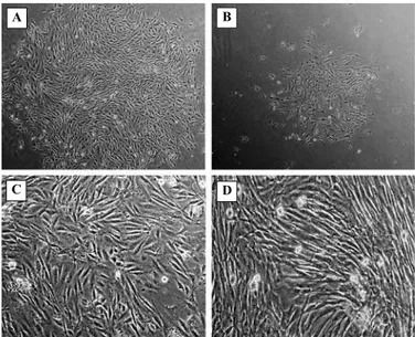
Primary culture of the cells from removed medium contained the large colonies consisting of the cells with spindle-shaped morphology (Fig 1A), this culture reached confluence in an average of 5.36±0.50 days; whereas the adherent cell culture had small colonies (Fig 1B) and approached confluence after an average of 8.09±0.70 days. The cells from either culture were also examined in terms of the time required to reach confluence during each subculture. In the present study, the culture considered as being confluence when all available surfaces of 75-cm2 flask were covered by
the monolayer (Fig 1C and D).
At this time, the culture of the cells from removed medium had average of 4.5×10⁶ cells, while that from the primarily adherent culture contained a little less than 4.5×10⁶ cells. According to our results, the rate of reaching confluence was comparatively high in the cell culture prepared from removed medium than adherent culture. The values of confluence rate of removed medium culture compared with that of primarily adherent cell culture were respectively 3/54±0.52 versus 5/54±0.52 at passage-1, 3.54±0.52 versus 5.63±0.50 at passage-2 and 3.27±0.46 versus 5/36±0.50 at passage-3. All these differences were statistically significant (Fig 2).
Fig 2: Comparison of the culture from removed medium and that from primarily adherent cells in terms of the confluence rate. *The cells from the removed medium possessed significantly more growth rate than those from the primarily adherent culture (n = 22, p<0.05).
Fig 1: The culture of primarily adherent cells and floating cells from removed medium. The primary culture of the cells from removed medium (A) contained large colonies of spindle-shaped cells (Magnification: ×40), while the culture from primarily adherent cells (B) had small colonies (Magnification: ×40). Part C and D are respectively showing the cells from removed medium and primarily adherent cells at confluence state.
*
*
*
0.00 2.00 4.00 6.00 8.00 10.00
Mean Day
Primary Culture Passage 1 Passage 2 Passage 3 Adherent culture Removed medium culture
A B
MSCs dimensions
According to our data, the length and width of MSCs from primarily adherent cultures appeared to be 166.4±7 and 16±0.4 mμ respectively, compared with 165.7±8 and 16±0.5 mμ that were measured as a length and width of the cells comprising the culture of removed medium.
Differentiation Adipogenic cultures
At both culture, 2-3 days after induction initiation, the first lipid droplets were
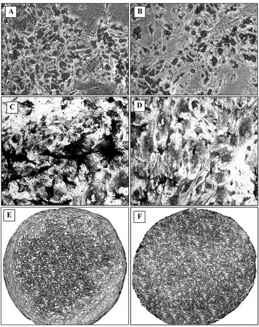
appeared inside the cells and increased in number as the time progressed. To ensure the lipid-nature of the droplet, the cultures were evaluated with oil red staining in 21 days, which as a result they were positively stained red, indicating the adipocytic nature of the differentiated cells (Fig 3A, B).
RT-PCR analysis were further indicated the expression of adipocyte marker genes including PPAR-alpha, PPAR-gamma2 and C/EBP-alpha in the cells from either cultures (Fig 4A).
Fig 3: Evaluation of differentiation potential. Similar to the cells from primarily adherent culture, those from the removed medium could easily differentiate towards skeletal cell lineages. Part A and B are respectively showing the oil red staining for adipocyte detection in the culture from removed medium and primarily adherent cells. At part C and D, alizarin red staining for mineralized matrix detection in the culture from removed medium (C) and primarily adherent cells (D) were shown. Toloidin blue staining for cartilage differentiation of the cells from removed medium and primarily adherent culture were respectively shown at part E and F (Magnification of all images: ×100). (This figures has also been printed in full-color at the end of the tissue)
A B
C D
Fig 4: RT-PCR analysis of differentiation into adipocyte (A), Bone cells (B) and cartilage cells (C).
Osteogenesis
At osteogenic culture of either cell, nodule-like aggregations were appeared. Upon
alizarin red staining, these nodules stained red indicating that they were mineralized during the induction period (Fig. 3C and 3D). RT-PCR analyses indicated that bone specific protein including osteocalcin, osteopontin and alkaline phosphatase was largely expressed in either cell (Fig 4B).
Chondrogenesis
The cells from removed medium similar to those from primarily adherent cells indicated methachromatic properties upon toluidin blue staining (Fig 3E, F). RT-PCR analysis revealed that the mRNA of collagen II, X and aggrecan macromolecules were largely produced in either differentiated cells (Fig 4C).
Discussion
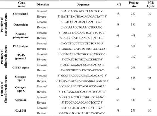
In this study, we replated the cells from removed medium of the rat’s marrow culture, which resulted in appearance of a population of the cells with fibroblastic morphology. Our further examination revealed that these cells possessed high growth rate than the primarily adherent cells and similar to them were able to easily differentiate into bone, cartilage and adipose cells. Former studies have shown that the cells capable of giving rise to three skeletal cell lineages as bone, cartilage and adipose cells could be considered as MSCs, therefore, the isolated Table 1: The primers used for RT-PCR analysis.
Gene
name SequenceDirection A.T
Product size
PCR Cycle
Forward
5´-AGCAGGAATACTAACTGC -3 Osteopontin
Reverse
5´-GATTATAGTGACACAGACTATT-3´ 48
287 30
Forward
5´-GTCCCACACAGCAACTCG-3´ Osteocalcin
Reverse
5´-CCAAAGCTGAAGCTGCCG-3´
58
380 30
Forward
5´-TGCCTTACCAACTCATTTGTG-3´
Primers of
osteogenic genes Alkaline
phosphatase 5´-ACGCGATGCAACACCACTC-3´Reverse 61
401 30
Forward
5´-CCCTGCCTTCCCTGTGAAC-3´ PPAR-alpha
Reverse
5´-GGGACTCATCTGTACTGGTGG-3´ 61
367 35
Forward
5´-GGTGAAACTCTGGGAGATCCC-3´ PPAR-
gamma2 5´-CCATCTCTGCCACGGGCT-3´Reverse
64
352 35
Forward
5´-ACGTGGAGACGCAGCAGAA-3´
Primers of
adipogenic genes C/EBP-alpha
Reverse
5´-AGGCGGTCATTGTCACTGG-3´
63
295 35
Forward
5´-GGCTTAGGGCAGAGAGAGAAG-3´ Collagen
type II 5´-TGGACAGTAGACGGAGGA Reverse AAGTC-3´ 63
315 35
Forward
5´-CAGCAGCATTACGACCCAAG-3´ Collagen
type X 5´-CCTGAGAAGGACGAGTGGAC-3´Reverse 61
334 30
Forward
5´-GGCAACCTCCTGGGTGTAAG-3´
Primers of
Chondrogenic genes Aggrecan 5´-TCGCACCACCAGGTCCTC-3´Reverse
63
444 30
Forward
5´-TCGGTGTGAACGGATTTG-3´ GAPDH
Reverse
5´-ACTCCACGACATACTCAGCAC-3´ 58
276 30
RT(-) RT(+) Primary Removed Medium MSCs
PPAR-alpha
PPAR-Gamma2
C/EBP-alpha
GAPDH
RT(-) RT(+) Primary Removed Medium
MSCs
GAPDH ALP Osteocalcin
Osteopontin
RT(-) RT(+) Primary Removed Medium
MSCs S
Collagen II
Collagen X
Aggrican
GAPDH
A
B
cells from rat’s marrow culture ,in this study, would be considered as MSCs described elsewhere (26-28).
In all protocols for MSCs isolation, a few days after culture initiation, the non adherent cells were discarded at first medium replacement, and those adherent cells on culture surfaces were maintained and expanded as MSCs populations. Indeed, researchers believe that MSCs existed in bone marrow samples are those that adhere on culture surfaces at early days and the cells remained floating in culture medium are non mesenchymal cells (1-5, 19-20). Based on our data, among the non adherent cells of bone marrow primary culture, there are MSCs with differentiation potential similar to the adherent cells. According to our results, these cells possessed comparatively even more growth rate than the adherent cells.
Our data indicated that MSCs are heterogeneous with respect to time of adherence on culture surfaces, while some attaches at early days upon plating, the others remained floating in medium for a while before establishing attachment. Indeed in current protocols, these cells were discarded with medium substitution which is an inevitable step in culture maintaining. Our findings strongly suggested that, these floating cells could survive, adhere and expand with replating, therefore, could be considered as additional source for MSCs population.
To ensure that whether or not the difference in the cell size influence the rate in which the culture became confluence, we measured the dimensions of the cells from either culture. According to the results, there was no significant differences between the two studied populations, indeed the cells of removed medium appeared a little smaller than the adherent cells, indicating that if the cell size is critical for confluence time , this could be in favor of the adherent population rather than non adherent cells from removed medium. Given this fact, the validity of the obtained data could easily be verified. The point that verifies the findings related to cell dimension is the cell counting data. According to these results, the number of the cells in confluence culture of the non adherent cells (about 4.5×10⁶) appeared to be a little more than that of primarily adherent cells.
Current study is not the first report on existence of MSCs in removed medium, indeed there is an experiment by Wan and
associates who first described these cells in human in the year 2006 (25). These authors reported that the cells from removed medium, similar to primarily adherent cells, could differentiate into bone, cartilage and adipose cell lineages. Our results are in agreement with Wan and co-worker’s findings. Additionally, these researchers indicated no difference among the either cells in terms of growth rate. In this regard, our data is different in that, we indicated that the cells from removed medium can grow in rapid rate than the primarily adherent cells. Given that we conducted the experiments using rat marrow cells and Wan and colleagues performed the research on human cells, the difference of growth rate could be attributed to differences in species.
The other finding of the current study was that, replating the removed medium from rat’s marrow culture can result in growing a MSCs population, hence providing a complementary source of MSCs for routine culture setup. This is of great importance in particular, in case when MSCs transplantation is the experiment objective. Taking biopsy from rat’s bone marrow can yield a maximum of 100 micro liters of bone marrow samples, according to our experience in the present study. This amount is not enough to obtain adequate cells to conduct the transplantation experimentation; therefore, a complementary source like the removed medium seemed to be valuable.
Conclusion
Taken together, it was concluded that replating the cells suspended in primary culture medium could yield the cells similar to those from primarily adherent culture with differentiation ability into bone, cartilage and adipose cells. Our data suggested that in bone marrow samples there are at least two subclasses of MSCs, the first are those that are able to adhere at early days of primary cultures and the other are those that attach on surface with some delay, the latter can expand with rapid rate than the former.
References
1. Friedenstein AJ, Petrakova KV. Osteogenesis in transplants of bone marrow cells. J embryol Exp Morph, 1966; 16: 381-390
3. Woodbury D, Schwaz E, Prockop DJ, Black IB. Adult rat and human bone marrow stromal cells differentiate into neurons. J Neuro Scien Res, 2000; 61:364-370 4. Chen L-B, Jiang Xb, Yang: Differentiation of rat marrow mesenchymal stem cells into pancreatic islet beta-cells. World J Gastroenternol, 2004; 10: 3016-3020
5. Oh S-H, Muzzonigro TM, Bae S-H, Laplante JM, Hatch HM, Petersen BE. Adult bone marrow-derived cells trans-differetiating into insulin-producing cells for the treatment of type I diabetes. Lab Invest, 2004; 84:607-617
6. Herrera MB, Bussolati B, Bruno S, Fonsato V, Romanazzi GM, Camussi G. Mesenchymal stem cells contribute to the renal repair of acute tubular epithelial injury. Int J Mol Med, 2004; 14: 1035-1041
7. Xu W, Zhang X, Qian H, Zhu W, Sun X, Hu J, et al. Mesenchymal stem cells from adult human bone marrow differentiate into a cardiomycyte phenotype in vitro. Exp Biol Med, 2004; 229: 623-631
8. Chapel A, Bertho JM, Bensidhoum M, Fouillard L, Young RG, Frick J, et al. Mesenchymal stem cells home to injured tissues when co-infused with hematopoietic cells to treat a radiation-induced multi-organ failure syndrome. J Gene Med, 2003; 5: 1028-1038
9. Piersma AH, Brockbank KG, Ploemacher RE, Van Vliet E, Brakel-van Peer KM, Visser PJ. 1985. Characterization of fibroblastic stromal cells from murine bone marrow. Exp Hematol, 1985; 13, 237-243
10. Owen M. Marrow stromal stem cells. J Cell Sci, 1988; 3: 63-76
11. Koc ON, Peters C, Aubourg P, Raghavan S, Dyhouse S, DeGasperi R, et al. Bone-marrow derived mesenchymal stem cells remained host-derived despite successful hematopoietic engraftment after allogenic transplantation in patient with lysosomal and peroxisomal storage diseases. Exp Hematol, 1999; 27:1675-1681
12. Digirolamo CM, Stokes D, Colter D, Phinney DG, Class R, Prockop DJ. Propagation and senescence of human marrow stromal cells in culture: a simple colony-forming assay identifies samples with the greatest potential to propagate and differentiate. Br J Haematol. 1999; 107:275–281
13. Javazon EH, Colter DC, Schwarz EJ, Prockop DJ. Rat marrow stromal cells are more sensitive to plating density and expand more rapidly from single-cell-derived colonies than human marrow stromal cells. Stem Cells, 2001; 19:219–225
14. Friedenstein AJ, Chailakhjan RK, Lalykina KS. The development of fibroblast colonies in monolayer cultures of guinea-pig bone marrow and spleen cells.
Cell Tissue Kinet, 1970; 3:393–403
15. Rickard DJ, Sullivan TA, Shenker BJ, Leboy PS, Kazhdan I. Induction of rapid osteoblast differentiation in rat bone marrow stromal cell cultures by dexamethasone and BMP-2. Dev Biol, 1994; 161:218–228
16. Awad HA, Butler DL, Boivin GP, Smith FN, Malaviya P, Huibregtse B, et al. Autologous mesenchymal stem cell-mediated repair of tendon. Tissue Eng, 1999; 5: 267–277
17. Eslaminejad MB, Nadri S, Hosseini RH. Expression of thy 1.2 surface antigen increases significantly during the murine mesenchymal stem cells cultivation period. Dev Growth Differ, 2007: 49: 351-364
18. Eslaminejad MB, Nikmahzar A, Taghiyar L, Nadri S, Massumi M. Murine mesenchymal stem cells isolated by low density primary culture system. Dev Growth Differ, 2006: 48, 361-370
19. Castro-Malaspina H, Gay RE, Resnick G, Kappor N, Meyers P, Chiareri D, et al. Characterization of human bone marrow fibroblast colony forming cells (CFC-F) and their progeny. Blood, 1980; 56:289-301 20. Friedenstein ZJ, Gorskaja JF, Kulagina NN.Fibroblast precursors in normal and irradiated mouse hematopoietic organs. Exp Hematol, 1976; 4:267-274
21. Zvaifler NJ, Marinova-Mutafchieva L, Adams G, Edwards CJ, Moss J, Burger JA, et al. Mesenchyamal precursor cells in the blood of normal individuals. Artheritis Res, 2000; 2:477-488
22. Campagnoli C, Roberts IA, Kumar S, Bennett PR, Bellantuono I, Fisk NM. Identification of mesenchymal stem/progenitor cells in human first trimester fetal blood, liver, and bone marrow. Blood, 2001; 98: 2396-2402
23. Erices A, Conget P, Miguell JJ. Mesenchymal progenitor cells in human umbilical cord blood. Br J Haematol, 2000; 109: 235-242
24. Baksh D, Davis JE, Zandstra PW. Adult human bone marrow-derived mesenchymal progenitor cells are capable of adhesion-independent survival and expansion. Exp Hematol, 2003; 31:723-732
25. Wan C, He Q, McCaigue M, Marsh D, Li Gang. Nonadherent cell population of human marrow culture is a complementary source of mesenchymal stem cells (MSCs). J Orthopaedic Res, 2006; 24: 21-28 26. Pittenger MF, Mackay AM, Beck SC, Jaiswal RK, Douglas R, Mosco JD. Multilineage potential of adult human mesenchymal stem cells. Science, 1999; 284:143–147