Retention period after treatment of posterior crossbite
with maxillary expansion: a systematic review
Julia Garcia Costa1, Thaís Magalhães Galindo1, Claudia Trindade Mattos2, Adriana de Alcantara Cury-Saramago2
Objective: The aim of this systematic review was to evaluate the duration of the retention period in growing patients
undergoing maxillary expansion and its relation with posterior crossbite stability. Methods: Search strategies were
ex-ecuted for electronic databases Cochrane Library, Web of Science, PubMed and Scopus, which were completed on January 15, 2016. The inclusion criteria included randomized, prospective or retrospective controlled trials in growing subjects with posterior crossbite; treated with maxillary expanders; retention phase after expansion; post-retention phase of at least 6 months. The exclusion criteria were anterior crossbite, craniofacial anomalies, surgery or another orthodontic intervention; case reports; author’s opinions articles, thesis, literature reviews and systematic reviews. The risk of bias of selected articles was assessed with Cochrane risk of bias tool for RCTs and Downs and Black checklist for non-RCTs. Results: A total of 156 titles/abstracts was retrieved, 44 full-texts were examined, and 6 articles were selected and as-sessed for their methodological quality. The retention period after maxillary expansion ranged between 4 weeks and 16 months. Fixed (acrylic plate, Haas, Hyrax and quad-helix) or removable (Hawley and Hawley expander) appliances were
used for retention. Conclusions: Six months of retention with either fixed or removable appliances seem to be enough
to avoid relapse or to guarantee minimal changes in a short-term follow-up.
Keywords: Crossbite. Maxillary expansion. Retainer.
1 Orthodontics department, Universidade Federal Fluminense, Niterói, Brazil. 2 Professor of Orthodontics, Dental Clinic department, Universidade Federal
Fluminense, Niterói, Brazil.
» The authors report no commercial, proprietary or financial interest in the products or companies described in this article.
Submitted: January 29, 2016 - Revised and accepted: September 12, 2016 DOI: http://dx.doi.org/10.1590/2177-6709.22.2.035-044.oar
How to cite this article: Costa JG, Galindo TM, Mattos CT, Cury-Sarama-go AA. Retention period after treatment of posterior crossbite with maxillary ex-pansion: a systematic review. Dental Press J Orthod. 2017 Mar-Apr;22(2):35-44. DOI: http://dx.doi.org/10.1590/2177-6709.22.2.035-044.oar
Contact address: Adriana de Alcantara Cury-Saramago
Rua Mário Santos Braga, 30, 2º andar, sl. 214 – Campus do Valonguinho Centro – Niterói/RJ – Brazil – CEP: 24.020-140
E-mail: [email protected]
Objetivo: o objetivo da presente revisão sistemática foi avaliar a duração do período de contenção e a estabilidade do tratamento
ortodôntico com expansão maxilar em pacientes em crescimento com mordida cruzada posterior. Métodos: foram realizadas
buscas estratégicas nas bases eletrônicas: Cochrane Library, Web of Science, PubMed e Scopus, até 15 de janeiro de 2016. Os critérios de inclusão foram: estudos clínicos controlados e randomizados, prospectivos ou retrospectivos, de pacientes em crescimento com mordida cruzada; tratados com aparelhos expansores maxilares, com fase de contenção pós-expansão e no mínimo seis meses de fase de pós-contenção. Os critérios de exclusão foram: mordida cruzada anterior, anomalias craniofaciais, cirurgia ou outro trata-mento ortodôntico; relato de casos; artigos de opinião; teses; revisões de literatura e revisões sistemáticas. O risco de viés dos artigos selecionados foi avaliado a partir do Cochrane risk of bias tool para ensaios clínicos randomizados e Downs and Black checklist para ensaios
clínicos não randomizados. Resultados: a busca resultou em 156 títulos/resumos, sendo 44 textos examinados na íntegra. Foram
selecionados 6 artigos para o acesso à qualidade metodológica. A duração do período de contenção ocorreu entre 4 semanas e 6 meses. Aparelhos fixos (aparelho em acrílico, Haas, Hyrax e quad-helix) ou removíveis (Hawley e Hawley com expansor) foram
utilizados na fase de contenção. Conclusão: parece que seis meses de contenção com aparelhos fixos ou removíveis são suficientes
para evitar a recidiva ou garantir mudanças mínimas em um curto período de acompanhamento pós-contenção.
INTRODUCTION
Posterior crossbite is a common malocclusion in the deciduous and mixed dentitions, with prevalence rates
of 7.5%1 to 22%,2 and in the permanent dentition with
rates of 10.2% to 14.4%.3
The etiology of this malocclusion may be dental,
skeletal and/or functional.4 Few studies have reported
the self-correction of posterior crossbite in the decidu-ous dentition, related to the discontinuation of
suck-ing habits and chronic respiratory childhood diseases.5,6
However, this condition is usually not self-corrected.4,7,8
Studies with adolescents and adults have revealed that patients presenting posterior crossbite have an increased risk to develop craniomandibular disorders, showing more signs and symptoms of these
condi-tions.2,5 Several authors suggest the early treatment of
crossbites to prevent mandibular dysfunction as well
as craniofacial asymmetry.7-10
Adults can be submitted to maxillary expansion, al-though there are controversies regarding the nonsurgi-cal treatment.11,12
Various methods have been suggested for correction and retention ater treatment of posterior crossbite in
growing patients: Haas,8,13-16 Hyrax,14,15,17,18 quad-helix
appliance (QDH),4,7,14,15,19-21 removable plates,4,7,9,20-22
grinding7,10 and edgewise ixed appliances.23
The successful treatment of a posterior crossbite is frequently reached not only by the expansion of the maxilla. In growing subjects, the treatment must also achieve the reestablishment of the normal growth rate
on a longitudinal basis,24 as well as improve the oral and
general health.25
No consensus among authors exists regarding the optimal retention period ater maxillary expansion. Some authors recommend that the retention phase
should last for 6 weeks,19 while others advocate 64,21 or 8
months.8 Thus, a systematic review of the literature was
deemed appropriate.
The aim of this systematic review was to evaluate the duration of the retention period in growing patients un-dergoing maxillary expansion and its relation with poste-rior crossbite stability. The PICOS is shown in Table 1.
MATERIAL AND METHODS
This systematic review was registered on the Na-tional Institute of Health Research Database:
www.crd.york.ac.uk/prospero.
The inclusion criteria were randomized controlled trials (RTCs) and controlled trials in human grow-ing subjects; experimental group presentgrow-ing posterior crossbite; treatment with maxillary expanders; retention phase ater expansion; and a minimum 6-month post-retention phase.
The exclusion criteria were subjects presenting an-terior crossbite, craniofacial anomalies, previous sur-gery or another orthodontic intervention; case reports; author’s opinions articles, thesis, literature reviews and systematic reviews.
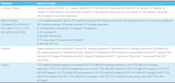
To identify the studies, detailed search strategies were developed and executed in the following elec-tronic databases: Cochrane Library, Web of Science, PubMed and Scopus (Table 2). All electronic searches were conducted between May 28, 2015 and January 15, 2016. No restrictions for language or publication date were used.
The results were compiled into a reference manager (EndNote X5, Thomson Reuters), and duplicate re-cords were excluded.
Two authors independently reviewed titles and ab-stracts according to the inclusion and exclusion criteria. Any disagreement was solved by consultation with two others authors until mutual agreement was reached and initial selection was completed.
Full texts of articles where it was not possible to de-cide for inclusion or exclusion only by reading the title and abstract were also screened to conirm their eligibil-ity. Two authors independently read the full texts of the articles previously selected.
Ater electronic searches and the initial selection process, a supplementary hand search was implemented by checking the references of each selected study. Af-terwards, two authors independently performed a struc-tured quality assessment of the selected articles based
on risk of bias. The Cochrane risk of bias tools26 was
used for randomized studies, and the Downs and Black
checklist27 for non-randomized studies. Any
disagree-ment on the risk of bias assessdisagree-ment was resolved ater consulting other two authors.
The following data from the included articles were ex-tracted and independently compiled by two researchers: author/year; sample description; crossbite type; expander/ activation time; activation rate; retainer appliance and re-tention time; measurements; follow-up time;
Table 1 - PICOS.
Table 2 - Search strategy in databases.
relapse ater follow-up time; crossbite correction stability ater follow-up; conclusion.
In order to verify the percentage of relapse for each transversal measure given by the authors, the diference between the measure immediately ater expansion (AE) and the measure ater 6-month follow-up (FU) was cal-culated following the equation: [(AE-FU)x100/AE].
RESULTS
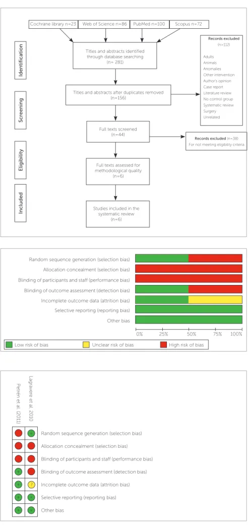
In the databases search, 281 articles were found. After duplicates were excluded, we screened 156 titles and abstracts; and 112 studies were excluded from this review; 44 full texts were screened, and 6 articles were selected according to the eligibility cri-teria. The search process is shown in the Prisma flow diagram (Fig 1).
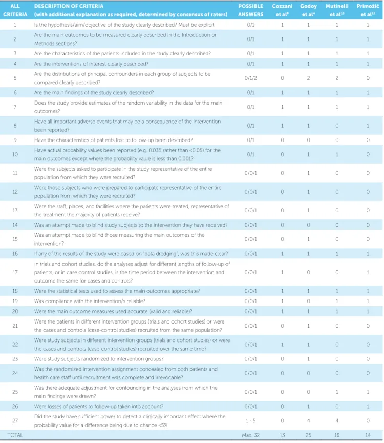
Two articles included, which are randomized con-trolled trials, were assessed with the Cochrane tool and the corresponding graphs are shown in Figures 2 and 3. The non-randomized studies were classi-fied according to their risk of bias, using the Downs
and Black checklist, as: low risk,4 medium16 and high
risk8,22 (Table 3).
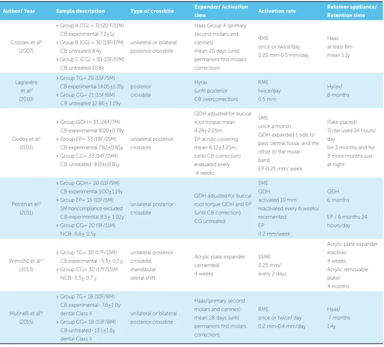
Data extracted from the included articles are dis-played in Tables 4A and 4B. The retention period
af-ter maxillary expansion ranged from ive22 to sixteen
months,16 and the appliances used were: ixed (acrylic
plate expander,22 Haas,8,16 Hyrax17 and quad-helix4,21) or
removable (hawley4,22 and Hawley expander4,21,22).
The follow-up of these patients ranged from 6
months4 to 6016 months, and the relapses of the
mea-surements described reached 0%4 to 27%17.
PICOS Description
Population Growing subjects presenting posterior crossbite
Intervention Treated with maxillary expansion
Comparison Another maxillary expansion procedure, untreated crossbite subjects or untreated subjects without posterior crossbite
Outcomes Duration of the retention period after maxillary expansion and its relation with posterior crossbite stability
Study design Randomized controlled trials (RTCs) and controlled trials in human growing subjects
Database Search strategy
Cochrane Library “palatal expansion technic” or “maxillary expansion” in Title, Abstract, Keywords and “retention” or “retainer” or “stability” or
“relapse” in Title, Abstract, Keywords and “crossbite” in Title, Abstract, Keywords not “case report” in Title, Abstract, Keywords (Word variations have been searched)
Web of Science
(Database=SCI-EXPANDED, SSCI, A&HCI, S, CPCI-SSH, BKCI-S, BKCI-SSH)
1) TS=(palatal expansion technic OR maxillary expansion OR maxillary disjunction OR palatal disjunction OR expansion appliance OR maxillary expander OR palatal expander OR maxillary expander)
2) TS=(retention* OR retainer* OR relapse* OR stability*) 3) TS=(crossbite*)
4) #1 AND #2 AND #3
5) TI=(case report OR case series OR adult*) 6) #4 AND NOT #5
PubMed (palatal expansion technique[MeSH Terms]) OR “maxillary expansion”[Title/Abstract]) OR “maxillary disjunction”[Title/Abstract])
OR “palatal disjunction”[Title/Abstract]) AND “retention”[Title/Abstract]) OR orthodontic retainer[MeSH Terms]) OR “stability”[Title/ Abstract]) OR “relapse”[Title/Abstract]) AND “crossbite”[Title/Abstract]) NOT “case report”[Title]) NOT “case series”[Title]) NOT adult[Title]
Scopus TITLE-ABS-KEY(palatal expansion technique) OR TITLE-ABS-KEY(“maxillary expansion”) OR TITLE-ABS-KEY(“maxillary
Figure 3 - Risk of bias summary for RCTs studies.
Figure 2 - Risk of bias graph for RCTs studies.
Figure 1 - Prisma flow diagram.
Cochrane library n=23 Web of Science n=86 PubMed n=100 Scopus n=72
Titles and abstracts identified through database searching
(n= 281)
Titles and abstracts after duplicates removed (n=156)
Full texts screened (n=44)
Full texts assessed for methodological quality
(n=6)
Studies included in the systematic review
(n=6)
Records excluded
(n=112)
Adults Animals Anomalies Other intervention Author’s opinion Case report Literature review No control group Systematic review Surgery Unrelated
Included
Scr
eening
Eligibility
Identifica
tion
Records excluded (n=38) For not meeting eligibility criteria
Random sequence generation (selection bias)
Random sequence generation (selection bias) Allocation concealment (selection bias)
Allocation concealment (selection bias) Blinding of participants and staff (performance bias)
Blinding of participants and staff (performance bias) Incomplete outcome data (attrition bias)
Incomplete outcome data (attrition bias) Other bias
Other bias
0% 25% 50% 75% 100%
Low risk of bias Unclear risk of bias High risk of bias Blinding of outcome assessment (detection bias)
Blinding of outcome assessment (detection bias) Selective reporting (reporting bias)
Selective reporting (reporting bias) ?
-+
+
+ + +
+ +
Lagraver
e et al. 2010
P
etr
Table 3 - Downs and Black checklist for non-randomized studies.
ALL
CRITERIA
DESCRIPTION OF CRITERIA
(with additional explanation as required, determined by consensus of raters)
POSSIBLE
ANSWERS
Cozzani
et al8
Godoy
et al4
Mutinelli
et al16
Primožič
et al22
1 Is the hypothesis/aim/objective of the study clearly described? Must be explicit 0/1 1 1 1 1
2 Are the main outcomes to be measured clearly described in the Introduction or
Methods sections? 0/1 1 1 1 1
3 Are the characteristics of the patients included in the study clearly described? 0/1 1 1 1 1
4 Are the interventions of interest clearly described? 0/1 1 1 1 1
5 Are the distributions of principal confounders in each group of subjects to be
compared clearly described? 0/1/2 0 2 2 0
6 Are the main indings of the study clearly described? 0/1 1 1 1 1
7 Does the study provide estimates of the random variability in the data for the main
outcomes? 0/1 1 1 1 1
8 Have all important adverse events that may be a consequence of the intervention
been reported? 0/1 1 1 0 1
9 Have the characteristics of patients lost to follow-up been described? 0/1 0 0 0 0
10 Have actual probability values been reported (e.g. 0.035 rather than <0.05) for the
main outcomes except where the probability value is less than 0.001? 0/1 0 1 1 0
11 Were the subjects asked to participate in the study representative of the entire
population from which they were recruited? 0/0/1 0 1 0 0
12 Were those subjects who were prepared to participate representative of the entire
population from which they were recruited? 0/0/1 0 1 0 0
13 Were the staf, places, and facilities where the patients were treated, representative of
the treatment the majority of patients receive? 0/0/1 0 1 0 0
14 Was an attempt made to blind study subjects to the intervention they have received? 0/0/1 0 0 0 0
15 Was an attempt made to blind those measuring the main outcomes of the
intervention? 0/0/1 0 1 0 0
16 If any of the results of the study were based on “data dredging”, was this made clear? 0/0/1 1 1 1 1
17
In trials and cohort studies, do the analyses adjust for diferent lengths of follow-up of patients, or in case control studies, is the time period between the intervention and outcome the same for cases and controls?
0/0/1 1 0 0 1
18 Were the statistical tests used to assess the main outcomes appropriate? 0/0/1 1 1 1 1
19 Was compliance with the intervention/s reliable? 0/0/1 1 0 1 1
20 Were the main outcome measures used accurate (valid and reliable)? 0/0/1 1 1 1 1
21 Were the patients in diferent intervention groups (trials and cohort studies) or were
the cases and controls (case-control studies) recruited from the same population? 0/0/1 0 1 0 0
22 Were study subjects in diferent intervention groups (trials and cohort studies) or were
the cases and controls (case-control studies) recruited over the same time? 0/0/1 1 1 0 0
23 Were study subjects randomized to intervention groups? 0/0/1 0 1 0 0
24 Was the randomized intervention assignment concealed from both patients and
health care staf until recruitment was complete and irrevocable? 0/0/1 0 0 0 0
25 Was there adequate adjustment for confounding in the analyses from which the
main indings were drawn? 0/0/1 0 0 1 1
26 Were losses of patients to follow-up taken into account? 0/0/1 0 1 0 1
27 Did the study have suicient power to detect a clinically important efect where the
probability value for a diference being due to chance <5% 1 - 5 0 4 4 0
TOTAL Max. 32 13 25 18 14
Table 4A - Characteristics and data of included studies.
TG= Treatment group; CG = Control group; F= female; M= male; PFM= Permanent first molar; PSM= Primary second molar; IC= Intercuspid canines; y= years; m= months; RME= Rapid maxillary expansion; QDH= Quad-Helix appliance;
EP= Expansion plate; NCB= Non crossbite group; CB= Crossbite; UPC= Unilateral posterior crossbite.
Author/ Year Sample description Type of crossbite Expander/ Activation
time Activation rate
Retainer appliance/
Retention time
Cozzani et al8
(2007)
» Group A (TG) = 31 (20 F/11 M) CB experimental 7.3 ± 1y » Group B (CG) = 30 (13 F/17 M) CB untreated 8.4y » Group C (CG) = 30 (13 F/17 M) CB untreated 10.8y
unilateral or bilateral posterior crossbite
Haas Group A (primary second molars and canines)
mean 20 days (until permanent irst molars correction)
RME
once or twice/day 0.25 mm-0.5 mm/day
Haas at least 8m mean 1.1y
Lagravère et al17
(2010)
» Group TG= 20 (15F/5M) CB experimental 14.05±1.35y » Group CG= 21 (15F/6M) CB untreated 12.86± 1.19y
posterior crossbite Hyrax (until posterior CB overcorrection) RME twice/day 0.5 mm Hyrax/ 6 months
Godoy et al4
(2011)
» Group QDH= 33 (26F/7M) CB-experimental 8.00±0.79y » Group EP= 33 (18F/15M) CB-experimental 7.82±0.85y » Group CG= 33 (14F/19M) CB-untreated 8.09±0.81y
unilateral posterior crossbite
QDH adjusted for buccal root torque mean 4.24±2.05m EP acrylic covering mean 6.12±3.25m (until CB correction) evaluated every 4 weeks
SME once a month QDH expanded 1 side to pass central fossa; and the other to the molar-band
EP-0.25 mm/ week
Plate placed/ To be used 24 hours/ day
for 3 months and for 3 more months just at night
Petrén et al21
(2011)
» Group QDH= 20 (11F/9M) CB-experimental 9.00±1.19y » Group EP= 15 (10F/5M) 5M noncompliance excluded CB-experimental 8.5± 1.02y » Group CG= 20 (9F/11M) NCB- 8.8± 0.5y
unilateral posterior crossbite
QDH adjusted for buccal root torque QDH and EP (until CB correction) CG untreated
SME QDH
activated 10 mm, reactivated every 6 weeks/ recemented
EP
0.2 mm/week
QDH 6 months
EP / 6 months 24 hours/day
Primožič et al22
(2013)
» Group TG= 30 (17F/13M) CB experimental - 5.3± 0.7 y » Group CG= 30 (17F/13)M NCB- 5.3± 0.7 y
unilateral posterior crossbite, mandibular lateral shift
Acrylic plate expander cemented/
4 weeks
SSME 0.25 mm/ every 2 days
Acrylic plate expander inactive/
4 weeks Acrylic removable plate/
4 months
Mutinelli et al16
(2015)
» Group TG= 18 (10F/8M) CB experimental- 7.6±1.0y dental Class II
» Group CG= 18 (10F/8M) CB-untreated- 13.1±1.6y dental Class II
unilateral or bilateral posterior crossbite
Haas/(primary second molars and canines) mean 28 days (until permanent irst molars correction)
RME
once or twice/ day 0.2 mm-0.4 mm/day
TG= Treatment group; CG = Control group; PFM= Permanent first molar; PSM= Primary second molar; IC= Intercuspid canines; DC= Dental cast; y= years; m= months; RME= Rapid maxillary expansion; QDH= Quad-Helix appliance; EP= Expansion plate; IMD= Intermolar distance; ICD= intercanine distance; GM = Gingival margin; MCT= Mesiobuccal cusp tips; BCT= Gingival margin and buccal cusp tips; NCB= Non crossbite group; CB= Crossbite; NS= Not signifi-cant; NR= Not reported.
Table 4B - Characteristics and data of included studies.
Author/ Year Measurements Follow-up
time
Overcorrec-tion
Experimental
group x Control
group (P value)
Relapse
measure-ments after
follow-up
Crossbite
corrected after
follow-up
Conclusion
Cozzaniet al8
(2007)
» Maxillary arch width: » PFM- center of the fossa » PSM-center of the fossa » IC-cusp tip
» DC minimum 1 y after appliance removal 2.4 ± 1.7y yes - primary teeth no - permanent irst molar PFM: ≤0.01 PSM: ≤0.01 IC: ≤0.05
PFM = 0.9% PSM = 6.0% IC = 5.5%
yes
Relapse: PFM < PSM Overexpand PSM
PFM was stable for 2y 4m after treatment
Lagravere et al17
(2010)
» PC- center of pulp chamber in molars and tip of premolars buccal pulp horn » MBA-mesiobuccal root apex of molars » BA-buccal root apex of premolars » AIB-outer cortex of alveolar bone at the vertical level of the root apex » mm » CBCT Before ixed bonding (12m) long-term post-relapse
yes all groups P<.001
PC16-PC26 = 27% PC14-PC24 = 39% MBA16-MBA26= 28% BA14-BA24 = 18% AIB16-AIB26 = 51% AIB14-AIB24 = 20%
yes
aprox 4mm (70%) expansion - at T4 at molars Dental expansion> skeletal expansion Midpalatal suture separation on TG. No signiicant changes at the level of the pterigoid plates TG=CG
Godoy et al4
(2011)
Maxillary arch width: PSM-center of the fossa IC- cusp tip
DC 6m after appliance removal no IMD: P<0.001 (QDH=EP; QDH≠ CG; EP≠ CG) ICD: P= 0.354
PSF QDH= 2.2% EP = 1.7%
IC
QDH = 0.3% EP = 0% yes
9.1% of the each sample showed relapse
QDH=EP for correct posterior crossbite
QDH> breakage EP> lost appliances QDH< treatment time Treatment may be performed in 1y for posterior CB correction and 6m for retention
Petrén et al21
(2011)
Maxillary arch width: PSM-gingival margin (GM) PSM-mesiobuccal cusp tip (MCT) IC-gingival margin (GM)
IC-buccal cusp tip (BCT) DC
QDH and EP group 4y after correction no IMD (MCT): P=NR (CG>QDH,EP) ICD (BCT): P=NR (CG>QDH)
PSM QDH = 1.6% EP = 5.6% IC (GM) QDH = 4.9% EP = 5.6% IC (BCT) QDH = 1.2% EP = 0.6%
yes
The long-term stability of crossbite correction in the mixed dentition is favorable.
Results: QDH=EP
Primozic et al22
(2013)
Palatal surface area (mm²) 3D digital
DC
12 months later 18 months later 30 months later
yes
Surface(mm²): P= NR NS (TG=CG)
Palatal surface area (TG) = - 0.5%
26.7% of the TG showed relapse
Treatment of unilateral CB in the deciduous dentition also create conditions for normal occlusal and craniofacial development.
Improves facial symmetry and increase palatal area and volume
Mutinelli et al16
(2015)
Maxillary arch width: PSM and IC (mm); 3D digital DC In the permanent dentition 5.3 ± 0.8y
yes - primary teeth no - permanent irst molar IC P= 0.02 PSM P= 0.001
PSM = 1%
IC = 5.1% yes
DISCUSSION
The duration of the steady retention ater maxillary expansion that guarantees the correction of posterior crossbite is not well established in the literature and this was the main reason that led to this systematic review.
The evidence collected in this systematic review combined low, medium and high risk of bias stud-ies. The main drawback in RCTs and non-RCTs was blinding, which is unfeasible in the assessed type of intervention. In non-RCTs, another main problem was the description of the characteristics of subjects lost to follow-up.
However, the heterogeneity among the studies made the comparison diicult. Dental and skeletal measures varied widely, as follows: intermolar distance measured between the center of the fossae of maxillary permanent
irst molars,4,8,16 measured between the mesiobuccal
cusp tips and gingival margin,21 distance between the
center of the fossae of maxillary primary second molars,8
intercanine distance measured between cusp tips,4,8,21
gingival margin,21 palatal surface area,22 and distance of
center of pulp chamber in molars and tip of premolar buccal pulp horn, mesial buccal root apex of molars, buccal root apex of premolars, outer cortex of alveolar
bone at the vertical level of the root apex.17
The appliances used for maxillary expansion in the
studies included were Haas,8,16 Hyrax,17 QDH,4,21
re-movable acrylic expansion plate,4,21 and cemented
acryl-ic plate.22 All authors used the same expander appliance
for retention of the maxillary expansion,4,8,16,17,21,22
ex-cept the quad-helix group in the study from Godoy et
al,4 who used a removable Hawley retainer for retention.
The control group also difered among the studies. In some studies, subjects presenting posterior crossbite
were included in the control group,4,8,16,17 while other
authors selected only patients with no posterior cross-bite (normal occlusion or a diferent malocclusion with
no transverse discrepancies) for the control group.21,22
When these studies featured more than one control group, it was taken into account only the group of
sub-jects with similar occlusion.17
Four studies4,8,16,17 where the control group
com-prised subjects with posterior crossbite were approved by ethics committees and the authors followed their
guidelines. Lagravere et al17 beneited from a treatment
control group with delay of 12 months, and there were no negative consequences for the treatment of patients.
However, that may be an ethical issue, since delaying the correction of a problem, which is known to be bet-ter solved as early as possible may be considered
unethi-cal. This was the reason why Petrén et al21 did not
in-clude a control group of crossbite untreated subjects as their follow-up reached three years ater treatment.
Overcorrection of the posterior crossbite is
recom-mended by some authors4,19,28,29 due to the tooth crown
buccal inclination, which is usually a consequence of
tooth-supported expanders.21 The physiology of the
relapse demonstrate that molars tend to return to their original buccolingual inclination ater retention is dis-continued, that would not allow relapse of the posterior
crossbite if overexpansion was performed.11 Four of the
included studies8,16,17,22 expanded the maxilla until the
crossbite was overcorrected in all groups, particularly it was performed only in primary teeth for Cozzani et al8 and Mutinelli et al.16 In two articles4,21 however, no overexpansion was produced.
Petrén et al21 claims that overcorrection might be
unnecessary, since their results without overexpansion were found to be stable in a long-term, the rate of re-lapse was 1.6% in the intermolar cusp distance, even so to avoid buccal tipping of the molars, the appliance was adjusted for buccal root torque.
Authors that used Haas as retainers for at least 7
months16 and 8 months8 presented a relapse of 1.0% and
0.9% respectively, in the intermolar distance. These re-sults may suggest that a longer time of retention ater maxillary expansion — that is, more than 7 months — would favor stability and less relapse. Moreover, the dif-ference of the mean relapse was only 0.1 mm, which may be clinically irrelevant.
Lagravere et al17 who used Hyrax as a retainer,
ob-served the highest relapse of measurements, 27% in the molar distance, probably related to patient age, since their sample of the treated group was 14 years. All
oth-ers authors4,8,16,21,22 presented younger samples, between
5.1 a 9.7 years old, in the mixed dentition.
When removable appliances were used as retainers
for 6 months, a relapse of 3.2%4 and 1.2%21 was found
in the intermolar distance. Godoy et al4 instructed the
patients to use the removable plate 24 hours a day for 3 months and just at night for 3 more months, while
Petrén et al21 recommended a 24-hour/day use for 6
The overall comparison among fixed and re-movable retainers when a six-month retention was used, showed a very small range of variation,
be-tween 1.2%21 and 3.2%4 in the intermolar distance.
When comparing treatment groups which had as their expander/retainer the QDH and EP, Petrén et
al21 observed similar results. According to Godoy et
al,4 the greatest disadvantage of EP was lost
appli-ances and subsequent laboratory costs, and QDH’s frequent breakage. In spite of this, one of the most cited disadvantages of removable appliances in the
literature is the need for patients’ compliance.4,30
Primozic et al22 assessed skeletal measures through
the palatal surface area. Considering a 30-month fol-low-up, there was no relapse in this skeletal measure. On the contrary, there was an increment of 6.38%. They found that increase in the experimental group to be similar to or greater than the increase observed in the control group of normal occlusion. According to the authors, that indicates the reestablishment of a normal growth rate and the condition for normal occlusion and craniofacial development.
However, relapse in dental and skeletal measures does not necessarily represent a relapse in the posterior crossbite. Four authors have reported recurrence of pos-terior crossbite. That relapse is expressed in percentage of patients as reported by authors or calculated according
to their data: 0%16,21 (Haas group for at least 7 months;
removable plate group, 6 months of retention), 5%21
(QDH group, 6 months of retention), 9.1%4 (QDH
and removable plate, 6 months of retention), 26.7%22
(acrylic cemented plate group, cemented as retention for 1 month and removable for 4 months). Relapse is not
a rare event ater correction of posterior crossbite.21,22,30
Primozic et al22 showed the biggest recurrence of
posterior crossbite ater the treatment, amounting of 8 participants, they suggest that part of this relapse could be explained because the subjects expressed a Class III growth trend, inverse overjet and facial asymmetries.
Limitations of this review are: not enough RCTs were found that were able to answer our question; ad-ditionally, no study speciically aimed at answering this question, nor did any study assessed or compared dif-ferent periods of retention in patients wearing the same kind of appliance. Our systematic review clearly shows the need for randomized controlled trials that specii-cally assess diferent periods of retention with the same appliances and the stability of correction of the posterior crossbite, so that a protocol may be created for success-ful treatment maintenance.
The clinical implication of this systematic review is that six months of retention of crossbite correction used 24 hours a day should be able to maintain the results obtained. However, the evidence for this con-clusion is moderate.
CONCLUSION
1. Keski-Nisula K, Lehto R, Lusa V, Keski-Nisula L, Varrela J. Occurrence of malocclusion and need of orthodontic treatment in early mixed dentition. Am J Orthod Dentofacial Orthop. 2003 Dec;124(6):631-8.
2. Tausche E, Luck O, Harzer W. Prevalence of malocclusions in the early mixed dentition and orthodontic treatment need. Eur J Orthod. 2004 June;26(3):237-44.
3. Jonsson T, Arnlaugsson S, Karlsson KO, Ragnarsson B, Arnarson EO, Magnusson TE. Orthodontic treatment experience and prevalence of malocclusion traits in an Icelandic adult population. Am J Orthod Dentofacial Orthop. 2007 Jan;131(1):8.e11-8.
4. Godoy F, Godoy-Bezerra J, Rosenblatt A. Treatment of posterior crossbite comparing 2 appliances: a community-based trial. Am J Orthod Dentofacial Orthop. 2011 Jan;139(1):e45-52.
5. Egermark-Eriksson I, Carlsson GE, Magnusson T, Thilander B. A longitudinal study on malocclusion in relation to signs and symptoms of cranio-mandibular disorders in children and adolescents. Eur J Orthod. 1990 Nov;12(4):399-407.
6. Thilander B, Wahlund S, Lennartsson B. The efect of early interceptive treatment in children with posterior cross-bite. Eur J Orthod. 1984 Feb;6(1):25-34.
7. Petrén S, Bondemark L, Söderfeldt B. A systematic review concerning early orthodontic treatment of unilateral posterior crossbite. Angle Orthod. 2003 Oct;73(5):588-96.
8. Cozzani M, Guiducci A, Mirenghi S, Mutinelli S, Siciliani G. Arch width changes with a rapid maxillary expansion appliance anchored to the primary teeth. Angle Orthod. 2007 Mar;77(2):296-302.
9. Defraia E, Marinelli A, Baroni G, Tollaro I. Dentoalveolar efects induced by a removable expansion plate. Prog Orthod. 2007;8(2):260-7.
10. Flores-Mir C. Grinding is efective in early orthodontic treatment of unilateral posterior crossbite. Evid Based Dent. 2005;6(1):24.
11. Handelman CS, Wang L, BeGole EA, Haas AJ. Nonsurgical rapid maxillary expansion in adults: report on 47 cases using the Haas expander. Angle Orthod. 2000 Apr;70(2):129-44.
12. Lee KJ, Park YC, Park JY, Hwang WS. Miniscrew-assisted nonsurgical palatal expansion before orthognathic surgery for a patient with severe mandibular prognathism. Am J Orthod Dentofacial Orthop. 2010 June;137(6):830-9. 13. Haas AJ. Long-term posttreatment evaluation of rapid palatal expansion.
Angle Orthod. 1980 July;50(3):189-217.
14. Huynh T, Kennedy DB, Joondeph DR, Bollen AM. Treatment response and stability of slow maxillary expansion using Haas, hyrax, and quad-helix appliances: a retrospective study. Am J Orthod Dentofacial Orthop. 2009 Sept;136(3):331-9.
15. Wong CA, Sinclair PM, Keim RG, Kennedy DB. Arch dimension changes from successful slow maxillary expansion of unilateral posterior crossbite. Angle Orthod. 2011 July;81(4):616-23
REFERENCES
16. Mutinelli S, Cozzani M. Rapid maxillary expansion in early-mixed dentition: efectiveness of increasing arch dimension with anchorage on deciduous teeth. Eur J Paediatr Dent. 2015 June;16(2):115-22.
17. Lagravère MO, Carey J, Heo G, Toogood RW, Major PW. Transverse, vertical, and anteroposterior changes from bone-anchored maxillary expansion vs traditional rapid maxillary expansion: a randomized clinical trial. Am J Orthod Dentofacial Orthop. 2010 Mar;137(3):304.e1-12; discussion 304-5. 18. Wangsrimongkol T, Manosudprasit M, Pisek P, Leelasinjaroen P. Correction
of complete maxillary crossbite with severe crowding using Hyrax expansion and ixed appliance. J Med Assoc Thai. 2013 Sept;96 Suppl 4:S149-56. 19. Bell RA, Le Compte EJ. The efects of maxillary expansion using a quad-helix
appliance during the deciduous and mixed dentitions. Am J Orthod. 1981 Feb;79(2):152-61.
20. Bjerklin K. Follow-up control of patients with unilateral posterior cross-bite treated with expansion plates or the quad-helix appliance. J Orofac Orthop. 2000;61(2):112-24.
21. Petrén S, Bjerklin K, Bondemark L. Stability of unilateral posterior crossbite correction in the mixed dentition: a randomized clinical trial with a 3-year follow-up. Am J Orthod Dentofacial Orthop. 2011 Jan;139(1):e73-81. 22. Primožič J, Richmond S, Kau CH, Zhurov A, Ovsenik M. Three-dimensional
evaluation of early crossbite correction: a longitudinal study. Eur J Orthod. 2013 Feb;35(1):7-13.
23. Gurel HG, Memili B, Erkan M, Sukurica Y. Long-term efects of rapid maxillary expansion followed by ixed appliances. Angle Orthod. 2010 Jan;80(1):5-9. 24. Kau CH, Zhurov A, Scheer R, Bouwman S, Richmond S. The feasibility
of measuring three-dimensional facial morphology in children. Orthod Craniofac Res. 2004 Nov;7(4):198-204.
25. McNamara JA Jr, Lione R, Franchi L, Angelieri F, Cevidanes LH, Darendeliler MA, et al. The role of rapid maxillary expansion in the promotion of oral and general health. Prog Orthod. 2015;16:33.
26. Higgins JPT, Green S, editors. Cochrane handbook for systematic reviews of interventions. 2009. version 5.1.0, updated March 2011.
27. Downs SH, Black N. The feasibility of creating a checklist for the assessment of the methodological quality both of randomised and non-randomised studies of health care interventions. J Epidemiol Community Health. 1998 June;52(6):377-84.
28. Mew J. Relapse following maxillary expansion. A study of twenty-ive consecutive cases. Am J Orthod. 1983 Jan;83(1):56-61.
29. Sandikçioğlu M, Hazar S. Skeletal and dental changes after maxillary expansion in the mixed dentition. Am J Orthod Dentofacial Orthop. 1997 Mar;111(3):321-7.