CLINICAL SCIENCE
I Department of Cardiac and Thoracic Surgery, The Chaim Sheba Medical Center, Tel Hashomer 52621, Israel.
II Department of Cardiothoracic Surgery, Jinling Hospital, School of Clinical Medicine, Nanjing University, Nanjing 210002, Jiangsu Province, People’s Republic of China.
Tel: 86 25 84801332
Received for publication on January 20, 2010 First review completed on February 08, 2010 Accepted for publication on February 25, 2010
THE BICUSPID AORTIC VALVE AND ITS RELATION TO AORTIC DILATION
Shi-Min Yuan,I,II Hua Jing,II Jacob Lavee I
doi: 10.1590/S1807-59322010000500007
Yuan SM, Jing H, Lavee J. The bicuspid aortic valve and its relation to aortic dilation. Clinics. 2010;65(5):497-505.
BACKGROUND: A bicuspid aortic valve (BAV) is a common congenital heart disease, which affects 1-2% of the population. However, the relationship between BAVs and aortic dilation has not been suficiently elucidated.
METHODS: A total of 241 BAV patients who were referred to this hospital for cardiac surgey over a 4.75-year period were in-cluded in this study. In addition to the clinical characteristics of the inin-cluded patients, the morphological features of the aortic valve and aorta, the length of the left main coronary artery, and the laboratory indings (the coagulation and hematological parameters as well as the total cholesterol concentration) were determined and compared with those of the tricuspid aortic valve (TAV) patients.
RESULTS: The BAV patients were younger than the TAV patients for a valve surgery in the last 3 months of the study period. The BAV patients were predominantly male. Most of the BAVs that were surgically treated were stenotic, regurgitant, or combined, and only 19 (7.88%) were normally functioning valves. According to echocardiography or operative records, 148 (78.31%) were type A, 31 (16.40%) were type B, and 10 (5.29%) were type C. The left main coronary artery was much shorter in the BAV patients than it was in the TAV patients. There was no signiicant difference between BAV and TAV patients in the total cholesterol concentra-tions; whereas differences were noted between patients receiving lipid-lowering therapy and those not receiving lipid-lowering therapy. The dimensions of the aortic root, sinotubular junction, and ascending aorta were beyond normal limits, while they were signiicantly smaller in the BAV patients than in the TAV patients. They were also much smaller in patients receiving statin therapy than those not receiving statin therapy in both groups. Moreover, the aortic dilation in the BAV group was found to be signiicantly associated with patient age.
CONCLUSIONS: The BAV patients developed aortic wall and aortic valve disorders at a younger age than the TAV patients and were predominantly male. Aortic dilation was observed in the aortic root, sinotubular junction, and ascending aortic segments in both the BAV and TAV patients undergoing surgical aortic valve replacement, although the BAV patients had a smaller degree of dilation than the TAV patients, and dilation was also signiicantly age-related in this group. The shorter left main coronary artery that the BAV patients possess may contribute to the progressive course of aortic dilation that these patients experience. Statin therapy did not affect the aortic annulus in either group, but did decrease the dimensions of the aortic root, sinotubular junction and ascend-ing aorta. In general, statin therapy had a better effect on the aortas of the TAV patients than it did on those of the BAV patients.
KEYWORDS: Aortic dilation; Aortic valve disorder; Bicuspid aortic valve; Cholesterol; Left main coronary artery.
INTRODUCTION
A bicuspid aortic valve (BAV) is a common congenital heart disease. It is observed in 1-2% of the entire population1
and in 31.9% of individuals with aortic valve disorders.2 A
BAV may be accompanied by aortic stenosis or insuficiency, infective endocarditis, an aortic aneurysm or an aortic dissection. It can also be associated with aortic coarctation and Marfan’s syndrome.1 Aortic dilation begins at an early
age in the BAV patients and is usually characterized by “mid-ascending”-type dilation.3 The dilation process can
be observed even in patients with normally functioning BAVs.4-7 The increased stress on the aortic wall that results
from the recirculation vortices produced by the abnormal opening of the BAVs has been recognized as a risk factor for aortic dilation.3 The intrinsic links between the BAVs
BAVs represent a disorder that affects both the aortic valve and the aorta3,6,7 and have led some authors to suggest
early replacement of the aorta instead of isolated valve replacement in such patients.8,9 In spite of multiple previous
morphological investigations, 2,10 little is known concerning
the relationship between the BAVs and aortic dilation.7 The
goal of this study is to describe the relation between the BAV and aortic dilation in addition to its clinical implications and morphological characteristics by comparing to those of the TAV.
PATIENTS AND METHODS
Patient selection. We performed a retrospective search for the BAV patients who were referred to this hospital for cardiac surgery between January 1, 2004 and September 30, 2008 using the Doctor’s Record Database by examining perfusionists’ records. Patients who came in for a repeated operation whose native aortic valve (bicuspid, tricuspid, or unknown) had been replaced in their primary operations within or outside of the study period were excluded from the present study. As a result, a total of 241 patients were included in the BAV group.
Controls. Patients with a tricuspid aortic valve (TAV) referred to this hospital for a cardiac operation for an isolated aortic dilation, a combined aortic dilation and aortic valve abnormality, coarctation of the aorta, Marfan’s syndrome, or aortic dissection during the entire study period as well as those with infective endocarditis or aortic valve disorders who were seen during the last three months of the study period were included in the study as control patients. As a result, a total of 225 patients were included in the control group.
Observations. The patients’ surgical indications, the characteristics of their BAVs, and the location of their aortic dilations were recorded. The pathological types of the BAVs were determined based on a short axis view of the pre- or intraoperative echocardiograms, which were either viewed on the Horizon Cardiology Web of this hospital or taken from the operative records. The dimensions of the aorta were measured at four levels on the long axis view of the archived echocardiogram: the aortic annulus, the aortic sinus, the sinotubular junction and the ascending aorta. The length of the left main coronary artery was measured on the right anterior oblique view of the archived coronary angiography images. Perioperative laboratory indings (the coagulation and hematological parameters as well as the total cholesterol concentration) were collected from the “Autolab” database of the hospital and analyzed. The laboratory values of the BAV group were compared to those of the control group.
Statistical analysis. Data were expressed as means±SDs.
Unpaired t-tests were used in comparative analyses. Two-tailed p-values <0.05 were considered to be statistically signiicant.
RESULTS
A total of 241 consecutive patients with a BAV were referred to this hospital for a cardiac operation during the study period, representing 6.71% of 3,594 adult patients during the same period. The group included 186 males and 55 females. The mean age of the BAV patients was 56.129 ± 15.096 (range, 16-85) years, which was much lower than that of the TAV patients (Table 1). Their surgical indications included diseases of the aorta (aortic dilation, combined aortic dilation and aortic valve disorder, aortic dissection, coarctation of the aorta, and Marfan’s syndrome) and valvular disorders (aortic valve disorder, infective endocarditis, and mitral valve regurgitation) (Table 2). The cardiopulmonary bypass time was 109.39±35.92 (range, 46-324) min (n=236), the aortic cross-clamp time was 83.74±28.15 (range, 29-225) min (n=236), the total circulatory arrest time was 16.292±5.385 (range, 8-35) min (n=24), and the minimal core temperature of the patients during total circulatory arrest was 18.138±2.225 (range, 15-20) °C (n=13). The postoperative morbidity rate was 4.98%. The postoperative complications included atrial ibrillation (n=2), cerebrovascular accident (n=2), prosthetic aortic valve leak (n=4), prosthetic valvular endocarditis (n=2), and spontaneous cerebral hemorrhage (n=1). One patient with poor left ventricular function who underwent aortic valve replacement and repeated coronary artery bypass died 27 days after the operation, yielding a mortality rate of 0.41%. The BAV was stenotic in 122 patients, regurgitant in 62 patients, combined stenotic and regurgitant in 3 patients, and normal in 19 patients, respectively (Table 3).
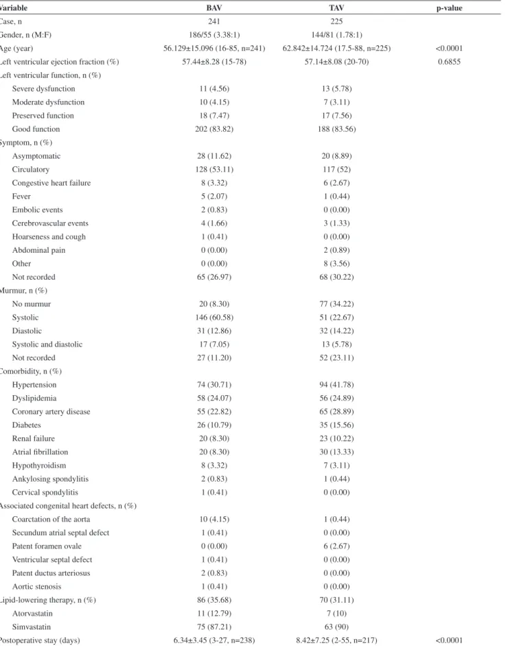
Table 1 - Clinical characteristics of included patients.
Variable BAV TAV p-value
Case, n 241 225
Gender, n (M:F) 186/55 (3.38:1) 144/81 (1.78:1)
Age (year) 56.129±15.096 (16-85, n=241) 62.842±14.724 (17.5-88, n=225) <0.0001
Left ventricular ejection fraction (%) 57.44±8.28 (15-78) 57.14±8.08 (20-70) 0.6855
Left ventricular function, n (%)
Severe dysfunction 11 (4.56) 13 (5.78)
Moderate dysfunction 10 (4.15) 7 (3.11)
Preserved function 18 (7.47) 17 (7.56)
Good function 202 (83.82) 188 (83.56)
Symptom, n (%)
Asymptomatic 28 (11.62) 20 (8.89)
Circulatory 128 (53.11) 117 (52)
Congestive heart failure 8 (3.32) 6 (2.67)
Fever 5 (2.07) 1 (0.44)
Embolic events 2 (0.83) 0 (0.00)
Cerebrovascular events 4 (1.66) 3 (1.33)
Hoarseness and cough 1 (0.41) 0 (0.00)
Abdominal pain 0 (0.00) 2 (0.89)
Other 0 (0.00) 8 (3.56)
Not recorded 65 (26.97) 68 (30.22)
Murmur, n (%)
No murmur 20 (8.30) 77 (34.22)
Systolic 146 (60.58) 51 (22.67)
Diastolic 31 (12.86) 32 (14.22)
Systolic and diastolic 17 (7.05) 13 (5.78)
Not recorded 27 (11.20) 52 (23.11)
Comorbidity, n (%)
Hypertension 74 (30.71) 94 (41.78)
Dyslipidemia 58 (24.07) 56 (24.89)
Coronary artery disease 55 (22.82) 65 (28.89)
Diabetes 26 (10.79) 35 (15.56)
Renal failure 20 (8.30) 23 (10.22)
Atrial ibrillation 20 (8.30) 30 (13.33)
Hypothyroidism 8 (3.32) 7 (3.11)
Ankylosing spondylitis 2 (0.83) 1 (0.44)
Cervical spondylitis 1 (0.41) 0 (0.00)
Associated congenital heart defects, n (%)
Coarctation of the aorta 10 (4.15) 1 (0.44)
Secundum atrial septal defect 1 (0.41) 0 (0.00)
Patent foramen ovale 0 (0.00) 6 (2.67)
Ventricular septal defect 1 (0.41) 0 (0.00)
Patent ductus arteriosus 2 (0.83) 0 (0.00)
Aortic stenosis 1 (0.41) 0 (0.00)
Lipid-lowering therapy, n (%) 86 (35.68) 70 (31.11)
Atorvastatin 11 (12.79) 7 (10)
Simvastatin 75 (87.21) 63 (90)
Postoperative stay (days) 6.34±3.45 (3-27, n=238) 8.42±7.25 (2-55, n=217) <0.0001
Aortic dilation was present in 97 (40.25%) of the 241 patients that had BAVs. The aortic root was involved in 9 (9.28%) patients, the ascending aorta was involved in 68 (70.10%) patients, both the aortic root and the ascending aorta were involved in 14 (14.43%) patients, both the ascending aorta and aortic arch were involved in 5 (5.15%) patients, and the aortic root, ascending aorta, and aortic arch were involved in 1 patient (1.03%). The BAV patients that had evidence of aortic dilation were younger than their TAV counterparts and exhibited male gender predominance. The aortic annulus, aortic sinus, sinotubular junction, and ascending aorta were dilated in both BAV and TAV patients, and the dimensions of the aortic root and ascending aorta were much smaller in the BAV than in the TAV patients who
were undergoing valvular surgery. In the absence of aortic dilation, signiicant differences were observed between the dimensions of the aortic root and the sinotubular junction in the BAV patients as compared to the TAV patients, with the former having larger dimensions (Table 4).
The length of the left main coronary artery could be measured in 86 patients with the BAVs and in 61 patients in the TAV group, all of whose coronary angiograms were available for viewing in the "Horizon Cardiology Web" of this hospital. A signiicant difference in the length of the left main coronary artery was noted between the patients with a BAV and those with a TAV (5.4669 ± 2.9332 mm vs. 7.4967 ± 6.7316 mm, p = 0.0141). The range of the left main coronary artery length was 1.1-15.2 mm in the former group and 0-30.8 mm in the latter group. The shortest left main coronary artery identiied among the BAV patients was 1.1 mm, while three patients with a TAV (who presented with type A aortic dissection, ascending aortic aneurysm, and ascending aortic aneurysm associated with aortic valve regurgitation, respectively) had absent left main coronary arteries and instead had left anterior descending coronary arteries and marginal arteries that bifurcated at the aortic sinus (Figure 3).
In the BAV group, no differences were noted in the length of the left main coronary artery with regard to the cardiac circulation dominance type of the patients [right vs. left dominance 5.4381±3.3298 (range, 2.3-12.1) mm (n=36) vs. 6.1050 ±2.7765 (range, 1.9-15.2) mm (n=12), p=0.5357; right vs. codominance 5.4381±3.3298 (range, 2.3-12.1) mm (n=36) vs. 5.3009±2.7479 (range, 1.92-14.6) mm (n=34), p=0.4512; and left vs. codominance 6.1050 ±2.7765 (range, 1.9-15.2) mm (n=12), vs. 5.3009±2.7479 (range, 1.92-14.6) mm (n=34), p=0.8519].
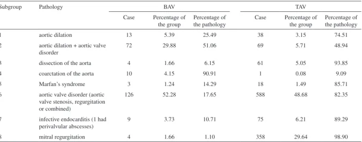
Table 2- A comparison of the constitutional percentages between BAV and TAV patients of the entire study period.
Subgroup Pathology BAV TAV
Case Percentage of the group
Percentage of the pathology
Case Percentage of the group
Percentage of the pathology
1 aortic dilation 13 5.39 25.49 38 3.15 74.51
2 aortic dilation + aortic valve disorder
72 29.88 51.06 69 5.71 48.94
3 dissection of the aorta 4 1.66 6.15 61 5.05 93.85
4 coarctation of the aorta 10 4.15 90.91 1 0.08 9.09
5 Marfan’s syndrome 3 1.24 14.29 18 1.49 85.71
6 aortic valve disorder (aortic valve stenosis, regurgitation or combined)
126 52.28 17.65 588 48.68 82.35
7 infective endocarditis (1 had perivalvular abscesses)
9 3.73 10.71 75 6.21 89.29
8 mitral regurgitation 4 1.66 1.10 358 29.64 98.90
BAV=bicuspid aortic valve; TAV=tricuspid aortic valve.
Table 3 - Functional parameters of the bicuspid aortic valve.
Valve function Case (%)
Aortic stenosis 122 (50.62%)
Severe 113 (92.62%)
Moderate-to-severe 4 (3.28%)
Moderate 3 (2.46%)
Mild 2 (1.64%)
Aortic regurgitation 62 (25.73%)
Severe 36 (58.06%)
Moderate-to-severe 6 (9.68%)
Moderate 8 (12.90%)
Mild-to-moderate 4 (6.45%)
Mild 7 (11.29%)
Minimal 1 (1.61%)]
Aortic stenosis + regurgitation 38 (15.77%)
Homological parameters were much lower than normal limits. In the BAV group, there was no signiicant difference
noted between the hemoglobin concentrations of patients without and with infective endocarditis [10.6947±1.1914 (n=231) (range, 7.6-14.9) vs. 9.9744±1.3726 (n=9) (range, 7.9-12.1), p= 0.0781].
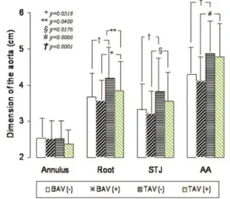
There were no inter-group differences in the total cholesterol concentrations. However, intra-group differences were noted between patients receiving lipid-lowering therapy and those not receiving lipid-lowering therapy (Figure 4). The baseline dimensions of the aortic root, sinotubular junction, and ascending aorta were greater than the upper limit of normal in both groups, although those of the TAV patients tended to be larger than those of the BAV patients (Figure 5). Multiple linear regression analysis did not show any strong correlations between the dimensions of the aorta and the length of the left main coronary artery or the total serum cholesterol concentration in the BAV group. However, aortic dimensions at the four levels that were measured were found to be signiicantly related to patient age (Y=0.823+0.748X1 -0.0253X2+0.383X3-0.00057X4; R = 0.749; R2 = 0.562;
adjusted R2 = 0.552; F = 56.693; p<0.0001).
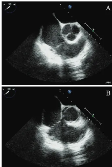
Figure 1 - Short axis views from intraoperative transesophageal
echocar-diography. (A) Type A bicuspid aortic valve with two commissures at 2 and 8 o’clock and a raphe (arrow) at the 5 o’clock position. The anterior lealet and the raphe are calciied. (B) Type B bicuspid aortic valve with two com-missures at 5 and 11 o’clock and a raphe (arrow) at the 7-8 o’clock position. Both lealets and the raphe are calciied. (C) Type C bicuspid aortic valve with two commissures at the 3-4 and 9 o’clock positions with no raphe. The lealets were free of calciication.
Figure 2 - A short axis view of the aorta, showing the aortic valve. (A) Three
DISCUSSION
Approximately 15-50% of the BAV patients retained their normal valvular function,11 while the others had
dysfunctional valves at the early age of 61.3±12.8 years as compared to 67.5±12.9 years in the TAV patients.2 The
BAV patients also exhibited a male gender predominance with regard to the occurrence of valvular dysfunction. Including the BAV patients who had infective endocarditis but normal functioning valves, 78.3% of the aortic valves of the BAV patients were stenotic, 15.6% were regurgitant, and 4% were both stenotic and regurgitant. In a previous report, Collins et al.2 found that among the
BAV patients included in their study, calciication of the base, calciication of the commissure, and ossiication were found in 39.1%, 43.3%, and 7% of the valves.
Aortic dilation and aortic dissection accounted for 50-70% and 7-13% of the BAV patients, respectively.7,12
About 40% of the patients with Marfan’s syndrome had aortic dissections, and the BAVs were present in 5% of the Marfan’s patients.12 Except for the complications
described in the literature, the presented BAV patient setting represented 1.66% of the patient population with mitral regurgitation, and one of them was associated with a large secundum atrial septal defect.13 TheBAVs can be associated
with other congenital cardiovascular lesions, including coarctation of the aorta, ventricular septal defects, patent ductus arteriosus, William's syndrome, Turner's syndrome, and Shone's syndrome.1 However, none of our BAV patients
had any of these conditions.
Brandenburg et al.10 deined the morphological types of
the BAVs based on the short axis view of the transthoracic echocardiogram. In type A BAVs, the posterior and anterior commissures and raphe are located at 4-5, 9-10, and 1-2 o'clock, and the anterior lealet is larger; in type B BAVs, the posterior and anterior commissures and raphe are located at
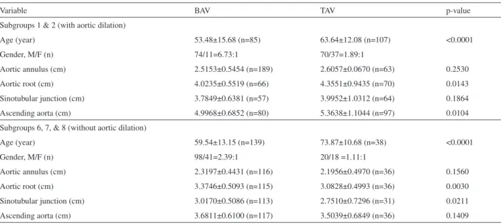
Table 4 - Age, gender, and aortic dimensions of patients with and without aortic dilation.
Variable BAV TAV p-value
Subgroups 1 & 2 (with aortic dilation)
Age (year) 53.48±15.68 (n=85) 63.64±12.08 (n=107) <0.0001
Gender, M/F (n) 74/11=6.73:1 70/37=1.89:1
Aortic annulus (cm) 2.5153±0.5454 (n=189) 2.6057±0.0670 (n=63) 0.2530
Aortic root (cm) 4.0235±0.5519 (n=66) 4.3551±0.9435 (n=70) 0.0143
Sinotubular junction (cm) 3.7849±0.6381 (n=57) 3.9952±1.0312 (n=64) 0.1864
Ascending aorta (cm) 4.9968±0.6852 (n=80) 5.3638±1.1044 (n=97) 0.0104
Subgroups 6, 7, & 8 (without aortic dilation)
Age (year) 59.54±13.15 (n=139) 73.87±10.68 (n=38) <0.0001
Gender, M/F (n) 98/41=2.39:1 20/18 =1.11:1
Aortic annulus (cm) 2.3197±0.4431 (n=116) 2.1956±0.4970 (n=36) 0.1560
Aortic root (cm) 3.3746±0.5093 (n=115) 3.0828±0.4993 (n=36) 0.0030
Sinotubular junction (cm) 3.0170±0.5086 (n=113) 2.7510±0.7296 (n=31) 0.0211
Ascending aorta (cm) 3.6811±0.6100 (n=117) 3.5039±0.6849 (n=36) 0.1409
BAV=bicuspid aortic valve; TAV=tricuspid aortic valve.
Table 5 - Laboratory indings of patients undergoing aortic valve replacement.
Variable BAV TAV p-value
Prothrombin time (%) 53.55±27.26 (n=240) (range 9-120) 71.58±29.61 (n=220) (range 17-128) <0.0001
International normalized ratio 1.6699±0.6841 (n=240) (range 0.9-4.9) 1.3847±0.5652 (n=222) (range 0.89-3.55) <0.0001
Partial thromboplastin time (sec) 33.4125±6.3080 (n=240) (range 20-59) 33.8308±13.4102 (n=220) (range 22-150) 0.6647
Cholesterol (mg/dL) 130.41±30.50 (n=239) (range 63-246) 126.95±30.97 (n=223) (range 65-222) 0.2266
Hemoglobin (g/dL) 10.6677±1.2033 (n=240) (range 7.6-14.9) 10.8509±1.3307 (n=223) (range 7.9-16.5) 0.1205
Red blood cell (M/µL) 3.6748±0.4501 (n=239) (range 2.87-5.44) 3.7287±0.4715 (n=222) (range 2.7-5.17) 0.2097
Hematocrit (%) 31.8633±3.6299 (n=239) (range 22.5-45.1) 32.4768±3.8058 (n=222) (range 22.8-42.7) 0.0772
Platelet (K/µL) 219.977±91.540 (n=240) (range 57-625) 212.117±98.219 (n=223) (range 38-609) 0.3733
6, 1-2, and 9-10 o'clock, and the right lealet is larger; and in type C BAVs, the commissures are at 3 and 9 o'clock, with no raphe, and the anterior and posterior lealets are equal in size. In our study, as in previous studies, type A BAVs were most common, followed by type B and type C BAVs.14,15
With echocardiography, it is possible to make a false positive diagnosis when the coaptation line of a TAV is faint, and a false negative diagnosis may be made if a prominent raphe is present in a BAV.16 However, only one patient in the
present series had a false negative diagnosis based on their
echocardiogram. In this patient, the aortic valve appeared to have three coaptations, but a speciic doming coniguration was present during systole. The diagnosis of a BAV was established intraoperatively.
A study on the aorta dimensions of patients with purely severely stenotic BAVs revealed that these patients developed a moderate dilation of the aorta at an early age, while controls that had TAVs did not.9 Yasuda et al.17 has
proposed two causes concerning the association between the dilation of the ascending aorta and the BAVs: hemodynamic burden caused by aortic valvular disorder and congenital aortic fragility. They compared the echocardiographic characteristics of patients with the BAVs and with the TAVs
Figure 3 - Images showing the absence of the left main coronary artery. (A)
A 3-D reconstruction of a computerized tomography scan of the heart of a 64-year-old male patient with an ascending aortic aneurysm and moderate-to-severe aortic regurgitation with a tricuspid aortic valve. (B) A right anterior oblique view of the coronary angiogram of a 75-year-old male patient with a chronic type A aortic dissection and a tricuspid aortic valve.
Figure 4 - Intra-group differences between patients receiving statin therapy
and those not receiving statin therapy. (+) = receiving statin therapy; (-) = not receiving statin therapy; BAV = bicuspid aortic valve; TAV = tricuspid aortic valve.
Figure 5 - Dimensions of the aorta of patients receiving statin therapy as
and found that, in addition to the fact that aortic dilation occurred more rapidly in patients with the BAVs, a similar aortic dilation rate was observed in the BAV patients who underwent aortic valve replacement as compared to those who did not. They concluded that aortic valve replacement could prevent aortic dilation in patients with the TAVs but that in patients with the BAVs, aortic valve replacement could not prevent aortic dilatation.
Scholz et al.18 reported that 24% of the BAV patients
in their study had a left dominant cardiac circulation. Additionally, they found that the incidence of this type of circulation was highest in patients with aortic coarctation regardless of whether these patients had a BAV or a TAV. Hutchins et al.19 found a higher incidence of left dominance,
29%, in postmortem angiography among the patients with the BAVs in their study. They postulated that the left dominance that they observed may have resulted from decreased blood low to the left side of the heart. In the BAV patients, the length of the left main coronary artery was 1.0 ± 0.3 cm (range, 0.6-1.3), 1.3 ± 0.2 cm (range, 0.6-1.8) and 0.9 cm in patients with a left, right and codominant coronary artery circulation, respectively. No signiicant difference in the length of the left main coronary artery was found between the BAV and TAV patients or between patients with different types of coronary artery circulation dominance.20
Collins et al.2 reported a prevalence of infective
endocarditis of 1.5% among patients with the BAVs in his study, while we observed a prevalence of 3.73%. Lamas and Eykyn11 reported that nearly one-third of the BAV patients
with infective endocarditis in their study had valvular or myocardial abscesses, whereas perivalvular abscesses were present in 1/9 of the infective endocarditis patients with the BAVs in our series. Lamas and Eykyn11 found that among 50
BAV patients with infective endocarditis, 86% had anemia with a mean hemoglobin level of 11.8±1.58 g/dL. Anemia can be a result of several disorders of the aorta and aortic valve. Diehm et al.21 reported that 30.7% of the abdominal
aortic aneurysm patients had anemia at baseline in their study and that anemia was independently associated with aneurysmal size and reduced long-term survival following endovascular repair. Intravascular hemolysis and hemolytic anemia are sometimes seen in patients with aortic valve stenosis.22 It has therefore been suggested that severe aortic
valve disorders may be associated with anemia, especially in non-survivors of such patients.23
An even younger age and a greater male gender predominance of the BAV patients were observed in this patient group than have been observed in previous studies. A previous study found that the BAVs were present in 1-15% of patients who suffered aortic dissections at <54 years of age.12 Those results were slightly different from our study,
in which we observed an aortic dissection rate of 1.66% occurring at a median age of 68.5 (range, 28-77) years. In the present study, anemia was noted in both the BAV and TAV patients with and without infective endocarditis based on their hemoglobin levels, red blood cell counts, and hematocrits. In the BAV patients, the prothrombin time was shorter and the international normalized ratio was higher than the upper limit of normal, but the partial thromboplastin time was normal.
An important result of this study that is contrary to reports in the literature24 is the disparity between the
aortas of the BAV and TAV patients, in that the aortic dilation observed in the aortic root, sinotubular junction, and ascending aorta segments of the BAV patients undergoing surgery was less severe than that of the TAV patients undergoing surgery. A possible explanation for this phenomenon is that aortic dilation is age-related, a inding that has been previously described by Cecconi and associates, 5,6 because in our study, the BAV patients were
much younger than the TAV patients. Recent retrospective trials 25,26 have demonstrated that it is possible to slow the
progression of aortic stenosis with statin therapy, and statin therapy was therefore recommended to all the patients with aortic valve disorders in order to slow or prevent dilation of the aorta.27 Our results are in agreement with those of
the previous study in that statin therapy has the potential to decrease the degree of aortic dilation that patients experience. In our study, statin therapy was associated with smaller dimensions of the aortic root, sinotubular segment, and ascending aorta in both groups. Interestingly, we found that statin therapy did not inluence the dimension of the aortic annulus and was more effective among the TAV patients.
REFERENCES
1. Braverman AC, Güven H, Beardslee MA, Makan M, Kates AM, Moon MR. The bicuspid aortic valve. Curr Probl Cardiol. 2005;30:470-522.
2. Collins MJ, Butany J, Borger MA, Strauss BH, David TE. Implications of a congenitally abnormal valve: a study of 1025 consecutively excised aortic valves. J Clin Pathol. 2008;61:530-6.
3. Della Corte A, Bancone C, Quarto C, Dialetto G, Covino FE, Scardone M, et al. Predictors of ascending aortic dilatation with bicuspid aortic valve: a wide spectrum of disease expression. Eur J Cardiothorac Surg. 2007;31:397-405.
4. Nistri S, Sorbo MD, Marin M, Palisi M, Scognamiglio R, Thiene G. Aortic root dilatation in young men with normally functioning bicuspid aortic valves. Heart. 1999;82:19-22.
5. Cecconi M, Manfrin M, Moraca A, Zanoli R, Colonna PL, Bettuzzi MG, et al. Aortic dimensions in patients with bicuspid aortic valve without signiicant valve dysfunction. Am J Cardiol. 2005;95:292-4.
6. Cecconi M, Nistri S, Quarti A, Manfrin M, Colonna PL, Molini E, et al. Aortic dilatation in patients with bicuspid aortic valve. J Cardiovasc Med. (Hagerstown) 2006;7:11-20.
7. Zeppilli P, Bianco M, Bria S, Palmieri V. Bicuspid aortic valve: an innocent finding or a potentially life-threatening anomaly whose complications may be elicited by sports activity? J Cardiovasc Med (Hagerstown). 2006;7:282-7.
8. Ergin MA, Spielvogel D, Apaydin A, Lansman SL, McCullough JN, Galla JD, et al. Surgical treatment of the dilated ascending aorta: when and how? Ann Thorac Surg. 1999;67:1834-9.
9. Morgan-Hughes GJ, Roobottom CA, Owens PE, Marshall AJ. Dilatation of the aorta in pure, severe, bicuspid aortic valve stenosis. Am Heart J. 2004;147:736-40.
10. Brandenburg RO Jr, Tajik AJ, Edwards WD, Reeder GS, Shub C, Seward JB. Accuracy of 2-dimensional echocardiographic diagnosis of congenitally bicuspid aortic valve: echocardiographic-anatomic correlation in 115 patients. Am J Cardiol. 1983;51:1469-73.
11. Lamas CC, Eykyn SJ. Bicuspid aortic valve--A silent danger: analysis of 50 cases of infective endocarditis. Clin Infect Dis. 2000;30:336-41.
12. Ward C. Clinical significance of the bicuspid aortic valve. Heart 2000;83:81-5.
13. Yuan SM, Shinfeld A, Raanani E. Mitral valve cleft associated with secundum atrial septal defect: case report and review of the literature. Monaldi Arch Chest Dis. 2007;68:48-51.
14. Fernandes SM, Sanders SP, Khairy P, Jenkins KJ, Gauvreau K, Lang P, Simonds H, Colan SD. Morphology of bicuspid aortic valve in children and adolescents. J Am Coll Cardiol. 2004;44:1648-51.
15. Sabet HY, Edwards WD, Tazelaar HD, Daly RC. Congenitally bicuspid aortic valves: a surgical pathology study of 542 cases (1991 through 1996) and a literature review of 2,715 additional cases. Mayo Clin Proc. 1999;74:14-26.
16. De Mozzi P, Longo UG, Galanti G, Maffulli N. Bicuspid aortic valve: a literature review and its impact on sport activity. Br Med Bull. 2008;85:63-85.
17. Yasuda H, Nakatani S, Stugaard M, Tsujita-Kuroda Y, Bando K, Kobayashi J, et al. Failure to prevent progressive dilation of ascending aorta by aortic valve replacement in patients with bicuspid aortic valve: comparison with tricuspid aortic valve. Circulation. 2003;108 Suppl 1:II291-4.
18. Scholz DG, Lynch JA, Willerscheidt AB, Sharma RK, Edwards JE. Coronary arterial dominance associated with congenital bicuspid aortic valve. Arch Pathol Lab Med. 1980;104:417-8.
19. Hutchins GM, Nazarian IH, Bulkley BH. Association of left dominant coronary arterial system with congenital bicuspid aortic valve. Am J Cardiol. 1978;42:57-9.
20. Virmani R, Chun PK, Robinowitz M, Goldstein RE, McAllister HA Jr. Length of left main coronary artery. Lack of correlation to coronary artery dominance and bicuspid aortic valve: an autopsy study of 54 cases. Arch Pathol Lab Med. 1984;108:638-41.
21. Diehm N, Benenati JF, Becker GJ, Quesada R, Tsoukas AI, Katzen BT, et al. Anemia is associated with abdominal aortic aneurysm (AAA) size and decreased long-term survival after endovascular AAA repair. J Vasc Surg. 2007;46:676-81.
22. Kawase I, Matsuo T, Sasayama K, Suzuki H, Nishikawa H. Hemolytic anemia with aortic stenosis resolved by urgent aortic valve replacement. Ann Thorac Surg. 2008;86:645-6.
23. Bruch C, Kauling D, Reinecke H, Rothenburger M, Scheld HH, Breithardt G, et al. Prevalence and prognostic impact of comorbidities in patients with severe aortic valve stenosis. Clin Res Cardiol. 2007;96:23-9.
24. Basso C, Boschello M, Perrone C, Mecenero A, Cera A, Bicego D, et al. An echocardiographic survey of primary school children for bicuspid aortic valve. Am J Cardiol. 2004;93:661-3.
25. Novaro GM, Tiong IY, Pearce GL, Lauer MS, Sprecher DL, Grifin BP. Effect of hydroxymethylglutaryl coenzyme a reductase inhibitors on the progression of calciic aortic stenosis. Circulation. 2001;104:2205-9.
26. Aronow WS, Ahn C, Kronzon I, Goldman ME. Association of coronary risk factors and use of statins with progression of mild valvular aortic stenosis in older persons. Am J Cardiol. 2001;88:693-5.