Page 284 VOJNOSANITETSKI PREGLED Vojnosanit Pregl 2016; 73(3): 284–287.
Correspondence to: Boris Džudović, Clinic for Emergency and Internal Medicine, Military Medical Academy, Crnotravska 17, 11 000 Belgrade, Serbia. E-mail: [email protected]
C A S E R E P O R T UDC: 616.12-073.75-06
DOI: 10.2298/VSP141208005D
Iatrogenic dissection of the left main coronary artery during elective
diagnostic procedures – A report on three cases
Jatrogena disekcija glavnog stabla leve koronarne arterije tokom elektivne
dijagnosti
č
ke procedure
Nemanja Djenić*, Boris Džudović*, Radoslav Romanović*†, Nenad Ratković*†, Zoran Jović‡, Boško Djukić*, Marijan Spasić‡, Siniša Stojković§, Slobodan
Obradović*†
*Clinic for Emergency and Internal Medicine, ‡Clinic for Cardiology, Military Medical Academy, Belgrade, Serbia; †Faculty of Medicine of the Military Medical Academy,
University of Defence, Belgrade, Serbia §Clinic for Cardiology, Clinical Center of Serbia, Belgrade, Serbia
Abstract
Introduction. Left main coronary artery dissection is a rare and potentially life-threatening complication of coronary an-giography and angioplasty which requests urgent revasculariza-tion. Case report. During the period between 2010 and No-vember 2014 at single healthcare center we did totally 8,884 coronary procedures, out of which 2,333 were percutaneous coronary interventions (PCI). In this period we had a total of 3 (0,03%) left main coronary artery dissections, and all of them were successfully treated by PCI. We presented three cases with iatrogenic dissection of the left main coronary artery, oc-curred during elective diagnostic procedures, successfully treated with PCI with different techniques. Conclusion. PCI could be fast and life-saving approach in iatrogenic dissections of the left main coronary artery.
Key words:
iatrogenic disease; coronary angiography; percutaneous coronary intervention; treatment outcome.
Apstrakt
Uvod. Disekcija glavnog stabla leve koronarne arterije je retka i potencijalno po život opasna komplikacija tokom koronarne angiografije i angioplastike i zahteva hitnu revaskularizaciju. Prikaz bolesnika. Tokom perioda između 2010. i novembra 2014. u našoj ustanovi sprovedene su ukupno 8 884 koronarne procedure, od kojih su 2 333 bile perkutane koronarne inter-vencije (PKI). U tom periodu imali smo ukupno 3 (0,03%) di-sekcije glavnog stabla leve koronarne arterije i sve su uspešno rešene putem PKI. U radu su prikazana tri bolesnika sa jatro-genom disekcijom glavnog stabla leve koronarne arterije, nasta-le tokom enasta-lektivnih dijagnostičkih procedura, koje su uspešno rešene pomoću PKI, korišćenjem različitih tehnika. Zaključak. PKI može biti brz pristup kojim se spasava život pri lečenju ja-trogenih disekcija glavnog stabla leve koronarne arterije.
Ključne reči:
jatrogena bolest; angiografija koronarnih arterija; perkutana koronarna intervencija; lečenje, ishod.
Introduction
Iatrogenic dissection of the left main coronary artery (LMCA) is a rare and potentially life-threatening complication of coronary angiography and angioplasty, which requires urgent revascularization, using percutaneous coronary intervention (PCI) or surgery revascularization, also known as coronary artery bypass graft (CABG) 1.
LMCA dissection often leads to abrupt occlusion causing a great deal of myocardial ischemia, which results in acute heart failure with hemodynamic collapse. Prior to 1993, when PCI of iatrogenic dissection of LMCA was first done, urgent CABG
surgery was the only treatment option 2. It has been shown that the above mentioned complications are significantly more likely to occur during PCI procedures comparing to diagnostic cathete-rization (0.10% vs. 0.06%) 3. When it comes to cardiac surgical care of these complications (for successful CABG) according to available data, 30-day mortality rate in this group of patients is slightly more than 26% 4.
Case report
interventi-Vol. 73, No. 3 VOJNOSANITETSKI PREGLED Page 285
Djenić N, et al. Vojnosanit Pregl 2016; 73(3): 284–287.
ons, out of which 2,333 were PCIs. In this period we had a to-tal of 3 (0.03%) LMCA dissections, and all of them were successfully treated by PCI (Figures 1–3). Common characte-ristics of all the patients were: they were women aged 61–72 years, with hypertension, angina pectoris complaints, and un-protected LMCA, and dissection occurred during elective dia-gnostic procedures as shown in Table 1.
Discussion
Left main coronary artery dissection is a rare but potentially fatal complication that requires emergency care and coordination of cardiologist, cardiac surgeon and cardiopulmonary resuscitation team. If these complications oc-cur, clinical picture, depending on the remaining anterograde blood flow, varies from asymptomatic in patients with preserved TIMI 3 flow, to clinical image of cardiogenic shock in patients with completely compromised flow behind the point of dissecti-on. However, even in cases with initially preserved TIMI 3 flow and hemodynamic stability, rapid deterioration can be quickly followed by progression of aortic dissection or thrombus forma-tion, which is always an urgent situation and requires immediate revascularization.
In patients with compromised hemodynamic status, an in-tra-aortic balloon pump could be a useful alternative for impro-ving blood flow, and increasing oxygen delivery of the patient 5. Dissection is the result of mechanical injury of the arteri-al warteri-all due to manipulation of the catheter, either diagnostic or
guided one, when the potential risk for dissection is much gre-ater, as we already stated. Catheter type also plays an impor-tant role. For example, Amplatz, or small Judkins catheters can go deep into LMCA, therefore should be avoided for deeper positioning within the LMCA. Also, attention should be paid when using wires with higher penetration index (because of the potential sub-intimal route) and concomitant contrast
injec-tion, with constant and careful monitoring of hemodynamic status. In addition, good judgment is needed regarding unusual anatomy of the left coronary artery (abnormal artery location or origin), as well as, atherosclerosis or potential calcification and plaque in the LMCA, or more expressed angulation of LMCA and LAD / circumflex artery (CXA) joint. Careful po-sitioning of the catheter in the artery coaxial level, contrast strength, and experience of the operator are also very impor-tant assumptions in the prevention of these complications.
Special group of patients like those with structural heart damage (bicuspid aortic valve) or the patients with hereditary connective tissue abnormalities (Marfan syndrome, cystic me-dial necrosis) are of special concern when performing elective diagnostic procedures 6.
Notwithstanding the foregoing, in certain cases it is im-possible to prevent LMCA dissection.
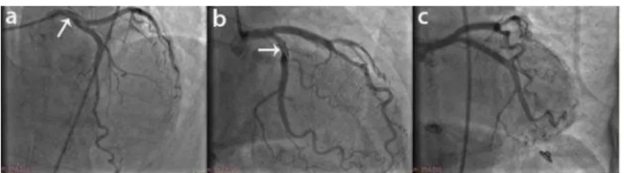
All the three cases of LMCA dissection caused by diag-nostic catheter, shown here, were successfully treated using PCI method, with the right selection of bifurcation techniques (Cullote, one stent or double stent technique), depending on Fig. 1 – a) (arrow) Spiral dissection line of the left main coronary artery (LMCA), and b) (arrow) Circumflex artery
(CXA); c) After stents implantation no dissection lines and coronary flow disturbance could be seen.
Fig. 2 – a) (arrow) Occlusive dissection of the left main coronary artery (LMCA); b) (arrow) Spreading down to significant stenosis in proximal left anterior descending artery (LAD) compromising coronary flow in mid and distal LAD; c) After
stent implantation in LMCA, complete left system coronary flow has been achieved.
Fig. 3 – a) Occlusive spiral dissection of the left main coronary artery (LMCA) with b) Complete flow obstruction in the entire left coronary artery system; c) After stent implantation in LMCA, complete left system coronary flow has been
Page 286 VOJNOSANITETSKI PREGLED Vol. 73, No. 3
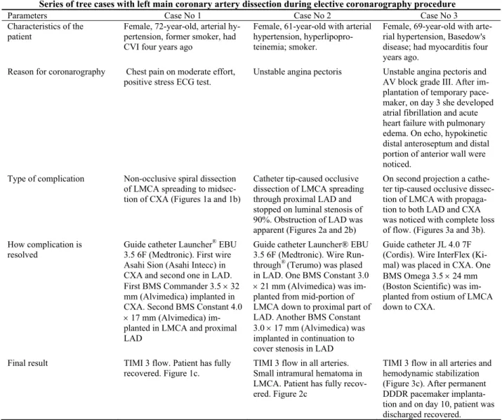
Djenić N, et al. Vojnosanit Pregl 2016; 73(3): 284–287. Table 1 Series of tree cases with left main coronary artery dissection during elective coronarography procedure
Parameters Case No 1 Case No 2 Case No 3
Characteristics of the patient
Female, 72-year-old, arterial hy-pertension, former smoker, had CVI four years ago
Female, 61-year-old with arterial hypertension, hyperlipopro-teinemia; smoker.
Female, 69-year-old with arte-rial hypertension, Basedow's disease; had myocarditis four years ago.
Reason for coronarography Chest pain on moderate effort, positive stress ECG test.
Unstable angina pectoris Unstable angina pectoris and AV block grade III. After im-plantation of temporary pace-maker, on day 3 she developed atrial fibrillation and acute heart failure with pulmonary edema. On echo, hypokinetic distal anteroseptum and distal portion of anterior wall were noticed.
Type of complication Non-occlusive spiral dissection of LMCA spreading to midsec-tion of CXA (Figures 1a and 1b)
Catheter tip-caused occlusive dissection of LMCA spreading through proximal LAD and stopped on luminal stenosis of 90%. Obstruction of LAD was apparent (Figures 2a and 2b)
On second projection a cathe-ter tip-caused occlusive dissec-tion of LMCA with propaga-tion to both LAD and CXA was noticed with complete loss of flow. (Figures 3a and 3b).
How complication is resolved
Guide catheter Launcher® EBU 3.5 6F (Medtronic). First wire Asahi Sion (Asahi Intecc) in CXA and second one in LAD. First BMS Commander 3.5 32 mm (Alvimedica) implanted in CXA. Second BMS Constant 4.0 17 mm (Alvimedica) im-planted in LMCA and proximal LAD
Guide catheter Launcher® EBU 3.5 6F (Medtronic). Wire Run-through® (Terumo) was plased in LAD. One BMS Constant 3.0 21 mm (Alvimedica) was im-planted from mid-portion of LMCA down to proximal part of LAD. Another BMS Constant 3.0 17 mm (Alvimedica) was implanted in continuation to cover stenosis in LAD
Guide catheter JL 4.0 7F (Cordis). Wire InterFlex (Ki-mal) was placed in CXA. One BMS Omega 3.5 24 mm (Boston Scientific) was im-planted from ostium of LMCA down to CXA.
Final result TIMI 3 flow. Patient has fully
recovered. Figure 1c.
TIMI 3 flow in all arteries. Small intramural hematoma in LMCA. Patient has fully recov-ered. Figure 2c
TIMI 3 flow in all arteries and hemodynamic stabilization (Figure 3c). After permanent DDDR pacemaker implanta-tion and on day 10, patient was discharged recovered. CVI – cerebrovascular insult; AV – atrioventricular; LMCA – left main coronary artery; LAD – left anterior descending;
CXA – circumflex artery. TIMI – thrombolysis in miocardial infarction flow grade; BMS – bare metal stent.
the estimated time, the patient’s hemodynamic status, coronary anatomy, dissection type (extensive or localized, occlusive or preserved with TIMI 3 flow), as well as, in accordance with the experience of operators in specific techniques.
Ending the bifurcation procedure with kissing technique is the recommended way and the final step in standard proce-dures. However, in the presented cases, we emphasize that those were patients with marked hemodynamic instability, but with the obtained adequate angiographic effect after placement of a stent in the left main branch. Due to the immediate excellent angiographic effect, after the state of extreme instability, operator’s evaluation was that additional methods as final kissing or morphological assessment as intravascular ultrasound (IVUS) or optical coherence tomography (OCT) could be done in the second act, and after stabilization of the
clinical status, with the aim to speed up the completion of the intervention, and to give as less as possible amount of contrast in the situations when the patient was extremely endangered.
Conclusion
If recognized in time, LMCA dissection can be successfully treated with stent implantation, resulting in a fa-vorable short-term and long-term outcome. PCI could be fast and life-saving approach in iatrogenic disseactions of the left main coronary artery.
Conflict of interest
Vol. 73, No. 3 VOJNOSANITETSKI PREGLED Page 287
Djenić N, et al. Vojnosanit Pregl 2016; 73(3): 284–287.
R E F E R E N C E S
1. Devlin G, Lazzam L, Schwartz L. Mortality related to diagnostic cardiac catheterization. The importance of left main coronary disease and catheter induced trauma. Int J Card Imaging 1997; 13(5): 379−84.
2. Onsea K, Kayaert P, Desmet W, Dubois CL. Iatrogenic left main coronary artery dissection. Neth Heart J 2011; 19(4): 192−5. 3. Eshtehardi P, Adorjan P, Togni M, Tevaearai H, Vogel R, Seiler C,
et al. Iatrogenic left main coronary artery dissection: incidence, classification, management, and long-term follow-up. Am Heart J 2010; 159(6): 1147−53.
4. Kovac JD, de Bono DP. Cardiac catheter complications related to left main stem disease. Heart 1996; 76(1): 76−8.
5. Cheng C, Wu C, Hsieh Y, Chen Y, Chen C, Chen S, et al. Percuta-neous coronary intervention for iatrogenic left main coronary artery dissection. Int J Cardiol 2008; 126(2): 177−82.
6. Awadalla H, Sabet S, el Sebaie A, Rosales O, Smalling R. Catheter-induced left main dissection incidence, predispositionand the-rapeutic strategies experience from two sides of the hemi-sphere. J Invasive Cardiol 2005; 17(4): 233−6.