Gonadotropin therapy in assisted reproduction: an
evolutionary perspective from biologics to biotech
Roge´rio de Barros F. Lea˜o, Sandro C. Esteves
Andrology & Human Reproduction Clinic (ANDROFERT), Referral Center for Male Reproduction, Campinas/SP, Brazil.
Gonadotropin therapy plays an integral role in ovarian stimulation for infertility treatments. Efforts have been made over the last century to improve gonadotropin preparations. Undoubtedly, current gonadotropins have better quality and safety profiles as well as clinical efficacy than earlier ones. A major achievement has been introducing recombinant technology in the manufacturing processes for follicle-stimulating hormone, luteinizing hormone, and human chorionic gonadotropin. Recombinant gonadotropins are purer than urine-derived gonadotropins, and incorporating vial filling by mass virtually eliminated batch-to-batch variations and enabled accurate dosing. Recombinant and fill-by-mass technologies have been the driving forces for launching of prefilled pen devices for more patient-friendly ovarian stimulation. The most recent developments include the fixed combination of follitropin alfa+lutropin alfa, long-acting FSH gonadotropin, and a new family of prefilled pen injector devices for administration of recombinant gonadotropins. The next step would be the production of orally bioactive molecules with selective follicle-stimulating hormone and luteinizing hormone activity.
KEYWORDS: Gonadotropins; Ovulation Induction; Assisted Reproductive Techniques; Systematic Review. Lea˜o RB, Esteves SC. Gonadotropin therapy in assisted reproduction: an evolutionary perspective from biologics to biotech. Clinics. 2014;69(4):279-293.
Received for publication onAugust 21, 2013;First review completed onAugust 30, 2013;Accepted for publication onAugust 30, 2013 E-mail: [email protected]
Tel.: 55 19 3295-8877
& INTRODUCTION
Gonadotropin therapy plays an integral role in ovarian stimulation for infertility treatments. It was introduced almost one century ago, and the last 25 years have yielded major advancements.
Treating anovulatory women through exogenous gona-dotropin administration began in the 1960s. It then expanded to ovulatory women undergoing treatment with assisted reproduction technology (ART) in the 1980s (1-3). In fact, the introduction of controlled ovarian stimulation (COS) for multiple follicular development significantly increased pregnancy rates inin vitrofertilization (IVF) (4). Such stimulation protocols have been developed and refined to maximize the beneficial effects of treatment while minimizing complications and risks (5).
In this review, we first describe the gonadotropin glycoprotein structure and actions. We then outline the landmark research that generated the currently used gonadotropins. Next, we critically discuss the quality,
safety, and clinical efficacy of commercially available gonadotropins, and last, we present an overview of the novel pharmaceutical preparations under investigation.
& GONADOTROPIN STRUCTURE AND FUNCTION
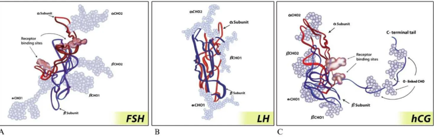
The three gonadotropins, follicle-stimulating hormone (FSH), luteinizing hormone (LH) and human chorionic gonadotropin (hCG), are glycoproteins composed of two non–covalently linked protein subunits, the alpha and beta subunits (6). The alpha subunit contains 92 amino acids (AA) and is identical in FSH, LH, and hCG. In contrast, the beta subunits are distinct and confer unique receptor specificity as well as differential biological properties (7). Its biological activity is provided by the attachment of carbohydrate moieties, forming heterodimers (3). The extent and pattern of glycosylation conveys the spectrum of different charges, bioactivities and half-lives for each glycoprotein (8). Glycoproteins have two basic types of glycosylation patterns: O-linked glycosylation, which is characterized by a carbohydrate N-acetylgalactosamine (GalNAc) attached to the hydroxyl group of an amino acid, serine or threonine, and N-linked glycosylation, which is characterized by an N-acetyl glucosamine (GlcNAc) attached to the amide group of asparagine (Asn) (9). The oligosaccharides often terminate with sialic acid and/or sulfonated b1–4-linked GalNAc (SO3-4GalNAc) (10,11).
Molecules with a large number of sulfonated Gal-NAcs disappear faster from the circulation than less-sulfonated isoforms due to their affinity for liver SO3-4GalNAc Copyrightß2014CLINICS– This is an Open Access article distributed under
the terms of the Creative Commons Attribution Non-Commercial License (http:// creativecommons.org/licenses/by-nc/3.0/) which permits unrestricted non-commercial use, distribution, and reproduction in any medium, provided the original work is properly cited.
No potential conflict of interest was reported.
receptors (10,12). On the other hand, more sialic acids enhance the half-life (10,13).
Follicle-stimulating hormone
The FSH beta subunit is composed of 111 AAs with four N-linked glycosylation sites, two on the alpha subunit (Asn52 and Asn78) and two on the beta subunit (Asn7 and Asn24) (9,14). Thus, each subunit is attached to two carbohydrate moieties with variable compositions that, in turn, create different isoforms with different plasma half-lives (ranging from 3 to 4 hours) and bioactivities (Figure 1) (3,9). Sialic acid residues are much more common in FSH than sulfonated residues (13). Increased sialylation enhances FSH metabolic stability by decreasing both glomerular filtration and clearance by liver sialoglycoprotein receptors, which is the major site for gonadotropin clearance (15,16).
FSH stimulates the recruitment and growth of early antral follicles (2-5 mm in diameter) by binding to the G protein-coupled receptors expressed exclusively on granulosa cells (GCs) (17,18). An adenylate cyclase-mediated signal is activated, followed by the expression of multiple mRNAs that encode proteins responsible for cell proliferation, differentiation, and function. FSH stimulates GC prolifera-tion and growth (mitogenic acprolifera-tion) and induces aromatase activity via P450 activation (19). Concomitantly, the number of FSH receptors increases as GCs respond to FSH. The regulation of GC FSH receptor activity involves not only a direct cAMP-mediated FSH influence on its own receptor gene but also estrogen and other inhibitory agents, includ-ing epidermal growth factor, fibroblast growth factor, and GnRH-like protein. Inhibin and activin, which are also produced by granulosa cells in response to FSH, have autocrine activity and stimulate FSH receptor production, thus enhancing FSH action (19,20).
Luteinizing hormone
The LH beta subunit is comprised of 121 AAs, which is a difference that confers specific biologic activity and facil-itates its interaction with the LH receptor (3). LHb-subunits contain a single site with N-linked glycosylation (Asn 30)
and fewer sialic acid residues (only 1 or 2); as such, LH has a short half-life of only 20 to 30 minutes (Figure 1) (15).
LH plays a key role in promoting steroidogenesis and developing the leading follicle; it has different functions at different stages of the cycle (19-22). During the early follicular phase, LH stimulates androgen production by theca cells. Cholesterol is converted into androgens (testos-terone and androstenedione) through the transcription of the cholesterol side-chain cleavage enzyme (P450scc), P450c17, and 3b-hydroxysteroid dehydrogenase (3b-HSD) genes (Figure 2). Androgens are then transferred to the GC and transformed into estrogens via aromatization (21). Finally, LH promotes final follicular maturation via its direct effects on the GC in the late follicular phase (22). Theca cells and GCs also secrete peptides, including insulin-like growth factor (IGF), inhibin, and activin, which act as both autocrine and paracrine factors and modulate LH-mediated androgen production in the thecal compartment as well as FSH-mediated aromatization in GCs (19,20,23).
Human chorionic gonadotropin
Although the beta subunit of hCG has an AA sequence similar to LH, a notable difference is the presence of a long carboxyterminal segment with 24 AAs containing four O-linked oligosaccharide sites (Figure 1) (3,9). In addition, hCG beta subunits contain two sites of N-linked glycosyla-tion compared with a single LH site. Due to the higher number of both glycosylation sites and sialic acid residues (approximately 20) compared with LH, hCG exhibits a markedly longer terminal half-life of 24 hours compared with approximately 30 minutes for LH (15).
Due to their similar structure, hCG binds the same receptor as LH. In gonadotropin therapy, hCG is used to promote the final follicular maturation stages and progres-sion of the immature oocyte at prophase I (the germinal vesicle stage) through meiotic maturation to metaphase II (3). The meiotic process requires approximately 36 hours to complete; a few hours later, ovulation occurs. As such, follicular aspiration upon oocyte retrieval is timed with hCG administration in IVF. In addition, hCG can be used to
maintain luteal function until placental steroidogenesis is well established (19).
& MILESTONES IN GONADOTROPIN DEVELOPMENT
Researchers began to develop gonadotropin preparations in 1910, when experimental evidence suggested that the pituitary has a role in regulating gonadal stems (2). Zondek, in collaboration with Ascheim (1927), demonstrated that blood and urine from pregnant women contained a gonad-stimulating substance capable of inducing both follicular
maturation and ovarian stromal luteinization when injected into immature mice. This substance was shown to be hCG, which is produced in the placental tissue of the syncytio-trophoblast (2,3,24). In vitro hCG production was then possible through placental tissue culture, and commercial hCG was first available in 1931 (2,3). Early observations revealed that hCG administered alone in the follicular phase failed to promote follicular development and ovulation, thus indicating that hCG had no effect in the absence of FSH (2,25). In contrast, a number of trials demonstrated that gonadotropins extracted from the blood of pregnant mares (PMSG) and from humans (post-mortem pituitary glands)
induced an ovarian response, but attempts to fully induce ovulation produced inconsistent results (3,26). Human pituitary extracts and PMSG were used in both Europe and the United States until the early 1960s, despite findings that such treatments produced neutralizing antibodies (anti-hormones) that rendered the ovaries unresponsive to repeated stimulation (2,3,27). By the mid-1980s, cases of dementia and death due to iatrogenic Creutzfeldt-Jacob disease (CJD) were identified in Australia, France, and the United Kingdom and linked to human pituitary gonado-tropin (hPG) use. As a consequence, hPG was banned from the market approximately 20 years after its introduction (2,3).
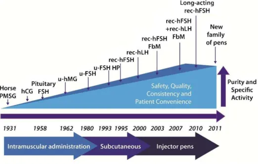
The recognition that animal gonadotropins induce anti-hormone antibody production, which neutralized not only the preparation administered but also endogenous gonado-tropins, was the driving force behind gonadotropin extrac-tion and purificaextrac-tion from human sources. In the 1940s, researchers began extracting gonadotropins from urine: hCG in 1940 and human menopausal gonadotropin (hMG) in 1949. A decade later, the first urinary forms of hCG and hMG became commercially available (2,3). Further improve-ments in the purification methods produced FSH-only products in the 1980s and highly purified urinary FSH (HP-hFSH) in 1993 (2,3). Advances in DNA technology enabled the development of recombinant human FSH (rec-hFSH), which became commercially available in 1995 (2,3,28). In 2000, recombinant human LH (rec-hLH) became available and, with the launch of recombinant hCG (rec-hCG) in 2001, the full recombinant gonadotropin portfolio was launched (2,3). The most recent developments include
the introduction of the filled-by-mass (FbM) rec-hFSH formulation in 2004, which improved batch-to-batch con-sistency compared with products quantified by the standard ratin vivobioassay; long-acting FSH gonadotropin in 2010; and novel pen injector devices to deliver precise recombi-nant FSH, LH, and hCG doses in 2011 (29-35) (Figure 3).
& PREPARATIONS CURRENTLY AVAILABLE FOR CLINICAL USE
Human menopausal gonadotropin (menotropin) Menotropin is extracted from the urine of postmenopau-sal women (2). Early preparations contained varying levels of FSH, LH, and hCG in only 5% pure forms (3). The purification techniques were improved, resulting in FSH and LH with activities standardized at 75 IU for each type of gonadotropin, as measured using a standard in vivo
bioassays (Steelman–Pohley assay). hMG preparations have both FSH and LH activity, but the latter is primarily derived from the hCG component in postmenopausal urine, which is concentrated during purification (2,36,37). Occasionally, hCG is added to induce a desired level of LH-like biological activity (2). In 1999, purified hMG gonadotropins were introduced, which facilitated its subcutaneous (SC) admin-istration (3,29). Currently, both conventional hMG and highly purified hMG (HP-hMG) are commercially available at an FSH:LH ratio of 151 (29).
Urinary FSH (urofollitropin)
Urinary FSH preparations are produced by removing LH with polyclonal antibodies. The production process is
passive because LH is separated from the bulk material, and FSH, together with certain other urinary proteins, is collected and lyophilized. Though they were biologically more pure, early preparations still contained high levels of other urinary proteins (38). Further technological advances facilitated the use of highly specific monoclonal antibodies to extract FSH and produce highly purified FSH (HP-hFSH). The latter has been commercially available since 1993 and contains,0.1 IU of LH and ,5% of unidentified urinary proteins. The specific activity of FSH is approximately 10,000 IU/mg protein, whereas that of the earlier urinary hMG preparations was 100–150 IU/mg protein (Table 1). Similar to HP-hMG, the enhanced purity of HP-hFSH enabled SC delivery (3). SC gonadotropin administration represented an important advance for patients. Consistently better tolerability (decreased pain at the injection site) was reported for SC injections compared with the intramuscular route (39,40). Moreover, SC administration allows self-administration, which is more convenient and less time consuming because patients require fewer visits to the clinic or hospital for injections (39,40).
Recombinant FSH
Recombinant technology has met the need for a more reliable FSH source. Under appropriate conditions, the genes that code for the human FSH alpha and beta subunits are incorporated into nuclear DNA of a host cell via a plasmid vector using spliced DNA strings containing the FSH gene and bacterial DNA segments (2,3,41). Early recombinant technology used Escherichia coli. However, due to the complex human gonadotropin structure and the need for post-translational glycosylation, which defines the degradation time and bioactivity, all recombinant gonadotropins are now produced using the Chinese hamster ovary (CHO) cell line. These cells are genetically stable, fully characterized, and easily transfected with foreign DNA. Furthermore, the cells can be grown in cell cultures on a large scale, and can produce adequate levels of biologically active recombinant gonadotropins (2,41).
Two types of recombinant FSH (rec-hFSH), the alfa and beta follitropins, are available for clinical use (2). In follitropin alfa, two separate vectors, one for each subunit, are used to construct the master cell bank for an FSH-producing cell line. Follitropin beta uses a single vector that contains the coding sequences for both subunit genes (41,42). The subsequent production steps are similar for both preparations. Nevertheless, in addition to a series of anion and cation exchange chromatography steps, hydro-phobic chromatography and size exclusion chromatography
used to produce follitropin beta, an immunoaffinity step with a specific monoclonal antibody similar to the antibody used for HP-hFSH production is used for follitropin alfa (Figure 4) (2,41). Due to the slight differences in their production and purification procedures, the preparations are not identical, with variations in posttranslational glycosylation that yield different sialic acid residue compo-sitions and different isoelectric coefficients (3,43,44). While follitropins alfa and beta are similar to the native FSH isoforms in the blood around mid-cycle (more basic isoforms), they differ slightly in charge heterogeneity, as follitropin alfa has slightly more acidic glycoforms than follitropin beta (43,45). The preparations include equivalent immunopotency,in vitrobiopotency, and internal carbohy-drate complexity (43,46). The initial and terminal half-lives after the administration of 150 IU recombinant FSH are 2 and 17 hours, respectively. Given their intrinsically similar structures, clinical efficacy is expected to be the same (3,43,46).
Long-acting recombinant FSH (corifollitropin alfa) Due to the relatively short half-life of FSH, daily FSH injections are used to prevent serum FSH levels from decreasing below the threshold that causes follicular growth arrest (47). After each injection, the peak serum FSH levels are reached within 10–12 hours; the FSH levels then decline until the next injection. Steady state levels are reached only after treatment for 3–5 days; thus, dose adjustments before day 5 of stimulation are not advised (48).
Recently, a novel long-acting gonadotropin molecule was developed by combining rec-hFSH with the hCG C-terminal peptide (CTP) using site-directed mutagenesis and gene transfer techniques (48). Its longer half-life is due to the hCG CTP, which includes four additional O-linked carbohydrate side chains, each with two terminal sialic acid residues (15,49,50). The new molecule was created using a chimeric gene containing the sequence that encodes CTP fused to the translated human FSH beta subunit sequence. The chimera was then transfected with the common glycoprotein alpha subunit and expressed in CHO cells. The CTP sequence does not significantly affect assembly or secretion of the intact dimer by stable cell lines. The chimeric recombinant molecule has similarin vitroreceptor binding and steroido-genic activity compared with wild-type FSH but exhibits significant enhancement of its in vivo activity and plasma half-life (48,51).
Corifollitropin alfa, initially produced in 2010, exclusively interacts with FSH receptors and has a plasma half-life of 65 hours (33,51). Clinical studies indicate that a single
Table 1 -Differences between gonadotropin formulations.
Purity (gonadotropin content)
Mean Specific Activity
(IU/mg protein) LH Activity (IU/vial) Injected Protein per 75 IU (mcg)
hMG ,5% ,100 75* ,750
HP-hMG ,70% 2,000-2,500 75* ,33
rec-hFSH
Follitropin beta .99% 7,000-10,000 0 8.1
Follitropin alfa .99% 13,645 0 6.1
Lutropin alfa (rec-hLH) .99% 22,000 75 3.7
rec-hFSH: recombinant human follicle-stimulating hormone; hMG: human menopausal gonadotropin; HP-hMG: highly purified human menopausal gonadotropin.
injection of corifollitropin alfa can replace the first seven daily standard gonadotropin injections and that stimulation could be continued with daily FSH injections until the final oocyte maturation had been reached (52).
Gonadotropin preparations with LH activity
Currently, three groups of commercially available gona-dotropin preparations contain LH activity: (i) urinary hMG, in which LH activity depends on hCG rather than pure LH glycoprotein; (ii) pure LH glycoprotein produced by recombinant technology (lutropin alfa), and (iii) a combina-tion of pure FSH and LH glycoproteins in a fixed ratio of 251, which is also manufactured through recombinant technology (Table 1) (3).
While hMG has been used for ovarian stimulation since 1960, lutropin alfa was introduced in 2000 for women with gonadotropin insufficiency. It is intended to be administered through subcutaneous daily injections. Recently, a new prefilled pen device was introduced for rec-hLH administra-tion (53). Currently, rec-hLH is used both to support follicular development during COS in hypogonadotropic hypogona-dal women and to offer LH supplementation to subsets of
women undergoing COS (2,54,55). Rec-hLH has three major differences compared to hMG. First, it has higher purity and specific activity due to the use of recombinant technology. Second, it is associated with better dose precision due to the vial/device filling method, which virtually eliminates batch-to-batch variation (2,30,56). Third, the LH activity is derived directly from pure LH glycoprotein, unlike hMG, in which hCG is concentrated during purification or added to achieve the desired level of LH-like biological activity (2). LH and hCG differ in their carbohydrate moiety compositions, which in turn, affect bioactivity and half-life. In serum, LH activity is 30-fold higher when hCG is used because it binds LH receptors with greater affinity. Rec-hLH is eliminated with a terminal half-life of 10-12 hours, in contrast to the 24-31 hours required for hMG preparations with hCG-driven LH activity (15,57-61).
A new combination of rec-hFSH and rec-hLH (follitropin alfa+lutropin alfa) at a 251 ratio was launched in 2007. This combination is advantageous for women who require LH supplementation because a single injection, rather than two, is used to deliver both preparations. The bioequivalence of
rec-hFSH and rec-hLH administered alone or in combina-tion is similar (32,62).
Human chorionic gonadotropin
hCG administration is the gold standard for promoting ovulation induction as a substitute for the mid-cycle LH surge (63). Due to structural and biological similarities, hCG and LH bind to and activate the same receptor (64). However, the luteotropic activity of hCG is markedly higher than that of LH due to its longer half-life and greater receptor affinity (57,65). Currently, urinary hCG prepara-tions are marketed in lyophilized vials with 5,000 or 10,000 IU for intramuscular use. In 2001, an hCG prepa-ration (choriogonadotropin alfa) was launched using recombinant technology. Recombinant hCG (rec-hCG) is available in prefilled syringes containing 250 mcg of pure hCG, which is equivalent to approximately 6,750 IU of urinary hCG (3). Due to its higher purity, rec-hCG is better tolerated and used subcutaneously, thus allowing patient self-administration (66). Nevertheless, the clinical efficacy of both urinary and recombinant preparations does not seem to differ (67). In a Cochrane meta-analysis including 11 randomized controlled trials (RCTs) of IVF with 1,187 women, Youssef et al. compared rec-hCG vs. urinary hCG for triggering final oocyte maturation. A significant difference was not detected in the main outcome measure-ments between the drugs: ongoing pregnancy/live birth rate (6 RCTs: odds ratio [OR] = 1.04, 95% confidence interval [CI]: 0.79 to 1.37; I2= 0%), incidence of ovarian
hyperstimulation syndrome (OHSS) (3 RCTs: OR = 1.5, 95% CI: 0.37 to 4.1; I2= 0%) and the number of retrieved oocytes
(9 RCTs: Mean difference = -0.04, 95% CI: -0.69 to 0.62; I2= 18%) (67).
Table 2 summarizes the gonadotropin preparations currently available for clinical use.
& QUALITY AND SAFETY PROFILES
Manufacturing urine-derived gonadotropins requires high levels of human urine as a primary source. In the 1960s and 1970s, when the demand for gonadotropins was low, the source material quality was controlled. However, the widespread availability of infertility treatments has rapidly increased demand since 1980. Unlike blood, human urine is not subject to specific regulations regarding collection. Because collected urine is pooled, the donor source cannot be traced, and quality cannot be checked throughout all manufacturing steps (38,68,69). However, extraneous urinary proteins may account for more than 30% of the protein levels in highly purified hMG products even with sophisticated purification techniques (56,69). Certain impurities have been identified as prion proteins, which have been associated with transmissible spongiform ence-phalopathy (TSE) diseases (70). Prion inactivation in urine-derived material may also denature other proteins, includ-ing FSH. In fact, several regulatory agencies limit urine-derived products (38,68). In contrast, each product batch of recombinant gonadotropin is routinely characterized and controlled using physicochemical techniques. The techni-ques include size exclusion high-performance liquid chro-matography (SE-HPLC), which facilitates assessment of both the integrity and the levels of glycoproteins, as well as isoelectric focusing (IEF) and glycan mapping, which are used to characterize the protein glycoforms in each preparation (71,72). Due to the linear relationship between recombinant gonadotropin mass and biological activity, a new method was developed to calibrate each batch of follitropin alfa, lutropin alfa, and choriogonadotropin. Although the Steelman–Pohley assay is used to quantify the protein levels in urinary preparations, which inherently vary up to 20%, recombinant products are filled and released based on protein mass (FbM) (30,73). FSH at
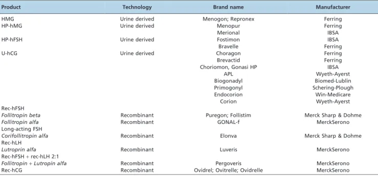
Table 2 -The most common gonadotropins available for clinical use.
Product Technology Brand name Manufacturer
HMG Urine derived Menogon; Repronex Ferring
HP-hMG Urine derived Menopur Ferring
Merional IBSA
HP-hFSH Urine derived Fostimon IBSA
Bravelle Ferring
U-hCG Urine derived Choragon Ferring
Brevactid Ferring
Choriomon, Gonasi HP IBSA
APL Wyeth-Ayerst
Biogonadyl Biomed-Lublin
Primogonyl Schering-Plough
Endocorion Win-Medicare
Corion Wyeth-Ayerst
Rec-hFSH
Follitropin beta Recombinant Puregon; Follistim Merck Sharp & Dohme
Follitropin alfa Recombinant GONAL-f MerckSerono
Long-acting FSH
Corifollitropin alfa Recombinant Elonva Merck Sharp & Dohme
Rec-hLH
Lutroprin alfa Recombinant Luveris MerckSerono
Rec-hFSH+rec-hLH 2:1
Follitropin+Lutropin alfa Recombinant Pergoveris MerckSerono
Rec-hCG Recombinant Ovidrel; Ovitrelle; Ovidrelle MerckSerono
75 IU was assessed using the Steelman-Pohley method, which corresponds to 5.0-5.5mg of follitropin alfa, with a dose variability of only 2% (3,30). This method ensures that a precise dose is delivered, thus maximizing the beneficial effects of gonadotropin therapy (3,29).
The pharmaceutical presentation of urinary gonadotro-pins consists of a freeze-dried lyospheres containing either 75 IU of FSH/hMG or 5,000/10,000 IU of hCG. The lyoph-ilized powder is then reconstituted using sterile water before injection (29). Higher gonadotropin purity yields higher specific activity, and therefore, less material is injected for the desired effect. Through these characteristics, highly purified urine-derived and recombinant gonadotro-pins can be administered subcutaneously (56). Given the high specific activity of recombinant gonadotropins, mini-mal volumes are injected, and injection devices have been developed to deliver the drug (40). The first injector was an adapted insulin pen. Early studies showed that drug delivery was more precise and better tolerated using the pens than syringe injections. Due to unavoidable losses during syringe filling and/or removing excess air, 18% of the FSH amount is lost in conventional syringe applications when compared with a ready-for-use solution in a pen device (40). Recently, novel devices were specifically developed for gonadotropin administration. The first gen-eration was released in 2004 followed by a second generation in 2011 (34,35). They are presented as ready-to-use, compact, and disposable pens, FbM with a fixed drug dose that can be administered in fractions over several days (30,56). In an RCT including 100 women, the efficacy, convenience, and local reactions after follitropin alfa administration were compared following the use of either the pen device or a conventional syringe. Outcomes, including self-administration and patient satisfaction (p,0.001), the overall incidence of local reactions (p= 0.04), the overall pain score (p,0.001), and burning sensation at the injection site (p= 0.04), clearly favored the pen device group (74). Later, in 2007, patients and their partners received nurse-led training on three gonadotropin presenta-tions: (i) powdered urofollitropin administered using con-ventional needles and syringes, (ii) follitropin beta in a premixed and prefilled cartridge with a reusable injection device, and (iii) follitropin alfa in a disposable, premixed, and prefilled injection device. One hundred twenty-three participants attended the training and were asked to complete a post-training questionnaire. More participants expressed a preference for using pen injectors compared with conventional syringes (84.6% vs. 5.7%;p,0.0001). Of the 94 participants who preferred a particular device, more preferred the follitropin alfa prefilled pen (68.1%) than either the follitropin beta cartridge and pen (24.5%;
p,0.0001) or urofollitropin with a needle-free reconstitution device and conventional syringe (7.4%; p,0.0001) (75). In conclusion, recombinant technology, the FbM method, and the use of pen devices for gonadotropin administration represent important advancements that have made inferti-lity treatments more patient friendly (40,74,76).
& CLINICAL EFFICACY OF GONADOTROPINS
The literature is rich in meta-analyses comparing efficacy for different gonadotropin products (77-81). The most recent studies are summarized in Table 3. Despite the hetero-geneity of several of these meta-analyses pertaining the
different stimulation protocols and choice of fertilization with standard in vitro fertilization or intracytoplasmatic sperm injection (ICSI), the overall conclusion is that both urinary gonadotropins, mainly hMG preparations, and recom-binant FSH have similar efficacy in terms of achieving a pregnancy or live birth per treatment cycle. While some of these studies were in favour of hMG preparations, albeit the lower confidence limits were 1% or less, others reported no differences in pregnancy outcomes between the two treat-ments. Furthermore, no significant differences were noted for spontaneous abortion, multiple pregnancy, cycle cancellation and OHSS rates. Notably, these studies did not stratify patients according to the need for LH supplementa-tion during COS. This is a relevant aspect given the fact rec-hFSH has solely FSH activity and recent evidence indicates that a subset of women benefit from LH supple-mentation during COS (55).
Several studies have also compared the potency of different gonadotropin formulations (29,60,82,83). In an RCT involving 629 women undergoing IVF/ICSI treatment with pituitary down-regulation, Hompes et al. compared HP-hMG and rec-hFSH. In their study, more oocytes were retrieved from patients treated with rec-hFSH (7.8 and 10.6, respectively; p,0.001), with no differences in pregnancy rates (82). Similarly, in an RCT involving 280 women undergoing IVF/ICSI with GnRH antagonists, Bosch et al. obtained more oocytes from patients who received rec-hFSH compared with those who received hMG (14.4¡8.1
vs. 11.3¡6.0, respectively; p,0.001). No significant differ-ences were observed in the ongoing pregnancy rate per initiated cycle (35.0vs. 32.1%, respectively; RR = 1.09; 95% CI: 0.78–1.51; risk difference [RD] = 2.9%) (60). Recently, in a large RCT involving more than 700 patients in a single blastocyst transfer IVF program, Devroey et al. confirmed that rec-hFSH results in more oocytes than HP-HMG when used at the same doses (10.6¡5.8vs. 9.1¡5.2;p,0.001) (83). We also compared different gonadotropin products in a large observational study involving 865 women undergoing IVF/ICSI with pituitary down-regulation, and found that the total gonadotropin dose was significantly lower in women who received rec-hFSH (2,268¡747 IU) compared with hMG (2,685¡720 IU) or HP-hMG (2,903¡867 IU;
p,0.001). In our study, the live birth rates per cycle initiated were the same in patients who received rec-hFSH (34.7%), hMG (35.5%), or HP-HMG (40%). However, the total gonadotropin dose required for a live birth was lower when rec-hFSH was compared with hMG (52% reduction) and HP-hMG (21% reduction) (29). The available data support the notion that recombinant FSH is more potent than hMG during COS (29,60,82,83).
The clinical efficacy of gonadotropins has also been assessed with regard to the vial-filling method. In a meta-analysis including four RCTs involving 1,055 women and two case-control studies with 272 patients undergoing IVF, the average rec-hFSH dose per patient was 230 IU lower when the drug was FbM compared with the filled-by-bioassay method (weighted mean difference [WMD] = -230.3; 95% CI: -326 to -134.5; p,0.001). In addition, the number of treatment days was reduced by 0.48 (WMD = -0.48; 95% CI: -0.69 to -0.27,p,0.001), whereas the numbers of oocytes retrieved (WMD = 0.84; 95% CI: 0.18 to 1.51;
to 1.82) and ovarian hyperstimulation syndrome (OHSS) incidence rates (OR = 0.78; 95% CI: 0.45 to 1.36) were the same for both formulations (84).
Similarly, long-acting and daily-use recombinant FSH preparations have been compared. A meta-analysis of four pharmaceutical industry-sponsored RCTs that included 2,377 participants evaluated the effectiveness, safety, and tolerability of corifollitropin alfa compared with follitropin beta in IVF/ICSI cycles with GnRH antagonists. The results favored corifollitropin alfa with regard to the number of oocytes retrieved (WMD = 1.99; 95% CI: 1.02 to 2.97;
p,0.0001), the number of mature oocytes (WMD = 1.92; 95% CI: 1.25 to 2.59; p,0.001), and the number of embryos formed (WMD = 1.09; 95% CI: 0.68 to 1.49;p,0.0001). The clinical pregnancy, live-birth, and miscarriage rates were similar regardless of the drug used for COS. The median duration of stimulation was 9 days in both groups, indicating that two additional single daily rec-hFSH injec-tions are required to complete the treatment regimen with corifollitropin alfa. Notably, corifollitropin alfa resulted in higher cycle cancellations due to an excessive response (OR = 5.67; 95% CI: 1.07 to 30.13; p =0.04), but the OHSS incidence was not significantly different between the groups (OR = 1.29; 95% CI: 0.78 to 2.26) (85). These results were further corroborated by a recent Cochrane review of the same studies (86). The main shortcoming of corifollitropin alfa is that the dose cannot be adjusted during ovarian stimulation, which is a particularly relevant limitation for patients at risk of developing OHSS. From the data available, corifollitropin alfa is likely efficacious and safe for COS in normal responders, but it is not recommended for women at risk of OHSS, such as women with polycystic ovaries (85).
Luteinizing hormone supplementation during COS The ‘‘LH window’’ concept outlined by Shoham in 2002 proposes that without a threshold level of serum LH, estradiol production is insufficient for follicular develop-ment, endometrial proliferation, and corpus luteum forma-tion. However, exposing the developing follicle to excessive
LH would suppress GC proliferation, induce follicular atresia of non-dominant follicles and premature luteiniza-tion, and impair oocyte development (87). Under this concept, optimal follicular development occurs when LH is above a threshold of 1.1 and below a ceiling of 5.1 IU/L (87,88). The validity of the LH threshold hypothesis has been demonstrated in patients with hypogonadotropic hypogonadism. These women do not achieve adequate steroidogenesis unless LH is added to the stimulation regimen (88).
Unlike pituitary insufficiency, most women undergoing COS for IVF have adequate endogenous LH levels and thus do not require LH supplementation (Table 4) (89-92). Indeed, only 1% of LH receptors must be occupied to drive adequate ovarian steroidogenesis (93,94). Nevertheless, the ovarian response to FSH-only gonadotropins is suboptimal in certain patient groups, including older women ($35 years old) (55,95) and women with a diminished ovarian reserve (54,81) or highly suppressed endogenous LH (96-100). Further, a subset of normogonadotropic women have a suboptimal response to FSH stimulation despite a normal ovarian reserve (101-105). All these patients share a similar trait, less sensitive ovaries, which can be explained by several factors, including reduced paracrine ovarian activity (106), LH receptor polymorphisms (105), reduced androgen secretory capacity (107), fewer functional LH receptors (108), and reduced LH bioactivity despite normal LH immunoreactivity (109-110).
It has been hypothesized that these women would benefit from preparations containing LH, which would act at the follicular level. An increase in androgen production for future aromatization into estrogens could restore the follicular milieu and thus positively impact oocyte quality (97,99,103,111,112). In fact, several studies have assessed the utility of LH supplementation during COS (Table 4). A recent meta-analysis by Hill et al., which included seven RCTs and 902 older women undergoing COS for IVF, demonstrated significantly higher embryo implantation (OR = 1.36; 95% CI: 1.05 to 1.78, I2= 12%) and clinical pregnancy rates (OR = 1.37; 95% CI: 1.03 to 1.83, I2= 28%) Table 3 -Meta-analyses comparing urinary and recombinant gonadotropins for controlled ovarian stimulation inin vitrofertilization.
Authors, Year Gonadotropins No. RCT No. Patients Main Findings
Coomarasamy et al., 2008
rec-hFSH; hMG 7 2,159 Higher clinical pregnancy (RR = 1.17, 95% CI: 1.03-1.34) and live birth rates (RR = 1.18, 95% CI: 1.02-1.38;p =0.03) with hMG. No significant differences in the spontaneous abortion, multiple pregnancy, cycle cancellation, or OHSS rates. Al Inany et al.,
2009
rec-hFSH; hMG; HP-hMG 6 2,371 Overall, no significant differences in the clinical, ongoing pregnancy, or live birth rates. Higher ongoing pregnancy/live-birth rates with HP-hMG (OR = 1.31, 95% CI: 1.02-1.68;p =0.03) after grouping the treatment cycles by method, ICSI and IVF. Jee et al., 2010 rec-hFSH; HP-hMG 5 2,299 No difference in ongoing pregnancy rate per initiated cycle (RR = 1.10; 95% CI:
0.96-1.26) or live birth rates per embryo transfer (RR = 1.14; 95% CI: 0.98-1.33). Van Wely et al.,
2010
rec-hFSH; hFSH-P; HP-hFSH; hMG; HP-hMG
28 7,339 Overall, no difference in live birth or OHSS rates.
Van Wely et al., 2012
rec-hFSH; hMG; HP-hMG 12 3,197 Fewer clinical pregnancies (OR = 0.85; 95% CI: 0.74-0.99; I2= 0%;p =0.03) and live births with rec-hFSH (OR = 0.84; 95% CI: 0.72-0.99; I2= 0%;p =0.04).
Gerli et al., 2013 rec-hFSH; hFSH-P; HP-hFSH 8 955 No difference in the clinical pregnancy (OR = 0.85, 95% CI: 0.68 to 1.07) or live birth rates (OR = 0.84; 95% CI: 0.63-1.11).
RCT: randomized controlled trial.
rec-hFSH: recombinant human follicle-stimulating hormone; hMG: human menopausal gonadotropin; HP-hMG: highly purified human menopausal gonadotropin; hFSH-P: purified urinary follicle-stimulating hormone; HP-hFSH: highly purified urinary follicle-stimulating hormone; COS: controlled ovarian stimulation.
RR: relative risk; CI: confidence interval; OR: odds ratio;
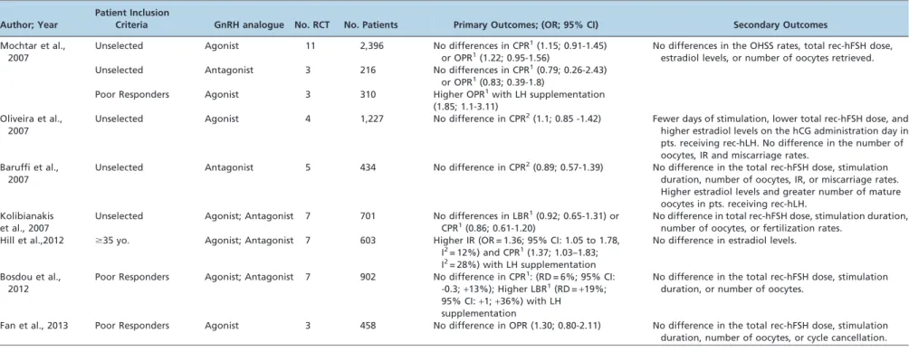
Table 4 -Meta-analyses comparing controlled ovarian stimulation with and without recombinant LH supplementation inin vitrofertilization.
Author; Year
Patient Inclusion
Criteria GnRH analogue No. RCT No. Patients Primary Outcomes; (OR; 95% CI) Secondary Outcomes
Mochtar et al., 2007
Unselected Agonist 11 2,396 No differences in CPR1(1.15; 0.91-1.45)
or OPR1(1.22; 0.95-1.56)
No differences in the OHSS rates, total rec-hFSH dose, estradiol levels, or number of oocytes retrieved. Unselected Antagonist 3 216 No differences in CPR1(0.79; 0.26-2.43)
or OPR1(0.83; 0.39-1.8)
Poor Responders Agonist 3 310 Higher OPR1with LH supplementation
(1.85; 1.1-3.11) Oliveira et al.,
2007
Unselected Agonist 4 1,227 No difference in CPR2(1.1; 0.85 -1.42) Fewer days of stimulation, lower total rec-hFSH dose, and
higher estradiol levels on the hCG administration day in pts. receiving rec-hLH. No difference in the number of oocytes, IR and miscarriage rates.
Baruffi et al., 2007
Unselected Antagonist 5 434 No difference in CPR2(0.89; 0.57-1.39) No difference in the total rec-hFSH dose, stimulation
duration, number of oocytes, IR, or miscarriage rates. Higher estradiol levels and greater number of mature oocytes in pts. receiving rec-hLH.
Kolibianakis et al., 2007
Unselected Agonist; Antagonist 7 701 No differences in LBR1(0.92; 0.65-1.31) or
CPR1(0.86; 0.61-1.20)
No difference in total rec-hFSH dose, stimulation duration, number of oocytes, or fertilization rates.
Hill et al.,2012 $35 yo. Agonist; Antagonist 7 603 Higher IR (OR = 1.36; 95% CI: 1.05 to 1.78, I2= 12%) and CPR1(1.37; 1.03–1.83;
I2= 28%) with LH supplementation
No difference in estradiol levels.
Bosdou et al., 2012
Poor Responders Agonist; Antagonist 7 902 No difference in CPR1: (RD = 6%; 95% CI:
-0.3;+13%); Higher LBR1(RD =+19%;
95% CI:+1;+36%) with LH supplementation
No difference in the total rec-hFSH dose, stimulation duration, or number of oocytes.
Fan et al., 2013 Poor Responders Agonist 3 458 No difference in OPR (1.30; 0.80-2.11) No difference in the total rec-hFSH dose, stimulation duration, number of oocytes, or cycle cancellation.
CPR: clinical pregnancy rate; OPR: ongoing pregnancy rate; LBR: live birth rate; IR: implantation rate. OR: odds ratio; CI: confidence interval; RD: risk difference.
OHSS: ovarian hyperstimulation syndrome.
rec-hFSH: recombinant human follicle-stimulation hormone; rec-hLH: recombinant human luteinizing hormone.
1per randomized woman;2per oocyte retrieval.
pins
in
assisted
reproducti
on
RB
and
Esteves
SC
CLINICS
2014;69
(4):279-293
when recombinant LH was added to the stimulation regimen (55). Along the same lines, Mochtar et al., while specifically studying poor responders, demonstrated the benefit of adding rec-hLH during COS. These authors pooled three RCTs, which included 310 participants, and showed higher ongoing pregnancy rates (OR = 1.85; 95% CI: 1.1 to 3.11) in patients treated with a combination of rec-hFSH and rec-hLH compared with rec-rec-hFSH alone (91). In contrast, Fan et al. also studied poor responders by pooling three RCTs and found no differences in ongoing pregnancy rates with LH supplementation (OR = 1.30; 95% CI: 0.80 to 2.11). Furthermore, a significant difference was not detected for the number of oocytes retrieved, the total rec-hFSH dose, the total stimulation duration and the cycle cancellation rate between the study and control groups (113). Finally, in a meta-analysis that included 7 RCTs and 603 patients, Bosdou et al. showed conflicting results following LH supplementation. Although the results were not statistically significant in their study, the magnitude of the size effect and width of the 95% CI for clinical pregnancy (RD =+6%;
95% CI: -0.3 to+13%;p =0.06) suggested a potential clinical
benefit of LH supplementation. Despite the heterogeinity of the studies included with regard to patient selection, stimulation protocol, and dose of rec-hLH, the results from a single RCT revealed significantly higher live birth rates after IVF upon LH supplementation (RD =+19%; CI:+1 to +36%) (54).
In summary, existing evidence suggests that LH supple-mentation could benefit select patient subgroups, but the results should still be interpreted with caution for several reasons. First, the definition of a poor ovarian response was not uniform among studies. Second, the ovarian stimulation protocols differed in dosing, the LH supplementation onset, and the duration of stimulation. Third, the number of completed trials remains low. Finally, many questions have not been fully answered, including how to identify patients who would benefit from LH supplementation, how much LH is necessary, when to begin LH supplementation, and which type of LH activity is best, i.e., recombinant LH or hCG derived (54,99,113).
Notably, an open-label RCT compared the clinical efficacy of LH supplementation using either recombinant LH (a combination of follitropin alfa + lutropin alfa at a fixed
ratio of 251) or hCG-driven LH activity (HP-hMG) in a small group of women with pituitary insufficiency. Although the proportion of patients who reached ovulation did not differ between the groups (70%vs. 88%, respectively), the pregnancy rate was significantly higher in the rec-hLH group (55.6%vs.23.3%; p =0.01) (114). Similarly, in an IVF RCT involving 106 women with a normal ovarian reserve and low endogenous LH levels (,1.2 IU/L), a shorter stimulation duration (10.9¡1.1vs.14.1¡1.6 days;p =0.013) and more retrieved oocytes (7.8¡1.1vs. 4.1¡12;p =0.002) were observed in patients who received follitropin alfa +
lutropin alfa 2:1 compared with HMG. At the end of stimulation, the estradiol level (1,987¡699 pg/mL vs. 2,056¡560 pg/mL), pregnancy rate per cycle (28.3% vs. 29.3%) and implantation rate (12.1%vs.12.2%) did not differ between the groups. However, a higher cancellation rate due to an excessive response was observed in women receiving follitropin + lutropin alfa (11.1% vs. 1.7%;
p =0.042) (115). Finally, a large matched case-control study involving 4,719 IVF patients showed that the probability of a clinical pregnancy was higher in patients who used the
fixed combination of rec-hFSH and rec-hLH at a 251 ratio (32%) when compared with patients who used hMG (26%;
p =0.02) (116). Not surprisingly, these limited data suggest that the fixed rec-hFSH plus rec-hLH combination is superior to hMG. Unlike rec-hLH, LH activity in hMG is derived from hCG, which has a markedly longer half-life and greater binding affinity for LH/hCG receptors com-pared with LH (57). Lower expression of the LH/hCG receptor gene as well as the genes involved in cholesterol and steroid biosynthesis has been observed in GCs from patients treated with hMG when compared with FSH-treated patients (58). Constant ligand exposure to hCG during the follicular phase likely produces these effects. In fact, LH receptor down-regulation for up to 48 h has been reported in animal models after hCG administration (59), but the clinical implications of these findings have not been fully elucidated in humans (61).
& FUTURE PERSPECTIVES
Low molecular weight (LMW) gonadotropins are cur-rently under investigation. These non-peptide molecules havein vivobioactivity when administered orally (117,118). The first LMW peptides with bioactivity for FSH receptors were described in 2002. In recent years, other compounds have been identified, including biaryl diketopiperazines, thienopyrimidines, dihydropyridines, and thiazolidinones. In the meantime, peptides with agonist activity for LH have also been identified (117,118). However, the clinical efficacy of these compounds has not been determined. FSH and LH receptors compose a subgroup of G protein-coupled receptors with seven transmembrane domains and a large N-terminal extracellular region, which is the predominant site for hormone binding (117,119,120). Receptor activation requires that the hormone binds the N-terminal region, thus leading to intramolecular signal transduction from the ligand-receptor complex to the transmembrane domains. Current LMW gonadotropins are allosteric compounds that presumably interact with the transmembrane domains instead of the N-terminal region. As such, the signaling pathways induced differ from those induced by the native, orthosteric ligands. Recently, a newly developed LMW agonist for the FSH (and LH) receptor has been shown to be orally bioactive in animal studies (118,119). In the future, gonadotropins could be taken orally and replace the injec-table forms currently available (117,118).
& REVIEW CRITERIA
& ACKNOWLEDGMENTS
The authors are grateful to Mrs. Fabiola C. Bento for the language revision.
& AUTHOR CONTRIBUTIONS
Both authors were involved in the data collection, critical analyses for factual and scientific content, and the drafting and revision of the manuscript.
& REFERENCES
1. Beall SA, DeCherney A. History and challenges surrounding ovarian stimulation in the treatment of infertility. Fertil Steril. 2012;97(4):785-801.
2. Lunenfeld B. Historical perspectives in gonadotrophin therapy. Human Reprod Update. 2004;10(6):453-67, http://dx.doi.org/10.1093/humupd/ dmh044.
3. Practice Committee of American Society for Reproductive Medicine, Birmingham, Alabama. Gonadotropin preparations: past, present, and future perspectives. Fertil Steril. 2008;90(5 Suppl):S13-20.
4. Trounson AO, Leeton JF, Wood C, Webb J, Wood J. Pregnancies in humans by fertilization in vitro and embryo transfer in the controlled ovulatory cycle. Science. 1981;212:681-2, http://dx.doi.org/10.1126/ science.7221557.
5. Bosch E, Ezcurra D. Individualised controlled ovarian stimulation (iCOS): maximising success rates for assisted reproductive technology patients. Reprod Biol Endocrinol. 2011;21:82, http://dx.doi.org/10. 1186/1477-7827-9-82.
6. Lapthorn AJ, Harris DC, Littlejohn A, Lustbader JW, Canfield RE, Machin KJ, et al. Crystal structure of human chorionic gonadotropin. Nature. 1994;369(6480):455-61, http://dx.doi.org/10.1038/369455a0. 7. Vaitukaitis JL, Ross GT, Braunstein GD, Rayford PL. Gonadotropins
and their subunits: basic and clinical studies. Recent Prog Horm Res 1976;32:289-331.
8. Ulloa-Aguirre A, Espinoza R, Damian-Matsumura P, Chappel SC. Immunological and biological potencies of the different molecular species of gonadotrophins. Hum Reprod. 1988;3(4):491-501.
9. Rozell TG, Okrainetz RJ. FSH: one hormone with multiple forms, or a family of multiples hormones. In: Chedrese PJ, Ed. Reproductive Endocrinology: A Molecular Approach. New York: Springer Science+
Business Media, LLC 2009; pp.145-60.
10. Wide L, Naesse´n T, Sundstro¨m-Poromaa I, Eriksson K. Sulfonation and sialylation of gonadotropins in women during the menstrual cycle, after menopause, and with polycystic ovarian syndrome and in men. J Clin Endocrinol Metab. 2007;92(11):4410-7, http://dx.doi.org/10.1210/jc. 2007-1342.
11. Green ED, Baenziger JU. Asparagine-linked oligosaccharides on lutropin, follitropin, and thyrotropin. I. Structural elucidation of the sulfated and sialylated oligosaccharides on bovine, ovine, and human pituitary glycoprotein hormones. J Biol Chem. 1988;263(1):25-35. 12. Fiete D, Srivastava V, Hindsgaul O, Baenziger JU. A hepatic
reticuloendothelial cell receptor specific for SO4-4GalNAc beta 1,4GlcNAc beta 1,2Man alpha that mediates rapid clearance of lutropin. Cell. 1991;67(6):1103-10, http://dx.doi.org/10.1016/0092-8674 (91)90287-9.
13. Wide L, Eriksson K, Sluss PM, Hall JE. Serum half-life of pituitary gonadotropins is decreased by sulfonation and increased by sialylation in women. J Clin Endocrinol Metab. 2009;94(3):958-64, http://dx.doi. org/10.1210/jc.2008-2070.
14. Fox KM, Dias JA, Van Roey P. Three-dimensional structure of human follicle-stimulating hormone. Mol Endocrinol. 2001;15(3):378-89, http://dx.doi.org/10.1210/mend.15.3.0603.
15. Campbell RK. Molecular pharmacology of gonadotropins. Endocrine. 2005;26(3):291-6, http://dx.doi.org/10.1385/ENDO:26:3:291. 16. Morell AG, Gregoriadis G, Scheinberg IH, Hickman J, Ashwell G.
The role of sialic acid in determining the survival of glycoproteins in the circulation. J Biol Chem. 1971;246(5):1461-7.
17. Hsueh AJ, Adashi EY, Jones PB, Welsh TH Jr. Hormonal regulation of the differentiation of cultured ovarian granulosa cells. Endocr Rev. 1984;5(1):76-127, http://dx.doi.org/10.1210/edrv-5-1-76.
18. Vegetti W, Alagna F. FSH and folliculogenesis: from physiology to ovarian stimulation. Reprod Biomed Online. 2006;12(6):684-94, http:// dx.doi.org/10.1016/S1472-6483(10)61080-2.
19. Speroff L, Fritz MA. Chapter 6: Regulation of the Menstrual Cycle. In Speroff L, Fritz MA. Clinical Gynecologic Endocrinology and Infertility. Philadelphia: Lippincott Williams & Wilkins, 2005: pp.348-83. 20. Speroff L, Fritz MA. Chapter 2: Hormone Biosynthesis, Metabolism,
and Mechanisms of Action. Stereidogenesis. In Speroff L, Fritz MA. Clinical Gynecologic Endocrinology and Infertility. Philadelphia: Lippincott Williams & Wilkins, 2005: pp.109-16.
21. Young JM, McNeilly AS. Theca: the forgotten cell of the ovarian follicle. Reproduction. 2010;140(4):489-504, http://dx.doi.org/10.1530/REP-10-0094.
22. Alviggi C, Mollo A, Clarizia R, De Placido G. Exploiting LH in ovarian stimulation. Reprod Biomed Online. 2006;12(2):221-33, http://dx.doi. org/10.1016/S1472-6483(10)60865-6.
23. Kol S, Adashi EY. Intraovarian factors regulating ovarian function. Curr Opin Obstet Gynecol. 1995;7(3):209-13, http://dx.doi.org/10. 1097/00001703-199506000-00010.
24. Midgley AR Jr, Pierce GB Jr. Immunohistochemical localization of human chorionic gonadotropin. J Exp Med. 1962;115:289-94, http://dx. doi.org/10.1084/jem.115.2.289.
25. Hamblen EC, Davis CD, Durham NC. Treatment of hypo-ovarianism by the sequential and cyclic administration of equine and chorionic gonadotropins—so-called one-two cyclic gonadotropic therapy Summary of 5 years’ results. Am J Obstet Gynecol. 1945;50:137-46. 26. Hamblen EC. The clinical evaluation of ovarian responses to
gonado-tropic therapy. Endocrinology. 1939;24(6):848-66, http://dx.doi.org/10. 1210/endo-24-6-848.
27. Maddock WO, Leach RB, Tokuyama I, Paulsen CA, Roy WR. Effects of hog pituitary follicle-stimulating hormone in women: antihormone formation and inhibition of ovarian function. J Clin Endocrinol Metab. 1956;16(4):433-48, http://dx.doi.org/10.1210/jcem-16-4-433.
28. Howles CM. Genetic engineering of human FSH (Gonal-F). Hum Reprod Update. 1996;2(2):172-91, http://dx.doi.org/10.1093/humupd/ 2.2.172.
29. Esteves SC, Schertz JC, Verza S Jr, Schneider DT, Zabaglia SF. A comparison of menotropin, highly-purified menotropin and follitropin alfa in cycles of intracytoplasmic sperm injection. Reprod Biol Endocrinol. 2009;7:111, http://dx.doi.org/10.1186/1477-7827-7-111. 30. Driebergen R, Baer G. Quantification of follicle stimulating hormone
(follitropin alfa): is in vivo bioassay still relevant in the recombinant age? Curr Med Res Opin. 2003;19(1):41-6, http://dx.doi.org/10.1185/ 030079902125001344.
31. Martinez G, Sanguineti F, Sepulveda J, Dorey J, Arici A, Patrizio P. A comparison between follitropinafilled by mass and follitropin a filled by bioassay in the same egg donors. Reprod Biomed Online. 2011;22(Suppl 1):S20-2, http://dx.doi.org/10.1016/S1472-6483(11) 60005-9.
32. Bosch E. Recombinant human follicular stimulating hormone and recombinant human luteinizing hormone in a 2:1 ratio combination. Pharmacological characteristics and clinical applications. Expert Opin Biol Ther. 2010;10(6):1001-9.
33. Verbost P, Sloot WN, Rose UM, de Leeuw R, Hanssen RG, Verheijden GF. Pharmacologic profiling of corifollitropin alfa, the first developed sustained follicle stimulant. Eur J Pharmacol. 2011;651(1-3):227-33. 34. Christen M, Schertz JC, Arriagada P, Keitel J, Mu¨ller H. The redesigned
follitropinapen injector for infertility treatment. Expert Opin Drug Deliv. 2011;8(6):833-9, http://dx.doi.org/10.1517/17425247.2011.581658. 35. Saunders H, Schertz JC, Hecker C, Lang B, Arriagada P. The recombinant human chorionic gonadotropin prefilled pen: results of patient and nurse human factors usability testing. Expert Opin Drug Deliv. 2012;9(8):893-900, http://dx.doi.org/10.1517/17425247.2012.698607.
36. Cole LA, Khanlian SA, Muller CY. Detection of perimenopause or postmenopause human chorionic gonadotropin: an unnecessary source of alarm. Am J Obstet Gynecol. 2008;198(3):275.
37. Cole LA, Khanlian SA, Muller CY. Normal production of human chorionic gonadotropin in perimenopausal and menopausal women and after oophorectomy. Int J Gynecol Cancer. 2009;19(9):1556-9. 38. Giudice E, Crisci C, Eshkol A, Papoian R. Composition of commercial
gonadotrophin preparations extracted from human post-menopausal urine: characterization of non-gonadotrophin proteins. Hum Reprod. 1994;9(12):2291-9.
39. Alviggi C, Revelli A, Anserini P, Ranieri A, Fedele L, Strina I, et al. A prospective, randomised, controlled clinical study on the assessment of tolerability and of clinical efficacy of Merional (hMG-IBSA) adminis-tered subcutaneously versus Merional adminisadminis-tered intramuscularly in women undergoing multifollicular ovarian stimulation in an ART programme (IVF). Reprod Biol Endocrinol. 2007;5:45, http://dx.doi. org/10.1186/1477-7827-5-45.
40. Platteau P, Laurent E, Albano C, Osmanagaoglu K, Vernaeve V, Tournaye H, et al. An open, randomized single-centre study to compare the efficacy and convenience of follitropin beta administered by a pen device with follitropin alpha administered by a conventional syringe in women undergoing ovarian stimulation for IVF/ICSI. Hum Reprod. 2003;18(6):1200-4, http://dx.doi.org/10.1093/humrep/deg234. 41. Howles CM. Genetic engineering of human FSH (Gonal-F). Hum
Reprod Update. 1996;2(2):172-91, http://dx.doi.org/10.1093/humupd/ 2.2.172.
43. de Leeuw R, Mulders J, Voortman G, Rombout F, Damm J, Kloosterboer L. Structure-function relationship of recombinant follicle stimulating hormone (Puregon). Mol Hum Reprod. 1996;2(5):361-9, http://dx.doi. org/10.1093/molehr/2.5.361.
44. Horsman G, Talbot JA, McLoughlin JD, Lambert A, Robertson WR. A biological, immunological and physico-chemical comparison of the current clinical batches of the recombinant FSH preparations Gonal-F and Puregon. Hum Reprod. 2000;15(9):1898-902, http://dx.doi.org/10. 1093/humrep/15.9.1898.
45. Anobile CJ, Talbot JA, McCann SJ, Padmanabhan V, Robertson WR. Glycoform composition of serum gonadotrophins through the normal menstrual cycle and in the post-menopausal state. Mol Hum Reprod. 1998;4(7):631-9, http://dx.doi.org/10.1093/molehr/4.7.631.
46. Orvieto R, Nahum R, Rabinson J, Ashkenazi J, Anteby EY, Meltcer S. Follitropin-alpha (Gonal-F) versus follitropin-beta (Puregon) in con-trolled ovarian hyperstimulation for in vitro fertilization: is there any difference? Fertil Steril. 2009;91(4 Suppl):1522-5, http://dx.doi.org/10. 1016/j.fertnstert.2008.08.112.
47. Fauser BC, van Heusden AM. Manipulation of human ovarian function: physiological concepts and clinical consequences. Endocr Rev. 1997; 18(1):71-106.
48. Fauser BC, Mannaerts BM, Devroey P, Leader A, Boime I, Baird DT. Advances in recombinant DNA technology: corifollitropin alfa, a hybrid molecule with sustained follicle-stimulating activity and reduced injection frequency. Hum Reprod Update. 2009;15(3):309-21, http://dx.doi.org/10.1093/humupd/dmn065.
49. Birken S, Canfield RE. Isolation and amino acid sequence of COOH-terminal fragments from the beta subunit of human choriogonadotro-pin. J Biol Chem. 1977;252(15):5386-92.
50. Kessler MJ, Mise T, Ghai RD, Bahl OP. Structure and location of the O-glycosidic carbohydrate units of human chorionic gonadotropin. J Biol Chem. 1979;254(16):7909-14.
51. Fares FA, Suganuma N, Nishimori K, LaPolt PS, Hsueh AJ, Boime I. Design of a long-acting follitropin agonist by fusing the C-terminal sequence of the chorionic gonadotropin beta subunit to the follitropin beta subunit. Proc Natl Acad Sci USA. 1992;89(10):4304-8, http://dx. doi.org/10.1073/pnas.89.10.4304.
52. Balen AH, Mulders AG, Fauser BC, Schoot BC, Renier MA, Devroey P, et al. Pharmacodynamics of a single low dose of long-acting recombinant follicle- stimulating hormone (FSH-carboxy terminal peptide, corifolli-tropin alfa) in women with World Health Organization group II anovulatory infertility. J Clin Endocrinol Metab. 2004;89(12):6297-304, http://dx.doi.org/10.1210/jc.2004-0668.
53. Dhillon S, Keating GM. Lutropin alfa. Drugs. 2008;68(11):1529-40, http://dx.doi.org/10.2165/00003495-200868110-00005.
54. Bosdou JK, Venetis CA, Kolibianakis EM, Toulis KA, Goulis DG, Zepiridis L, et al. The use of androgens or androgen-modulating agents in poor responders undergoing in vitro fertilization: a systematic review and meta-analysis. Hum Reprod Update. 2012;18(2):127-45, http://dx.doi.org/10.1093/humupd/dmr051.
55. Hill MJ, Levens ED, Levy G, Ryan ME, Csokmay JM, DeCherney AH, et al. The use of recombinant luteinizing hormone in patients under-going assisted reproductive techniques with advanced reproductive age: a systematic review and meta-analysis. Fertil Steril. 2012;97(5):1108-14.e1, http://dx.doi.org/10.1016/j.fertnstert.2012.01.130.
56. Bassett RM, Driebergen R. Continued improvements in the quality and consistency of follitropin alfa, recombinant human FSH. Reprod Biomed Online. 2005;10(2):169-77, http://dx.doi.org/10.1016/S1472-6483(10)60937-6.
57. le Cotonnec JY, Porchet HC, Beltrami V, Munafo A. Clinical pharmacology of recombinant human luteinizing hormone: Part I. Pharmacokinetics after intravenous administration to healthy female volunteers and comparison with urinary human luteinizing hormone. Fertil Steril. 1998;69(2):189-94, http://dx.doi.org/10.1016/S0015-0282(97) 00501-3.
58. Grøndahl ML, Borup R, Lee YB, Myrhøj V, Meinertz H, Sørensen S. Differences in gene expression of granulosa cells from women under-going controlled ovarian hyperstimulation with either recombinant follicle-stimulating hormone or highly purified human menopausal gonadotropin. Fertil Steril. 2009;91(5):1820-30, http://dx.doi.org/10. 1016/j.fertnstert.2008.02.137.
59. Menon KM, Munshi UM, Clouser CL, Nair AK. Regulation of luteinizing hormone/human chorionic gonadotropin receptor expres-sion: a perspective. Biol Reprod. 2004;70(4):861-6.
60. Bosch E, Vidal C, Labarta E, Simon C, Remohi J, Pellicer A. Highly purified hMG versus recombinant FSH in ovarian hyperstimulation with GnRH antagonists–a randomized study. Hum Reprod. 2008;23 (10):2346-51, http://dx.doi.org/10.1093/humrep/den220.
61. Venetis CA, Kolibianakis EM, Papanikolaou E, Bontis J, Devroey P, Tarlatzis BC. Is progesterone elevation on the day of human chorionic gonadotrophin administration associated with the probability of pregnancy in in vitro fertilization? A systematic review and meta-analysis. Hum Reprod Update. 2007;13(4):343-55, http://dx.doi.org/10. 1093/humupd/dmm007.
62. Picard M, Rossier C, Papasouliotis O, Lugan I. Bioequivalence of recombinant human FSH and recombinant human LH in a fixed 2:1 combination: two phase I, randomised, crossover studies. Curr Med Res Opin. 2008;24(4):1199-208, http://dx.doi.org/10.1185/030079908X291949. 63. Humaidan P, Kol S, Papanikolaou EG, Copenhagen GnRH Agonist Triggering Workshop Group. GnRH agonist for triggering of final oocyte maturation: time for a change of practice? Hum Reprod Update. 2011;17(4):510-24, http://dx.doi.org/10.1093/humupd/dmr008. 64. Kessler MJ, Reddy MS, Shah RH, Bahl OP. Structures of N-glycosidic
carbohydrate units of human chorionic gonadotropin. J Biol Chem. 1979;254(16):7901-8.
65. Yen SS, Llerena O, Little B, Pearson OH. Disappearance rates of endogenous luteinizing hormone and chorionic gonadotropin in man. J Clin Endocrinol Metab. 1968;28(12):1763-7, http://dx.doi.org/10. 1210/jcem-28-12-1763.
66. Driscoll GL, Tyler JP, Hangan JT, Fisher PR, Birdsall MA, Knight DC. A prospective, randomized, controlled, double-blind, double-dummy comparison of recombinant and urinary HCG for inducing oocyte maturation and follicular luteinization in ovarian stimulation. Hum Reprod. 2000;15(6):1305-10, http://dx.doi.org/10.1093/humrep/15.6. 1305.
67. Youssef MA, Al-Inany HG, Aboulghar M, Mansour R, Abou-Setta AM. Recombinant versus urinary human chorionic gonadotrophin for final oocyte maturation triggering in IVF and ICSI cycles. Cochrane Database Syst Rev. 2011;(4):CD003719.
68. van de Weijer BH, Mulders JW, Bos ES, Verhaert PD, van den Hooven HW. Compositional analyses of a human menopausal gonadotrophin preparation extracted from urine (menotropin). Identification of some of its major impurities. Reprod Biomed Online. 2003;7(5):547-57, http:// dx.doi.org/10.1016/S1472-6483(10)62071-8.
69. Kuwabara Y, Mine K, Katayama A, Inagawa T, Akira S, Takeshita T. Proteomic analyses of recombinant human follicle-stimulating hormone and urinary-derived gonadotropin preparations. J Reprod Med. 2009; 54(8):459-66.
70. Bassett R, Lispi M, Ceccarelli D, Grimaldi L, Mancinelli M, Martelli F, et al. Analytical identification of additional impurities in urinary-derived gonadotrophins. Reprod Biomed Online. 2009;19(3):300-13, http://dx.doi.org/10.1016/S1472-6483(10)60163-0.
71. Gervais A, Hammel YA, Pelloux S, Lepage P, Baer G, Carte N, et al. Glycosylation of human recombinant gonadotrophins: characterization and batch-to-batch consistency. Glycobiology. 2003;13(3):179-89, http:// dx.doi.org/10.1093/glycob/cwg020.
72. Hugues JN, Barlow DH, Rosenwaks Z, Ce´drin-Durnerin I, Robson S, Pidoux L, et al. Improvement in consistency of response to ovarian stimulation with recombinant human follicle stimulating hormone resulting from a new method for calibrating the therapeutic prepara-tion. Reprod Biomed Online. 2003;6(2):185-90, http://dx.doi.org/10. 1016/S1472-6483(10)61709-9.
73. Steelman SL, Pohley FM. Assay of follicle stimulating hormone based on the augmentation with human chorionic gonadotropin. Endocrinology. 1953;53(6):604-16, http://dx.doi.org/10.1210/endo-53-6-604.
74. Aghssa MM, Azargoon A, Ramezanzadeh F, Bagheri M. A comparison of the efficacy, tolerability, and convenience of two formulations of follitropin-alpha in Iranian woman undergoing intracytoplasmic sperm injection cycles. Fertil Steril. 2008;90(4):1043-8, http://dx.doi.org/10. 1016/j.fertnstert.2007.08.001.
75. Weiss N. Gonadotrophin products: empowering patients to choose the product that meets their needs. Reprod Biomed Online. 2007;15(1):31-7, http://dx.doi.org/10.1016/S1472-6483(10)60688-8.
76. Craenmehr E, Bontje PM, Hoomans E, Voortman G, Mannaerts BM. Follitropin-beta administered by pen device has superior local tolerance compared with follitropin-alpha administered by conventional syringe. Reprod Biomed Online. 2001;3(3):185-9, http://dx.doi.org/10.1016/ S1472-6483(10)62033-0.
77. Coomarasamy A, Afnan M, Cheema D, van der Veen F, Bossuyt PM, van Wely M. Urinary hMG versus recombinant FSH for controlled ovarian hyperstimulation following an agonist long down-regulation protocol in IVF or ICSI treatment: a systematic review and meta-analysis. Hum Reprod. 2008;23(2):310-5.
78. Al-Inany HG, Abou-Setta AM, Aboulghar MA, Mansour RT, Serour GI. Highly purified hMG achieves better pregnancy rates in IVF cycles but not ICSI cycles compared with recombinant FSH: a meta-analysis. Gynecol Endocrinol. 2009;25(6):372-8, http://dx.doi.org/10.1080/ 09513590802630120.
79. van Wely M, Kwan I, Burt AL, Thomas J, Vail A, Van der Veen F, et al. Recombinant versus urinary gonadotrophin for ovarian stimulation in assisted reproductive technology cycles. Cochrane Database Syst Rev. 2011;(2):CD005354.
80. Jee BC, Suh CS, Kim YB, Kim SH, Moon SY. Clinical efficacy of highly purified hMG versus recombinant FSH in IVF/ICSI cycles: a meta-analysis. Gynecol Obstet Invest. 2010;70(2):132-7, http://dx.doi.org/10. 1159/000308458.