Eicacy, eiciency and efectiveness of gonadotropin
therapy for infertility treatment
Sandro C. EstevesI
DOI: 10.5935/MedicalExpress.2015.03.02
I ANDROFERT, Centro de Referência para Reprodução Humana, Av. Dr. Heitor Penteado, 1464, Campinas 13075-460, SP, Brazil.
Gonadotropin therapy is an essential element in infertility treatments involving assisted reproductive technology. In recent years there have been outstanding advances in the development of new gonadotropins, particularly with the production of gonadotropins using biotechnological resources. Recombinant gonadotropins have higher speciic activity compared with urinary counterparts, thus allowing subcutaneous administration of minimal amounts of glycoprotein. As a result, recombinant formulations have a better safety proile despite an overall similarity in terms of eicacy for pregnancy, as reported in many randomized controlled trials and meta-analyses. Gonadotropins stimulate the ovaries to develop follicles and oocytes, which are the raw material for fertilization and embryo production. The resulting embryos are transferred (fresh or frozen-thawed) to achieve pregnancy. The eiciency of a gonadotropin should therefore measured by the amount of drug used, the number of oocytes/embryos produced, and the number of pregnancies achieved by transferring fresh and/or frozen-thawed embryos to the uterus (cumulative pregnancy). Comparisons between diferent gonadotropin preparations should also take into account other important quality indicators in reproductive medicine, such as safety and patient-centeredeness. Altogether, the aforementioned quality indicators favor biotech gonadotropins over biologic products in infertility therapy.
KEYWORDS: Gonadotropins; Controlled ovarian stimulation; Assisted reproductive technology biotechnology; Infertility.
Esteves, SC. Eicacy, eiciency and efectiveness of gonadotropin therapy for infertility treatment. MedicalExpress (São Paulo, online). 2015;2(3):M150302
Received for Publication on March 15, 2015; First review on April 21, 2015; Accepted for publication on April, 26, 2015
E-mail: [email protected]
■
INTRODUCTIONGonadotropin therapy has a central role in ovarian stimulation for infertility treatment. Its introduction in medical practice dates from almost one century ago, and represents a major upgrade in the treatment of infertility. Follicle stimulating hormone (FSH) was originally derived from animal (pregnant mare serum) or human (post-mortem pituitary glands) sources, but these preparations were abandoned because of safety concerns.1-3 Gonadotropins were first extracted from urine in the 1940s, more exactly, human chorionic gonadotropin
(hCG) in 1940 and human menopausal gonadotropin
(hMG) in 1949. Over a decade later, the first urinary forms
of hCG and hMG became commercially available.2,3
Further improvements in the purification methods
led to the production of FSH-only products in the 1980s,
and subsequent development of highly-purified urinary
FSH (HP-hFSH), which became available 10 years later, in 1993.2,3 In the 1970s and 1980s, advances in DNA
technology enabled the development of recombinant human FSH (rec-hFSH), which became commercially available in 1995.2-4 In 2000, recombinant human
luteinizing hormone (rec-hLH) became available and, with the launching of recombinant human hCG (rec-hCG) in 2001, the complete recombinant gonadotropin portfolio became available.2,3
In this article, I critically analyze the effectiveness
and efficiency of commercially available gonadotropins, especifically recombinant FSH and LH, and human
Gonadotropins are further modified in vivo by the
addition of a sialic acid (sialylation) or a sulfonic group (sul-fonation) to the carbohydrate moieties. Both sialylation and sulfonation are physiological processes with major roles in gonadotropin biological activity modulation.9,10 The
oligosac-charides often terminate with sialic acid and/or sulfonated
β1-4-linked GalNAc (SO3-4GalNAc).
9,11 Molecules with an
incre-ased number of sulfonated Gal-NAc disappear faster from the
circulation than less sulfonated isoforms, due to their affinity to specific SO3-4GalNAc receptors in the liver.
9,12 On the other
hand, an increased number of sialic acids enhances half-life.9,13
Removal of the carbohydrate moieties of either subunit diminishes gonadotropic activity; however,
experimental data indicate that carbohydrate chains
have no role in gonadotropin binding to their receptors.14
Nevertheless, carbohydrate components affect the biologic
activity of the hormone-receptor complex after binding,
thus playing a critical role in activation (coupling) of the adenylate cyclase system.15
Follicle-Stimulating Hormone (FSH)
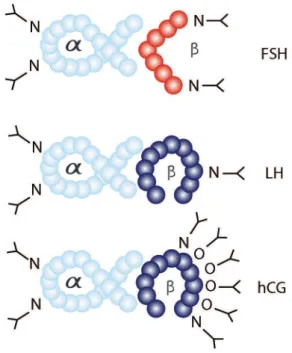
The alpha subunit of FSH contains 92 amino acids, as is the case for LH and hCG. The beta subunit, however, is unique: it is composed of 111 amino acids with four N-linked glycosylation sites, two on the alpha subunit, added to Asn52 and Asn78, and two on the beta subunit (Asn7 and Asn24).8,16 Thus, each subunit is attached to two
carbohydrate moieties with variable compositions that, in turn, create different isoforms, as shown in Figure 1.3,8 These
multiple isoforms of FSH differ in their plasma half-lives (ranging from 3 to 4 hours) and their bioactivity.3
Although both sialic acid and sulfonated GalNAc resi-dues may modulate the half-lives of human gonadotropins, sialic acid residues are much more common in FSH than sulfonated residues.13 Increased sialylation enhances FSH metabolic stability by decreasing both glomerular filtration
and clearance by sialoglycoprotein receptors in the liver, which is the major site for gonadotropin clearance.17,18 It
means that the greater the sialic acid content, the longer the hormone remains in circulation.9-11,13,17,18
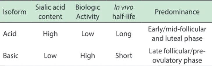
Production of different isoforms is controlled by a combination of steroidal feedback and gonadotropin-releasing hormone (GnRH).10,19 The higher the estradiol
levels, the lower the FSH sialylation, as shown in Table 1.3,20
Therefore, the pattern of circulating FSH during the menstrual cycle is dynamic with respect not only to its quantity (concentration) but also to isoform distribution (quality).21 The isoform profile is more acidic during early follicular to
midfollicular phase, and become more basic shortly before ovulation.3,21,22 These dynamic changes in sialylation are not mimicked by exogenous gonadotropin formulations, and it is unknown whether the absence of such fluctuations during
controlled ovarian stimulation would affect oocyte quality.3 Understanding the structure of gonadotropins
FSH, LH and hCG are proteins covalently linked to a carbohydrate (glycoproteins). They are composed of two non-covalently linked protein subunits, the alpha and beta. The three-dimensional structure and the active conformation of the subunits are maintained by internal disulfide bonds.5 The alpha subunit contains
92 amino acids and is identical in FSH, LH and hCG. In contrast, beta subunits are distinct and confer unique receptor specificity as well as differential biological and immunological properties.6 Protein subunits alone have no
biologic activity;the latter is provided by the attachment of carbohydrate moieties forming heterodimers.3 In
general, protein glycosylation plays a very important regulatory role in determining protein activity and
function. The extent and pattern of glycosylation convey
the differential spectrum of charges, bioactivities and half-lives of each glycoprotein.7 Glycoproteins have two
basic types of glycosylation patterns, the O-linked and the N-linked. O-linked glycosilation is characterized by attachment of carbohydrate N-acetylgalactosamine
(GalNAc) to the hydroxyl group of an amino acid, serine
or threonine. N-linked glycosilation involves attachment of N-acetyl glucosamine (GlcNAc) to the amide group of asparagine (Asn), as shown in Figure 1.8
Improvements in the purification techniques standardized
FSH and LH activities to 75 IU for each type of gonadotropin in 1963, as measured by standard in vivo bioassays (Steel-man-Pohley assay). The first hMG preparation was registe -red in Italy in 1950, but clinical trials only started ten years later.3 Human menopausal gonadotropin preparations have
both FSH and LH activity, but the latter is primarily derived from the hCG component present in postmenopausal urine
and concentrated during purification.2,18,19 Sometimes, hCG
is added to achieve the desired amount of LH-like biological activity.2 In 1999, purified hMG gonadotropins were intro
-duced, allowing its subcutaneous (SC) administration.3,20,21 Both conventional and highly-purified hMG (HP-hMG) are
commercially available in an FSH:LH ratio of 1:1.21 Urinary FSH
In the 1980s, pure urinary FSH preparations were produced by removing LH with polyclonal antibodies.The production process was essentially passive, because LH was separated from the bulk material, and FSH, together with some other urinary proteins, was collected and lyophilized. Despite being a biologically purer urinary
gonadotropin, urofollitropin or purified urinary
follicle-stimulating hormone (hFSH-P) still contained high amounts of urinary proteins.21,22 Further technological advances made it possible to use highly specific monoclonal antibodies to extract FSH and produce HP-hFSH. The latter
became commercially available in 1993 and is available to date. Such preparations contain < 0.1 IU of LH and < 5%
of unidentified urinary proteins. FSH specific activity is approximately 10,000 IU/mg protein compared to
100-150 IU/mg protein in the earlier urinary hMG preparations (Table 3). Similarly to hMG, the enhanced purity of HP-hFSH enabled subcutaneous administration.3 Subcutaneous
gonadotropin administration represented an important gain for patients. Consistently better tolerability (less pain at injection site) was reported with subcutaneous injections compared with the intramuscular route. More importantly, it allowed self-administration which is more convenient and less time-consuming, as patients require fewer visits to the clinic or hospital for the injections.23
Recombinant FSH preparations
Recombinant technology has fulfilled the need for a
more reliable source of FSH. The procedures use the genes coding for the human FSH alpha subunit and beta-subunit which are incorporated into the nuclear DNA of a host cell via a plasmid vector, using spliced DNA strings containing the FSH gene and segments of bacterial DNA.2,3,25 The Chinese hamster
ovary cell line has been chosen to produce gonadotropins because it is genetically stable, fully characterized and easily transfected with foreign DNA. Furthermore, it can be grown in cell cultures on a large scale and produce adequate levels of biologically active rec-hFSH.2,25
Table 1 - Characteristics of native FSH isoforms
Isoform Sialic acid content
Biologic Activity
In vivo
half-life Predominance
Acid High Low Long Early/mid-follicular
and luteal phase
Basic Low High Short Late
follicular/pre-ovulatory phase
Luteinizing Hormone
Although the LH alpha subunit is identical to that of FSH, the beta subunit contains more amino acids (121
amino acids) than FSH, a difference that confers its specific
biologic activity and is responsible for its interaction with the LH receptor.3 Elimination of LH from circulation is
modulated by the number of both SO3-GalNAc and sialic acid residues attached to the carbohydrate moieties.17 LH β-subunits contain a single site of N-linked glycosylation
(Asn 30) and less sialic acid residues (only 1 or 2); as such, native LH has a short half-life of only 20 to 30 minutes.17
Furthermore, LH molecules with increased number of SO3-4GalNAc disappear faster from the circulation due
to binding of sulfonic groups to specific SO3-4GalNAc
receptors at the hepatic endothelial cells.9,12
Similar to FSH, LH shows fluctuations in isoform profile during the menstrual cycle. More basic LH isoforms
are seen at midcycle due to considerably decreased sulfonation, concomitant with slightly increased sialylation. Both changes increase LH half-life in the circulation, thus
explaining the increased levels of serum LH at this period. This change in isoform profile seems to be physiologically
important for the triggering of ovulation.13
Human Chorionic Gonadotropin
As aforementioned, the alpha subunit of hCG is identical to those of LH and FSH. Although hCG amino acid sequence is similar to that of LH, a notable difference is the
presence of a long carboxyterminal segment with 24 amino
acids containing four sites of O-linked oligosaccharides, as shown in Figure 1.3,8 In addition, hCG beta subunits contain
two sites of N-linked glycosylation, compared with a single site in LH. Due to the higher number of both glycosylation
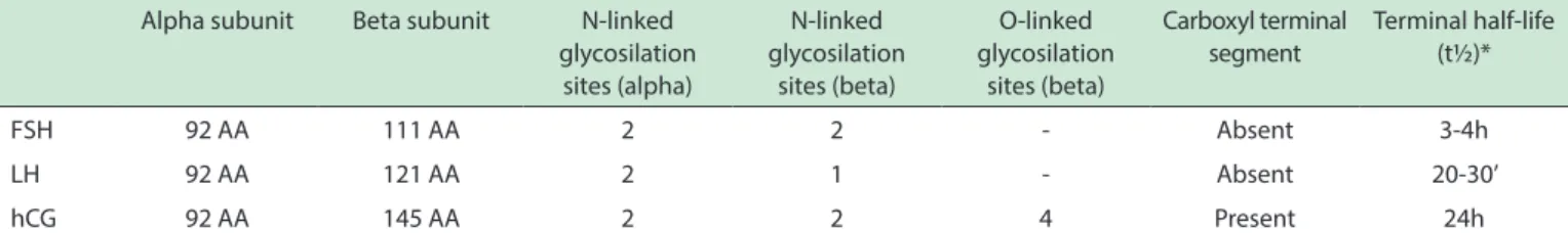
sites and sialic acid residues (approximately 20) than LH, native hCG exhibit a markedly longer terminal half-life of
24 hours after intravenous injection in comparison with
approximately 30 minutes for LH, as shown in Table 2.17
Gonadotropin preparations used for controlled ovarian stimulation
Human menopausal gonadotropin
Human menopausal gonadotropin (hMG), or
meno-tropin, was first extracted from the urine of postmenopausal
women in 1949.2 Urine was originally obtained from an
Table 2 - Structural characteristics of native FSH, LH and hCG molecules
Alpha subunit Beta subunit N-linked glycosilation
sites (alpha)
N-linked glycosilation
sites (beta)
O-linked glycosilation
sites (beta)
Carboxyl terminal segment
Terminal half-life (t½)*
FSH 92 AA 111 AA 2 2 - Absent 3-4h
LH 92 AA 121 AA 2 1 - Absent 20-30’
hCG 92 AA 145 AA 2 2 4 Present 24h
AA: Amino acids; t½ = time that it takes for the concentration in blood plasma of a substance to reach one-half of its steady-state value; * Following intravenous infusion.
Table 3 - Diferences between hMG and FSH formulations
Purity (FSH content) Mean speciic FSH activity
U/mg protein) LH activity (IU/vial)
Injected protein per 75 IU (mcg)
hMG < 5% ~100 75 ~750
HP-hMG < 70% 2,000-2,500 75 ~33
rec-hFSH
Follitropin beta > 99% 7,000-10,000 0 8.1
Follitropin alfa > 99% 13,645 0 6.1
rec-hFSH: Recombinant human follicle-stimulating hormone; hMG: Human menopausal gonadotropin; HP-hMG: Highly puriied human menopausal gonadotropin.
In 1995, the first rec-hFSH (follitropin alfa) was
licensed for clinical use in the European Union. One year later, a similar rec-hFSH (follitropin beta) was made available.2 In the manufacturing process of follitropin
alfa, two separate vectors, one for each subunit, are used to build the master cell bank of FSH-producing cell line, unlike follitropin beta in which a single vector contains the coding sequences of both subunit genes.21,25 The subsequent
production steps are similar for both preparations. First, a working cell bank is established by growing cells from a single vial that contains identical cell preparations. An aliquot from the selected clone of Chinese hamster ovary
cells is grown in T-flasks, then subcultured into roller bottles and allowed to expand for up to 36 days. The cells are then mixed with a suspension of microcarrier beads
and transferred to a bioreactor vessel with continuous culture media infusion for an average of 34 days. The cell culture supernatant medium, containing the proteins secreted by the cells, is collected from the bioreactor. The harvested ‘crude FSH’ is stored at 48 oC until purification.2 Lastly, the protein is purified by chromatography, followed by ultrafiltration. The downstream purification process
differs for the two commercially available recombinant FSH preparations. The follitropin beta process uses a
series of anion and cation exchange chromatography steps, hydrophobic chromatography and size exclusion
chromatography. A similar series of chromatography steps are used in the production of follitropin alfa, in addition
to an immunoaffinity step with a specific monoclonal
antibody that is similar to the one used in the production of HP-hFSH.2,25 Each purification step is rigorously controlled in order to ensure batch-to-batch consistency of the final purified product.2 While the production of urine-derived
gonadotropins is often performed in open, non-sterile
environments, the production of rec-hFSH takes place in closed, sterile environments, such as a bioreactor. Both the
production and the purification of rec-hFSH are subject to
continuous quality control assessments, ensuring a pure, consistent and high-quality product.25 These same concepts,
highlighted above, are now used in the manufacturing process of other recombinant gonadotropins including LH and hCG.2,3,21
Recombinant LH preparations
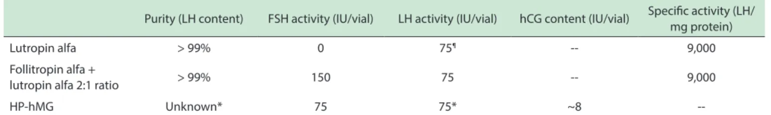
Currently, there are three groups of commercially available gonadotropin preparations containing LH activity, namely, (i) urinary hMG, in which LH activity is dependent on hCG rather than on pure LH glycoprotein, (ii) pure LH glycoprotein produced by recombinant technology (lutropin alfa), and (iii) a combination of pure FSH and LH
glycoproteins in a fixed ratio of 2:1 also manufactured by
recombinant technology, as shown in Table 4.3
Recombinant LH (rec-hLH;lutropin alfa) was intro-duced in the market in the year 2000 for use in women with
gonadotropin insufficiency. The manufacturing process of
rec-hLH is similar to rec-hFSH. Lutropin alfa is highly pure and has high biological activity.3 It is intended to be used
subcutaneously in daily injections. Up to date, lutropin alfa is the only recombinant form of human LH developed for use in ovarian stimulation. It is presented in vials of 82.5 IU of liophylized pure glycoprotein powder to be reconstituted with diluent before administration, using a conventional syringe and needle (75 IU of lutropin alfa is delivered per vial).
Table 4 - Diferences in LH activity of gonadotropins commercially available
Purity (LH content) FSH activity (IU/vial) LH activity (IU/vial) hCG content (IU/vial) Speciic activity (LH/ mg protein)
Lutropin alfa > 99% 0 75¶ -- 9,000
Follitropin alfa +
lutropin alfa 2:1 ratio > 99% 150 75 -- 9,000
HP-hMG Unknown* 75 75* ~8
--¶ 1 µg of lutropin alfa = 22 IU; * Derives primarily from the hCG component, which is preferentially concentrated during the puriication process but sometimes added to achieve the desired amount of LH-like biological activity; HP-hMG: highly puriied human menopausal gonadotropin
women over > 35 years of age and hypo-responders.2,21,26-28
Recombinant LH has three major differences compared to urinary products containing LH activity: (i) it has higher
purity and specific activity because it is manufactured using
recombinant technology; (ii) it is associated with better
dose precision due to filled-by-mass (FbM) technology that
virtually eliminates batch-to-batch variation;2,29,30 (iii) LH
activity is derived directly from pure LH glycoprotein, unlike
hMG, in which hCG is concentrated during purification or
added to achieve the desired amount of LH-like biological activity.2 LH and hCG differ in the composition of their
carbohydrate moieties; this, in turn, affects bioactivity and half-life. As mentioned earlier, LH activity in serum is 30 times higher when hCG is used due to its higher binding
affinity to LH receptors. After subcutaneous administration,
recombinant human LH is eliminated with a terminal
half-life of approximately 24 hours in contrast to 5-7 days
of hCG.17,31 It has been shown that the expression of the
LH/hCG receptor gene, as well as genes involved in the biosynthesis of cholesterol and steroids in granulosa cells, are lower in patients treated with hMG preparations.32 Such effects are caused by a constant ligand exposure during the
follicular phase due to longer half-life and higher binding
affinity of hCG compared with rec-hLH. In animal models,
down-regulation of LH receptors is maintained for up to 48h after hMG administration.33 These findings indicate that the
granulosa cells have lower LH-induced cholesterol uptake, a decrease in the novo cholesterol synthesis and a decrease
in steroid synthesis, thus explaining the observed lower
serum progesterone levels achieved in patients treated with hMG.32,33 The clinical implications of these findings,
however, have not been fully elucidated.
In 2007, a new fixed combination of rec-hFSH
and rec-hLH at 2:1 ratio was launched (follitropin alfa + lutropin alfa) as an alternative for those women who need LH supplementation.34 The 2:1 ratio of FSH and LH in a fixed dose combination was obtained by recombinant technology and vial filling using FbM. The use of FbM as opposed of filled-by-bioassay was possible because the specific activity, isoform distribution and sialylation profile of both gonadotropins are highly consistent among
manufactured batches.29 The bioequivalence of rec-hFSH
and rec-hLH administrated alone or in combination has been similar.34,35
Comparison of gonadotropin formulations Clinical efficacy, efficiency and effectiveness Efficacy, efficiency and effectiveness are frequently
used as synonymous, but technically they represent
different concepts. Efficacy in relation to medication can be defined as the extent to which a drug has the ability to bring
about its intended effect under controlled situations, such as in randomized controlled trials.36 To establish efficacy,
a drug should be at least as good as other available ones
to which it has been compared. Efficiency, on the other
hand, is the capability of a drug to effectivelly produce
a specific outcome with a minimum amount of quantity while effectiveness estimates the extent to which a drug
achieves its intended effect in the usual clinical setting.36 Effectiveness measures the degree of beneficial effect under
“real world” clinical settings, i.e., based on conditions of routine clinical practice and on outcomes essential for clinical decisions.37
A number of meta-analyses of randomized
controlled trials have compared the efficacy of different
gonadotropin products, as can be seen in Table 5.38-45
Despite the heterogeneity of several of these meta-analyses pertaining the different stimulation protocols and choice of fertilization with standard in vitro fertilization (IVF)
or intracytoplasmatic sperm injection (ICSI), the overall conclusion is that both urinary gonadotropins, mainly hMG preparations, and recombinant gonadotropins have
similar efficacy in terms of achieving a pregnancy or live
birth per treatment cycle. While some of these studies were in favour of hMG preparations,38,39,41 in spite of the fact that the confidence limits were just 1% lower, others reported
no differences in pregnancy outcomes between the two treatments.40,43-45 Furthermore, no significant differences
were noted for spontaneous abortion, multiple pregnancy, cycle cancellation and ovarian hyperstimulation syndrome (OHSS) rates.38-45
Although live birth per started cycle is an important endpoint in assisted reproductive technologies (ART), it is confounded by many variables, apart from the stimulation
protocol itself, which are difficult to ascertain. The intended
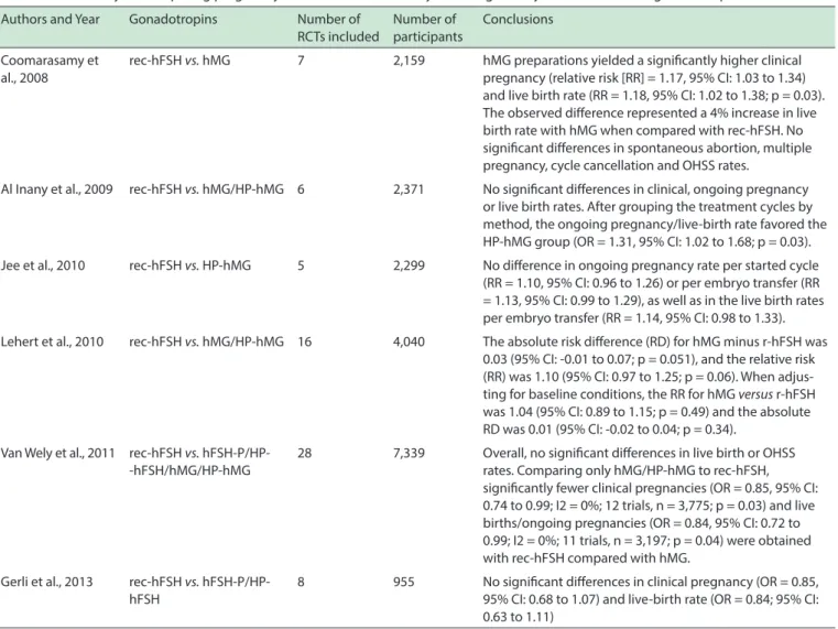
Table 5 - Meta-analyses comparing pregnancy/live birth rates in ART cycles using urinary and recombinant gonadotropins
Authors and Year Gonadotropins Number of RCTs included
Number of participants
Conclusions
Coomarasamy et al., 2008
rec-hFSH vs. hMG 7 2,159 hMG preparations yielded a signiicantly higher clinical
pregnancy (relative risk [RR] = 1.17, 95% CI: 1.03 to 1.34) and live birth rate (RR = 1.18, 95% CI: 1.02 to 1.38; p = 0.03). The observed diference represented a 4% increase in live birth rate with hMG when compared with rec-hFSH. No signiicant diferences in spontaneous abortion, multiple pregnancy, cycle cancellation and OHSS rates.
Al Inany et al., 2009 rec-hFSH vs. hMG/HP-hMG 6 2,371 No signiicant diferences in clinical, ongoing pregnancy
or live birth rates. After grouping the treatment cycles by method, the ongoing pregnancy/live-birth rate favored the HP-hMG group (OR = 1.31, 95% CI: 1.02 to 1.68; p = 0.03).
Jee et al., 2010 rec-hFSH vs. HP-hMG 5 2,299 No diference in ongoing pregnancy rate per started cycle
(RR = 1.10, 95% CI: 0.96 to 1.26) or per embryo transfer (RR = 1.13, 95% CI: 0.99 to 1.29), as well as in the live birth rates per embryo transfer (RR = 1.14, 95% CI: 0.98 to 1.33).
Lehert et al., 2010 rec-hFSH vs. hMG/HP-hMG 16 4,040 The absolute risk diference (RD) for hMG minus r-hFSH was
0.03 (95% CI: -0.01 to 0.07; p = 0.051), and the relative risk (RR) was 1.10 (95% CI: 0.97 to 1.25; p = 0.06). When adjus-ting for baseline conditions, the RR for hMG versus r-hFSH was 1.04 (95% CI: 0.89 to 1.15; p = 0.49) and the absolute RD was 0.01 (95% CI: -0.02 to 0.04; p = 0.34).
Van Wely et al., 2011 rec-hFSH vs.
hFSH-P/HP--hFSH/hMG/HP-hMG
28 7,339 Overall, no signiicant diferences in live birth or OHSS
rates. Comparing only hMG/HP-hMG to rec-hFSH, signiicantly fewer clinical pregnancies (OR = 0.85, 95% CI: 0.74 to 0.99; I2 = 0%; 12 trials, n = 3,775; p = 0.03) and live births/ongoing pregnancies (OR = 0.84, 95% CI: 0.72 to 0.99; I2 = 0%; 11 trials, n = 3,197; p = 0.04) were obtained with rec-hFSH compared with hMG.
Gerli et al., 2013 rec-hFSH vs.
hFSH-P/HP-hFSH
8 955 No signiicant diferences in clinical pregnancy (OR = 0.85,
95% CI: 0.68 to 1.07) and live-birth rate (OR = 0.84; 95% CI: 0.63 to 1.11)
RCT: Randomized controlled trial; rec-hFSH: Recombinant human follicle-stimulating hormone; hMG: Human menopausal gonadotropin; HP-hMG: Highly puriied human meno-pausal gonadotropin; hFSH-P: Puriied urinary follicle-stimulating hormone; HP-hFSH: Highly puriied urinary follicle-stimulating hormone; COS: Controlled ovarian stimulation; OHSS: Ovarian hyperstimulation syndrome.
pregnancy. The number of oocytes retrieved is therefore a highly relevant endpoint directly resulting from ovarian stimulation, and where the drug effect may be estimated with the best sensitivity. The higher the number of retrieved oocytes, the higher is the likelihood of having more embryos available both for transfer and cryopreservation, which ultimately impact on the cumulative pregnancy rates. The use of cumulative pregnancy rates per treatment instead of rates per fresh transfers is also becoming an important
endpoint to assess differences in efficacy and effectiveness. Regarding the efficacy of gonadotropins to yield
oocytes following ovarian stimulation with hMG and recombinant FSH, Lehert et al., in a meta-analysis that
included sixteen randomized controlled trials and 4,040
patients, showed that treatment with hMG resulted in fewer oocytes (mean difference [MD]: -1.54; 95% CI: -2.53 to -0.56; p < 0.0001) compared to rec-hFSH. When adjusting for baseline conditions, the MD estimate was
-2.10 (95% CI: -2.83 to -1.36; p < 0.001). Moreover, a higher total dose of hMG is necessary to achieve the intended effects (MD: 235.46 IU; 95% CI: 16.62 to 454.30; p = 0.03).45 Devroey et al. in a RCT involving more than 700
patients compared HP-hMG (n = 374) with rec-hFSH (n = 375) in antagonist cycles with single blastocyst transfer. The authors retrieved more oocytes in the group treated with rec-hFSH (10.6 ± 5.8 vs. 9.1 ± 5.2; p < 0.001).46
Although pregnancy outcomes did not differ between the treatment methods in both studies, none of them reported on the cumulative pregnancy rates associated with the transfer of fresh and frozen embryos. In a recent
study, Wex and Abou-Setta added to the literature by
pooling randomized controlled trials and meta-analyses
that compared highly-purified hMG with rec-hFSH in
[CI] 0.66-1.01), but a greater number of oocytes with rec-hFSH (mean difference [MD] 1.96, 95% CI 1.02-2.90). Using a model for economic evaluation that accounted for embryo availability, survival following thawing, and patient dropout, the investigators showed that rec-hFSH was cost-saving compared with HP-HMG in a combination of fresh and frozen cycles. Differences were equivalent to €315 per patient starting treatment or 6.4% of the total treatment cost.47
We have compared the effectiveness of different gonadotropin products for controlled ovarian stimulation in one of the largest observational studies to date.20 Our
study was intended to assess treatment effectiveness, which cannot be measured in controlled trials because the act of inclusion in a study is a distortion of usual practice. Effectiveness can be evaluated through observational studies of real practice, thus allowing practice to be assessed in qualitative as well as quantitative terms. In our aformentioned study, we compared normogonadotropic down-regulated women undergoing ICSI by type of gonadotropin, follitropin alfa FbM (n = 236), hMG (n = 299) and HP-hMG (n = 330). Overall, pregnancy rates did not differ among the three treatment groups. The clinical pregnancy rate per initiated cycle was 34.7%, 35.5% and 40% for rec-hFSH, hMG and HP-hMG, respectively while the live birth rate per initiated cycle was 30.1%, 24.4% and 32.4%, respectively. However, the total dose of
gonadotropin used for ovarian stimulation was significantly
lower in women who received rec-hFSH (2,268 ± 747 IU) compared with those who received hMG (2,685 ± 720 IU) or HP-hMG (2,903 ± 867 IU; p < 0.001). The difference
in favor of rec-hFSH was also reflected in the amount of gonadotropin needed per live birth, as significantly less
rec-hFSH was required compared with hMG (52% reduction) and HP-hMG (21% reduction).20
In summary, when comparing gonadotropins it is important to take into account the amount of drug used, the number of oocytes/embryos produced, and the number of pregnancies obtained by transferring fresh and frozen-thawed embryos. Assuming a similar quality, the quantity of oocytes and embryos produced, including those available for cryopreservation, will impact the chances of a patient to have a baby.
Quality indicators of infertility care
Quality indicators are measurable elements for which there is evidence that they assess the quality of health care.48,49 While the majority of quality indicators
includes safety and effectiveness of care,49,50 it is argued
that indicator sets should fully represent healthcare
quality and, therefore, cover the six dimensions of quality of care, including effectiveness, safety, efficiency,
timeliness, equity and patient centeredness.49,51 In a recent
study involving doctors, nurses and infertility patients, the
relative importance of the six aforesaid quality dimensions
of care was analyzed by the Delphi method. The workgroup agreed that safety, effectiveness and patient-centerdness were the most important quality dimensions for infertility care.51
Safety
Urine-derived gonadotropins require large amounts of human urine as a primary source for manufacturing. In the 1960s and 1970s, when the demand for gonadotropins was still low, most urine was collected in Italy and the Netherlands. However, the development of new clinical
indications for gonadotropins combined with the expansion
of infertility treatment on a worldwide basis led to a rapid
increase in demand. Urine collection was expanded to
countries such as Spain, China, Brazil and Argentina. As
the demand for gonadotropins began to rise exponentially
in the 1980s, the ability to control the source material
became more difficult. Unlike blood collection, human urine collection is not subject to specific collection regulations.
Moreover, urine is collected at home from tens of thousands of individual donors and pooled. A medical questionnaire is usually the only source of health information. Because urine is pooled, the donor source cannot be traced. As the
pool is constantly changing, standardization is difficult
to ascertain. Transportation (from urine collection sites to processing facility) is poorly monitored and therefore quality cannot be checked throughout all manufacturing steps.22,52,53 Although sophisticated purification techniques
are currently available, which allow the safe use of urinary
formulations, extraneous urinary proteins may account for more than 30% of the protein content in high-purified hMG
products even, as demonstrated by high-performance liquid chromatography analysis.30Following protein identification
by two-dimensional sodium dodecyl sulfate-polyacrylamide gel electrophoresis (SDS-PAGE) and mass spectrometry, a total of 23 non-gonadotropin-related proteins have been
identified at variable levels in different batches of the
urine-derived preparations.54 In other studies, two-dimensional
gel analysis demonstrated that protein impurities were composed of leukocyte elastase inhibitor, protein C inhibitor and zinc-a2-glycoprotein. These proteins are involved in receptor activity, immune response, protein metabolism, and cell growth. Tumor necrosis factor-binding protein I, transferrin and immunoglobulin-related proteins were also present in both hMG and urinary FSH preparations. Lastly, recent data has shown that some of these impurities are prion proteins, which are a matter of great concern for health regulatory agencies because of their association with transmissible spongiform encephalopathy diseases.55
to recognize and break down the abnormally folded protein. As a result, prions accumulate in the central nervous system, interfering with normal brain function. Conversion of normal cellular protein into the abnormal form can occur spontaneously or following infection. Abnormal prions include PrPsc, the protein associated with scrapie, and PrPres, the protein resistant to enzyme degradation found in patients with Creutzfeldt-Jakob disease (CJD).55
Inactivation of prions in urine-derived material may denature proteins, including FSH. For instance, urea, a commonly used denaturant, destroys the dimeric structure of proteins. This is one reason why urine-derived gonadotropins cannot be as pure as recombinant ones. Cross-contamination is another concern in urine-derived products. A rogue element in one individual donation of urine may spread through a complete batch and potentially
cause problems in the final product. Quality control is only
possible through stringent donor collection, transportation and production. In fact, several regulatory agencies have set limitations to urine-derived products.22,52 Although the clinical significance of most protein contamination from
urinary gonadotropins is unknown, it is certain that these contaminants are not needed to induce optimal follicle
development. More importantly, these findings underscore
the poor quality of urinary sources and stress the need for more reliable proteins.22,52 Many of the risks associated with biologically extracted proteins are avoided when the
protein is produced synthetically.
In the manufacturing process of recombinant gonadotropins, vectors are used to build the master cell
bank of a specific gonadotropin-producing cell line.21,25 A
working cell bank is established by growing cells from a single vial that contains identical cell preparations. The cell culture supernatant medium, containing the proteins secreted by the cells, is collected from the bioreactor, and
the protein is purified.21,25 Both the production and the purification of recombinant gonadotropins are subject
to continuous quality control assessments, ensuring a pure, consistent and high-quality glycoprotein as demonstrated by SDS-PAGE and western blot analysis.54
Given its high purity, each product batch of recombinant gonadotropin is routinely characterized and controlled
using physicochemical techniques, including size exclusion
high performance liquid chromatography (SE-HPLC), which allows assessment of both the integrity and the amount of glycoproteins;and isoeletric focusing and glycan mapping, which characterize protein glycoforms present in each preparation.56,57
Patient-centeredness
Recombinant gonadotropins are presented in ready-to-use solutions; they include pen devices, so that the gonadotropin can be self-administered subcutane-ously. In contrast, urinary gonadotropins consist of a
freeze-dried lyosphere containing either 75 IU of FSH/ hMG or 5,000/10,000 IU of hCG that need to be dissolved in sterile water before injection.12 Intramuscular
administra-tion is the route of choice for most urinary gonadotropins,
although it possible to apply highly-purified urine-derived
gonadotropins subcutaneously.30
Earlier studies have shown that drug delivery by pen devices was bioequivalent to that of conventional syringe injections. However, due to unavoidable losses
during syringe filling and/or removing excess air, about
18% of the FSH amount is lost in the conventional syringe application when compared to a ready-for-use solution with a pen device.35
Studies addressing satisfaction indicate that patients clearly prefer pen injections. Outcomes including self-ad-ministration and patient satisfaction, the overall incidence of local reactions, overall pain score, and burning sensation at the injection site clearly favor the pen device group.35,58
In a study focussing on patient-centeredness, patients and their partners received nurse-led training on three gonadotropin presentations: (i) powdered urofollitropin with conventional needles and syringes for administration,
(ii) follitropin beta in a premixed and prefilled cartridge
with a reusable injection device, and (iii) follitropin alfa in a
disposable, premixed and prefilled injection device. A total
of 123 participants attended the training and were asked to complete a post-training questionnaire. More participants
expressed a preference for using pen injectors compared
with conventional syringes (84.6% versus 5.7%; p < 0.0001).
Of the 94 participants who preferred a particular device,
more preferred the follitropin alfa prefilled pen (68.1%)
than the follitropin beta cartridge and pen (24.5%; p < 0.0001) or urofollitropin with needle-free reconstitution device and conventional syringe (7.4%; p < 0.0001).59
Recently in 2011, a new family of pen (FoP) devices has been approved in the European Union, Canada and Australia, intended to deliver recombinant preparations.60,61
The new device is like the pen used to administer follitropin
alfa, but explaining its use to patients is simpler and quicker,
which reduces the risk of errors. In addition, these novel devices offer dosing increments in very small amounts,
thus allowing the dosage to be fine-tuned at any point after
treatment has started.60,61 In summary, the introduction of recombinant technology, filling method by mass, and
pen devices for gonadotropin administration represent an important step forward in terms of patient-centeredness in infertility treatments.
CONCLUSIONS
recombinant products using biotechnology. Recom-binant gonadotropins are purer than urinary-derived gonadotropins, and the introduction of FbM technology virtually eliminated batch-to-batch variations and ena-bled accurate dosing in small increments; all of which allowed the introduction of individualized controlled ovarian stimulation. Biotech gonadotropins have better clinical efficiency and effectiveness compared with uri-nary products, owed to the production of more oocytes using lower amounts of the drug. An increased number of oocytes/embryos produced will positively impact the chances of patients to have a baby, by offering them the highest cumulative pregnancy rate. Biotech gonadotro-pins are also in line with important quality indicators of infertility care, including safety and patient-centerdness. Recombinant gonadotropin formulations have a better safety profile owing to purity, and the introduction of prefilled pen devices simplified infertility treatment for patients, doctors and nurses alike.
EFICÁCIA, EFICIÊNCIA E EFETIVIDADE DA TERAPIA GONADOTRÓFICA PARA O TRATAMENTO DA IN-FERTILIDADE
RESUMO: A terapia gonadotrófica é elemento essen
-cial nos tratamentos de infertilidade que envolvem tecnologia de reprodução assistida. Nos últimos anos houve avanços
notáveis no desenvolvimento de novas gonadotrofinas, prin
-cipalmente com a produção de gonadotrofinas via recursos biotecnológicos. As gonadotrofinas recombinantes têm maior actividade específica em comparação com os suas homólogas
urinárias, permitindo, assim, a administração subcutânea de quantidades mínimas de glicoproteína. Como resultado,
as formulações recombinantes tem um melhor perfil de segurança, apesar de semelhança em termos de eficácia
para a gravidez, como relatado em diversos ensaios clínicos
randomizados e meta-análises. As gonadotrofinas estimu -lam os ovários a desenvolver folículos e ovócitos, que são a
matéria-prima para a fertilização e produção de embriões.
Os embriões resultantes são transferidos (frescos ou conge-lados/descongelados) para produzir gravidez. Comparações
entre as gonadotrofinas devem, portanto, ser medidas não somente pela eficácia clínica de produzir gravidezes pela transferência de embriões a fresco, mas sobremaneira pela eficiência na produção de ovócitos e embriões em relação à
quantidade de droga administrada, e efetividade na
obten-ção de gravidezes pela transferência de embriões frescos e congelados/descongelados (taxa de gravidez cumulativa).
As comparações entre diferentes preparações de
gonado-trofinas também devem levar em conta outros indicadores
importantes de qualidade em medicina reprodutiva, como a segurança e o interesse do paciente. Estes indicadores de
qualidade favorecem as gonadotrofinas biotecnológicas em
relação aos produtos biológicos na terapia da infertilidade.
UNITERMOS: gonadotrofinas; estimulação ovariana
controlada; tecnologia reprodutiva assistida
■
REFERENCES1. Beall SA, DeCherney A. History and challenges surrounding ovarian stimulation in the treatment of infertility. Fertil Steril. 2012;97(4):795-801. 2. Lunenfeld B. Historical perspectives in gonadotrophin therapy. Human
Reprod Update. 2004;10(6):453-67.
3. Practice Committee of American Society for Reproductive Medicine, Birmingham, Alabama. Gonadotropin preparations;past, present, and future perspectives. Fertil Steril. 2008;90(5 Suppl):S13-20.
4. Howles CM. Genetic engineering of human FSH (Gonal-F). Hum Reprod Update. 1996;2(2):172–91.
5. Lapthorn AJ, Harris DC, Littlejohn A, Lustbader JW, Canfield RE, Machin KJ,et al. Crystal structure of human chorionic gonadotropin. Nature. 1994;369(6480):455-61.
6. Vaitukaitis JL, Ross GT, Braunstein GD, Rayford PL. Gonadotropins and their subunits: basic and clinical studies. Recent Prog Horm Res. 1976;32:289-331.
7. Ulloa-Aguirre–A, Espinoza R, Damian-Matsumura P, Chappel SC. Immu-nological and biological potencies of the different molecular species of gonadotrophins. Hum Reprod. 1988;3(4):491-501.
8. Rozell TG, Okrainetz RJ. FSH: one hormone with multiple forms, or a family of multiples hormones. In: Chedrese PJ, Ed. Reproductive Endocrinology: A Molecular Approach. New York: Springer Science + Business Media, LLC 2009; pp. 145-160.
9. Wide L, Naessén T, Sundström-Poromaa I, Eriksson K. Sulfonation and sialylation of gonadotropins in women during the menstrual cycle, after menopause, and with polycystic ovarian syndrome and in men. J Clin Endocrinol Metab. 2007;92(11):4410-7.
10. Ulloa-Aguirre A, Midgley AR Jr, Beitins IZ, Padmanabhan V. Follicle--stimulating isohormones: characterization and physiological rele-vance. Endocr Rev. 1995;16(6):765-87.
11. Green ED, Baenziger JU. Asparagine-linked oligosaccharides on lutro-pin, follitrolutro-pin, and thyrotropin. I. Structural elucidation of the sulfated and sialylated oligosaccharides on bovine, ovine, and human pituitary glycoprotein hormones. J Biol Chem. 1988;263(1):25-35.
12. Fiete D, Srivastava V, Hindsgaul O, Baenziger JU. A hepatic reticulo-endothelial cell receptor specific for SO4-4GalNAc beta 1,4GlcNAc beta 1,2Man alpha that mediates rapid clearance of lutropin. Cell. 1991;67(6):1103-10.
13. Wide L, Eriksson K, Sluss PM, Hall JE. Serum half-life of pituitary go-nadotropins is decreased by sulfonation and increased by sialylation in women. J Clin Endocrinol Metab. 2009;94(3):958-64.
14. Combarnous Y. Molecular basis of the specificity of binding of glyco
-protein hormones to their receptors. Endocr Rev. 1992;13(4):670-91. 15. Galway AB, Hsueh AJ, Keene JL, Yamoto M, Fauser BC, Boime I. In vitro and in vivo bioactivity of recombinant human follicle-stimulating hormone and partially deglycosylated variants secreted by transfected eukaryotic cell lines. Endocrinology. 1990;127(1):93-100.
16. Fox KM, Dias JA, Van Roey P. Three-dimensional structure of human follicle-stimulating hormone. Mol Endocrinol 2001;15(3):378-89. 17. Campbell RK. Molecular pharmacology of gonadotropins. Endocrine.
2005;26(3):291-6.
18. Cole LA, Khanlian SA, Muller CY. Detection of perimenopause or post-menopause human chorionic gonadotropin: an unnecessary source of alarm. Am J Obstet Gynecol. 2008;198(3):275. e1-7.
19. Cole LA, Khanlian SA, Muller CY. Normal production of human chorionic gonadotropin in perimenopausal and menopausal women and after oophorectomy. Int J Gynecol Cancer. 2009;19(9):1556-9.
21. Leão RdeB, Esteves SC. Gonadotropin therapy in assisted reproduction: an evolutionary perspective from biologics to biotech. Clinics (Sao Paulo). 2014;69(4):279-93.
22. Giudice E, Crisci C, Eshkol A, Papoian R. Composition of commercial gonadotrophin preparations extracted from human post-menopausal urine: characterization of non-gonadotrophin proteins. Hum Reprod. 1994;9(12):2291-9.
23. Alviggi C, Revelli A, Anserini P, Ranieri A, Fedele L, Strina I, et al. A prospective, randomised, controlled clinical study on the as-sessment of tolerability and of clinical efficacy of Merional (hMG--IBSA) administered subcutaneously versus Merional administered intramuscularly in women undergoing multifollicular ovarian stimulation in an ART programme (IVF). Reprod Biol Endocrinol. 2007;5;45.
24. Platteau P, Laurent E, Albano C, Osmanagaoglu K, Vernaeve V, Tournaye H, et al. An open, randomized single-centre study to compare the efficacy and convenience of follitropin beta administered by a pen device with follitropin alpha administered by a conventional syringe in women undergoing ovarian stimulation for IVF/ICSI. Hum Reprod. 2003;18(6):1200-4.
25. Howles CM. Genetic engineering of human FSH (Gonal-F). Hum Reprod Update. 1996;2(2):172–91.
26. Dhillon S, Keating GM. Lutropin alfa. Drugs. 2008;68(11):1529-40. 27. Bosdou JK, Venetis CA, Kolibianakis EM, Toulis KA, Goulis DG, Zepiridis
L, et al. The use of androgens or androgen-modulating agents in poor responders undergoing in vitro fertilization: a systematic review and meta-analysis. Hum Reprod Update. 2012;18(2):127-45.
28. Hill MJ, Levens ED, Levy G, Ryan ME, Csokmay JM, DeCherney AH, et al. The use of recombinant luteinizing hormone in patients undergoing assisted reproductive techniques with advanced reproductive age: a systematic review and meta-analysis. Fertil Steril. 2012;97(5):1108-14.e1.
29. Driebergen R, Baer G. Quantification of follicle stimulating hormone (follitropin alfa): is in vivo bioassay still relevant in the recombinant age? Curr Med Res Opin. 2003;19(1):41-6.
30. Bassett RM, Driebergen R. Continued improvements in the quality and consistency of follitropin alfa, recombinant human FSH. Reprod Biomed Online. 2005;10(2):169-77.
31. le Cotonnec JY, Porchet HC, Beltrami V, Munafo A. Clinical pharmacology of recombinant human luteinizing hormone: Part I. Pharmacokinetics after intravenous administration to healthy female volunteers and comparison with urinary human luteinizing hormone. Fertil Steril. 1998;69(2):189-94.
32. Grøndahl ML, Borup R, Lee YB, Myrhøj V, Meinertz H, Sørensen S. Differences in gene expression of granulosa cells from women undergoing controlled ovarian hyperstimulation with either recombinant follicle-stimulating hormone or highly purified human menopausal gonadotropin. Fertil Steril. 2009;91(5):1820-30.
33. Menon KM, Munshi UM, Clouser CL, Nair AK. Regulation of luteinizing hormone/human chorionic gonadotropin receptor expression: a perspective. Biol Reprod. 2004;70(4):861-6.
34. Bosch E, Vidal C, Labarta E, Simon C, Remohi J, Pellicer A. Highly
purified hMG versus recombinant FSH in ovarian hyperstimula
-tion with GnRH antagonists--a randomized study. Hum Reprod. 2008;23(10):2346-51.
35. Picard M, Rossier C, Papasouliotis O, Lugan, I. Bioequivalence of recombinant human FSH and recombinant human LH in a fixed 2:1 combination: two phase I, randomised, crossover studies. Curr Med Res Opin. 2008;24(4):1199-208.
36. Haynes B. Can it work? Does it work? Is it worth it? The testing of healthcareinterventions is evolving. BMJ. 1999;319;652-3.
37. Gartlehner G, Hansen RA, Nissman D, Lohr KN, Carey TS. Criteria for Distinguishing Effectiveness From Efficacy Trials in Systematic Reviews. Rockville (MD): Agency for Healthcare Research and Quality (US) 2006 Apr. Report No.: 06-0046. AHRQ Technical Reviews.
38. Coomarasamy A, Afnan M, Cheema D, van der Veen F, Bossuyt PM, van Wely M. Urinary hMG versus recombinant FSH for controlled ovarian hyperstimulation following an agonist long down-regulation protocol in IVF or ICSI treatment: a systematic review and meta-analysis. Hum Reprod. 2008;23(2):310-5.
39. Al-Inany HG, Abou-Setta AM, Aboulghar MA, Mansour RT, Serour GI. Highly purified hMG achieves better pregnancy rates in IVF cycles but not ICSI cycles compared with recombinant FSH: a meta-analysis. Gynecol Endocrinol. 2009;25(6):372-8.
40. Jee BC, Suh CS, Kim YB, Kim SH, Moon SY. Clinical efficacy of highly
purified hMG versus recombinant FSH in IVF/ICSI cycles: a meta
--analysis. Gynecol Obstet Invest. 2010;70(2):132-7.
41. van Wely M, Kwan I, Burt AL, Thomas J, Vail A, Van der Veen F, et al. Recombinant versus urinary gonadotrophin for ovarian stimulation in assisted reproductive technology cycles. Cochrane Database Syst Rev. 2011;(2):CD005354.
42. Al-Inany HG, Abou-Setta AM. Are all human-derived follicle-stimulating hormone products the same? A systematic review and meta-analysis using direct and adjusted indirect analyses, to determine whether
Fostimon® is more efficient than Metrodin-HP®. Gynecol Endocrinol.
2012;28(2):94-101.
43. Gerli S, Bini V, Favilli A, Di Renzo GC. Clinical efficacy and
cost--effectiveness of HP-human FSH (Fostimon®) versus rFSH
(Gonal--F®) in IVF-ICSI cycles: a meta-analysis. Gynecol Endocrinol.
2013;29(6):520-9.
44. van Wely M, Kwan I, Burt AL, Thomas J, Vail A, Van der Veen F, et al. Recombinant versus urinary gonadotrophin for ovarian stimulation in assisted reproductive technology cycles. A Cochrane review. Hum Reprod Update. 2012;18(2):111.
45. Lehert P, Schertz JC, Ezcurra D. Recombinant human follicle-stimulating hormone produces more oocytes with a lower total dose per cycle in assisted reproductive technologies compared with highly purified human menopausal gonadotrophin: a meta-analysis. Reprod Biol Endocrinol. 2010;8:112.
46. Devroey P, Pellicer A, Nyboe Andersen A, Arce JC; Menopur in GnRH an-tagonist cycles with single embryo transfer trial group. A randomized assessor-blind trial comparing highly purified hMG and recombinant FSH in a GnRH antagonist cycle with compulsory single-blastocyst transfer. Fertil Steril. 2012;97(3):561-71.
47. Wex J, Abou-Setta AM. Economic evaluation of highly purified human menopausal gonadotropin versus recombinant human follicle-stimu-lating hormone in fresh and frozen in vitro fertilization/intracyto-plasmic sperm-injection cycles in Sweden. Clinicoecon Outcomes Res. 2013;5:381-97.
48. Donabedian A. The quality of care. How can it be assessed? JAMA. 1988;260(12):1743-48.
49. Mainz J. Defining and classifying clinical indicators for quality impro
-vement. Int J Qual Health Care. 2003;15:523-30.
50. Copnell B, Hagger V, Wilson SG, Evans SM, Sprivulis PC, Cameron PA. Measuring the quality of hospital care: an inventory of indicators. Intern Med J. 2009;39(6):352-60.
51. Dancet EA, D’Hooghe TM, Spiessens C, Sermeus W, De Neubourg D, Karel N, et al. Quality indicators for all dimensions of infertility care quality: consensus between professionals and patients. Hum Reprod. 2013;28(6):1584-97.
52. van de Weijer BH, Mulders JW, Bos ES, Verhaert PD, van den Hooven HW. Compositional analyses of a human menopausal gonadotrophin preparation extracted from urine (menotropin). Identification of some of its major impurities. Reprod Biomed Online. 2003;7(5):547-57.
53. Kuwabara Y, Mine K, Katayama A, Inagawa T, Akira S, Takeshita T. Proteomic analyses of recombinant human follicle-stimulating hor-mone and urinary-derived gonadotropin preparations. J Reprod Med. 2009;54(8):459-66.
55. Johnson RT, Gibbs CJ Jr. Creutzfeldt-Jakob disease and related transmis-sible spongiform encephalopathies. N Engl J Med. 1998;339(27):1994-2004.
56. Gervais A, Hammel YA, Pelloux S, Lepage P, Baer G, Carte N, et al. Glycosylation of human recombinant gonadotrophins: characterization and batch-to-batch consistency. Glycobiology. 2003;13(3):179-89.
57. Hugues JN, Barlow DH, Rosenwaks Z, Cédrin-Durnerin I, Robson S, Pidoux L, et al. Improvement in consistency of response to ovarian stimulation with recombinant human follicle stimulating hormone resulting from a new method for calibrating the therapeutic prepara-tion. Reprod Biomed Online. 2003;6(2):185-90.
58. Aghssa MM, Azargoon A, Ramezanzadeh F, Bagheri M. A comparison of the efficacy, tolerability, and convenience of two formulations of follitropin-alpha in Iranian woman undergoing intracytoplasmic sperm injection cycles. Fertil Steril. 2008;90(4):1043-8.
59. Weiss N. Gonadotrophin products: empowering patients to choose the product that meets their needs. Reprod Biomed Online. 2007;15(1):31-7. 60. Christen M, Schertz JC, Arriagada P, Keitel J, Müller H. The redesigned follitropin α pen injector for infertility treatment. Expert Opin Drug Deliv. 2011;8(6):833-9.