M
ABSTRACT
Hybrid fixation in the bilateral sagittal split
osteotomy for lower jaw advancement
Felipe Ladeira PEREIRA1, Marcos JANSON2, Eduardo SANT’ANA3
1- DDS, Oral and Maxillofacial Surgeon, Brazilian Army, Juiz de Fora General Hospital (HGeJF), Juiz de Fora, MG, Brazil. 2- DDS, MS. Orthodontist, Private Practice, Bauru, SP, Brazil.
3- DDS, PhD, Associate Professor of Oral and Maxillofacial Surgery, Bauru School of Dentistry, University of São Paulo, Bauru, SP, Brazil.
Corresponding address: Felipe Ladeira Pereira - Av. Dr. Paulo Japiassú Coelho, 209/701 - Cascatinha - 36033-310 - Juiz de Fora, MG - Brasil - Phone: +55-32-3236-3150/9111-3900 - e-mail: [email protected]
Received: September 10, 2008 - Modification: May 18, 2009 - Accepted: June 02, 2009
iniplate and screw fixation has been widely used in bilateral sagittal split osteotomy, but some issues remain unclear concerning its lack of rigidity when compared to Spiessl’s bicortical technique. This paper demonstrates the hybrid fixation technique in a case report. A 34-year-old female patient underwent a double jaw surgery with counter-clockwise rotation of the mandible fixed using the hybrid fixation technique. The patient evolved well in the postoperative period and is still under follow up after 14 months, reporting satisfaction with the results and no significant deviation from the treatment plan up to now. No damage to tooth roots was done, maxillomandibular range of motion was within normality and regression of the inferior alveolar nerve paresthesia was observed bilaterally. The hybrid mandibular fixation is clearly visible in the panoramic and cephalometric control radiographs. It seems that the hybrid fixation can sum the advantages of both monocortical and bicortical techniques in lower jaw advancement, increasing fixation stability without significant damage to the mandibular articulation and the inferior alveolar nerve. A statistical investigation seems necessary to prove its efficacy.
Key words: Bilateral sagittal split osteotomy. Orthognathic surgery. Monocortical fixation. Bicortical fixation. Mandibular advancement. Condylar torque.
INTRODUCTION
Bilateral sagittal split osteotomy (BSSO) is commonly used to treat mandibular discrepancies7,11,28. The ability to rigidly and properly fix the fractured segments at the time of surgery may facilitate healing in the immediate postoperative period and reduce the displacement possibility of the bony segments, particularly the condylar proximal segment27. The technique that uses bicortical compressive screws was first described by Spiessl25 (1974) while the technique that uses miniplates and monocortical screws was introduced by Luhr14 (1986).
Monocortical osteosynthesis has been widely
used in the fixation of BSSO8, leading to stable results according to the literature3,8,15,19-21, in spite of being considered as a semi-rigid fixation21. Since monocortical fixation is not as rigid as bicortical osteosynthesis, the excessive shear force stress, produced by the compressive action of the masseter muscle to the osteotomy line, may transform the mandibular shape postoperatively9,18. On the other hand, other authors have found no differences in the stability promoted by both techniques8,11,26.
techniques: the positional bicortical screws and the monocortical plate osteosynthesis.
CASE REPORT
A 34-year-old female patient searched Dr. E.S. complaining the “lack of chin”, “reversed lower lip” and gummy smile. The patient had previous orthodontic treatment with dental compensation, having both maxillary first premolars already extracted and the gap closed. Clinically, the patient presented symmetric dolicocephalic face with maxillomandibular retrusion; maxillary vertical excess; chin deficiency and an accentuated facial convexity. In addition, healthy periodontal tissues and temporomandibular joints (TMJs), tension of the orbicularis oris and mental muscles during function and lower lip incompetency when relaxed were also observed. In order to acquire proper positioning of the mandibular incisors, the right and left first
general anesthesia, the mandible was managed and fixed as described below, followed by usual maxillary Le Fort I.
After BSSO according to Epker’s6 (1977) modified technique, the condyle was properly positioned1 and the osteotomy fixed with a 2.0 mm miniplate and two monocortical screws in each segment. An inset bend was made at the plate to maintain the gap and avoid condylar torque1,19,20. Having the 4 screws in position, a transorally oblique perforation was drilled in the retromolar region (visualising the proximal end of the distal fragment), the hole was tapped and a 2.0 mm diameter x 16 mm long screw was inserted. A second screw was placed distally from the first in the same manner. These screws were positional and do not exert pressure between segments (Figure 3). The same fixation was done on the other side. After completion of the osteosynthesis, maxillomandibular immobilization (MMI) was removed and occlusion and mouth
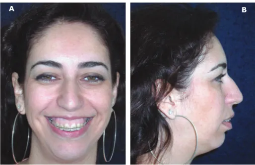
Figure 1- Preoperative frontal aspect (A) and facial profile (B) of the patient (patient signed informed consent authorizing the publication of these pictures)
Figure 2- Preoperative intraoral view of the left side (A) and right side (B)
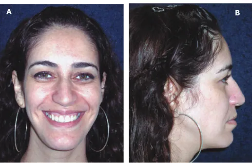
Figure 4- Postoperative frontal aspect (A) and facial profile (B) of the patient (patient signed informed consent authorizing the publication of these pictures)
Figure 3- Schematic illustration of the hybrid fixation in the BSSO. Note the inset bend at the plate
A B
Figure 5- Postoperative intraoral view of the left side (A) and right side (B)
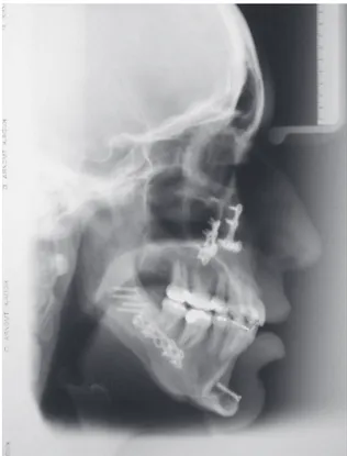
Figure 6- Panorex showing hybrid fixation in mandibular advancement
opening were checked. The wounds were then sutured as usual.
The patient evolved well in the postoperative period and is still under follow up after 14 months, reporting satisfaction with the results (Figures 4 to 5). No damage to tooth roots was done, maxillomandibular range of motion is within normality and regression of the inferior alveolar nerve (IAN) paresthesia is observed bilaterally. The hybrid mandibular fixation is clearly visible in the panoramic and cephalometric control radiographs (Figure 6 and 7). Figure 8 shows cephalometric tracing of the preoperative position and postoperative changes along the 16 months of treatment, including the removal of brackets.
DISCUSSION
The hybrid fixation technique in the BSSO with one 2.0 mm miniplate, four monocortical and two positional screws was initially described for management of cases where the lingual cortical plate of the distal segment had fractured27,28. The purpose of the suggested technique is to be the routine fixation method in mandibular advancements in order to increase stability of the single 4-hole miniplate while maintaining its advantages, such as: lower possibility of IAN compression, absence of skin scars (since the
retromolar screws are placed transorally) and passive condyle accommodation at the glenoid fossa. Moreover, the earlier release of the elastic MMI in the postoperative period, the sooner the patient will start soft diet ingestion (while using guiding elastics for intercuspation) without significant increase of the total fixture cost.
Among the drawbacks of the bicortical screw technique are IAN compression, scars in the face or neck made by the transcutaneous perforation and rotation of the mandibular condyles1,3,9,11,19,20,23. In the method proposed for this case, mandibular third molar extraction prior to surgery is necessary for bicortical screw placement. Since extractions are not absolutely necessary when miniplates are the only fixation method, it can be considered an advantage of this technique18.
Concerning the miniplate advantages, the three-dimensional relationship between the segments is established by the miniplate, with the condyle in the glenoid fossa and the proximal and distal segments in their initial contact point21. No compression is made between the segments and the result is immediate and functionally stable3,8,15,19-21. The miniplate applied in the anterior border of the buccal osteotomy facilitates manipulation of the proximal segment and seating of the condyle; after fixed it is stable
also conceivable that the fragments fixed by miniplates, which are bent to accommodate the step at the buccal surface, would tend to cause less harm to the IAN20,23, and also reduces the risk of damaging the roots11.
After monocortical osteosynthesis have been applied, small forces directed across the osteotomy can still change the relative positions of the segments. This can lead to occlusal changes when patients return to function early or when they are noncompliant21. The unpredictable fixation provided by the miniplates alone may compromise the clinical outcome if the patient is restored to early function23. Therefore, if the patient’s postsurgical occlusion is unstable, monocortical osteosynthesis will lead to too much rotation of the mandible and may cause delayed union and breakage of the miniplates4,8. Therefore, especially in these patients or those who underwent an overcorrection, osteosynthesis should be performed bicortically8.
The fixation relapse rate in the bicortical screws technique ranges from 8 to 11%12,13, while for the miniplates this value ranges from 5.2 to 15%3,19,20. According to a recent literature review10, bicortical screws show only slight differences regarding skeletal stability compared to miniplates in short-term, but a large number of studies with higher skeletal long-term relapse rates were seen in patients treated with bicortical screws instead of miniplates. By advancing the mandible, the submandibular soft tissue drape is stretched together with the suprahyoid and infrahyoid muscles. As a consequence, the hyoid, fixed by these muscles, is pulled forward, but will return to its original position several months postoperatively. Stretching of theses tissues gives rise to a constant force opposite to the vector of the mandibular advancement1,3,11,16. Relapse
in the early postoperative time (6 weeks to 6 months)5,16,29. A correlation between the amount of advancement and relapse may occur only when the advancement exceeds 7 mm3,10,16,19,29. Furthermore, the MMI period in the first weeks seems to reduce the relapse rate5,16, without great significant risks of muscular atrophy if this time is short16.
postoperatively, showing a direct connection between the amount of advancement and the risk of PCR20. In fact, the endorotation movement that happens in the TMJ in mandibular advancements has major potential to cause dysfunction in the TMJ than the exorotation observed in surgeries for mandibular prognathism19.
Shetty, et al.24 (1994), in a study using a biomechanical model of the BSSO, showed that, within a physiologic range of loading, their hybrid technique using one positional screw (different from the method reported in this paper), produced stability that was comparable with or superior to that produced by conventional methods of rigid internal fixation. Because the plate and the monocortical screws are placed first, a bone clamp is never applied across the osteotomy21. The segment clamping negates advantage of the positional screw, and makes it function as a lag screw21. This could produce lingual or rotational movement of the proximal segment, with consequent condylar displacement, occlusal changes and TMJ problems, in addition to the possibility of IAN compression between the segments, producing numbness or paresthesia1,12,13,21,23. Since the positional bicortical screw from the technique is placed far anteriorly than those normally placed in the Spiessl’s25 (1974) technique, it can be placed transorally, avoiding a transcutaneous stab incision and thus an unfavorable scar21,22. In the technique described for the present case, the two non-compressive bicortical screws are easily placed transorally, by angulating the drill and inserting the screw while visualizing if the gap between the fragments stays still. If the gap starts to increase, the screw should be removed, reangulated and reinserted. When the gap is greater than 2 mm, particulated bone graft are usually used.
The screw applied bicortically in the retromolar region inhibits the displacement tendencies through its resistance to axial and shear forces9,23. A second screw, as proposed in our technique, would guarantee the immobility of the segments. The use of 2 miniplates on each side to increase fixation stability in great mandibular
advancements has been described7,15,23. In the technique proposed here, stability can be increased without the expense of these additional two plates and eight screws usually necessary in these cases, but using four bicortical screws, two on each side, lowering the total fixture costs. Within the anatomic limits imposed by the BSSO, it is known that the greater the separation between the retromolar screw and the miniplate, the better the expected functional stability23.
The goal of the technique proposed in this paper is to associate the rigidity of the bicortical positional screws with the advantages of the monocortical miniplates in lower jaw advancement, without increasing the treatment cost or TMJ damage, allowing early release of the MMI and probably reducing the relapse rate. Further investigations comparing clinical relapse rate of the suggested technique to other technique are still necessary.
REFERENCES
1- Arnett GW. A redefinition of bilateral sagittal osteotomy (BSO) advancement relapse. Am J Orthod Dentofacial Orthop. 1993;104:506-15.
2- Arnett GW, Jelic JS, Kim J, Cummings DR, Beress A, Worley CM Jr, et al. Soft tissue cephalometric analysis: Diagnosis and treatment of dentofacial deformity. Am J Orthod Dentofac Orthop. 1999;116:239-53.
3- Borstlap WA, Stoelinga PJW, Hoppenreijs TJM, van’t Hof MA.
Stabilization of sagittal split advancement osteotomies with miniplates: a prospective study with two-year follow-up Part II. Radiographic parameters. Int J Oral Maxillofac Surg. 2004;33:535-42.
4- Champy M, Pape HD, Gerlach K, Lode JP. Oral and maxillofacial traumatology. In: Schilli W, Kruger E, editors. The strasbourg miniplate osteosynthesis. v. 2. Mandibular fractures. Chicago: Quintessence; 1986. p.19-43.
5- Ellis E, Reynolds S, Carlson DS. Stability of the mandible following advancement: a comparison of three postsurgical fixation techniques. Am J Orthod Dentofacial Orthop. 1988;94:38-49. 6- Epker BN. Modifications in sagittal osteotomy for the mandible. J Oral Surg. 1977;35:157-9.
7- Erkmen E, ªimºek B, Yücel E, Kurt A. Comparison of different fixation methods following sagittal split ramus osteotomies using three-dimensional finite elements analysis. Part 1: advancement surgery - posterior loading. Int J Oral Maxillofac Surg. 2005;34:551-8.
8- Fujioka M, Fujii T, Hirano A. Comparative study of mandibular stability alter sagittal split osteotomies: bicortical versus monocortical osteosynthesis. Cleft Palate Craniofac J. 2000;37:551-5.
15- McDonald WR, Stoelinga PJW, Blijdorp PA. Champy bone plate fixation in sagittal split osteotomies for mandibular advancement. Int J Adult Orthodon Orthognath Surg. 1987;2:89-97.
16- Perrot DH, Lu YF, Pogrel MA, Kaban LB. Stability of sagittal osteotomies. A comparison of three stabilization techniques. Oral Surg Oral Med Oral Pathol. 1994;78:696-704.
17- Proffit WR, White RP Jr, Sarver DM. Mandibular deficiency. In: Sarver DM, Proffit WR, Ackerman Jl, editors. Contemporary treatment of dentofacial deformity. St. Louis: Mosby; 2003. p.418-63.
18- Righi E, Carta M, Bruzzone AA, Lonardo PM, Marinaro E, Pastorino A. Experimental analysis of internal rigid fixation osteosynthesis performed with titanium bone screw and plate systems. J Craniomaxilofac Surg, 1996;24:53-7.
19- Rubens BC, Stoelinga PJW, Blijdorp PA, Schoenaers JHA, Politis C. Skeletal stability following sagittal split osteotomy using monocortical miniplate internal fixation. Int J Oral Maxillofac Surg. 1988;17:371-6.
20- Scheerlinck JPO, Stoelinga PJW, Blijdorp PA, Brouns JJA, Nijs MLL. Sagittal split advancement osteotomies stabilized with miniplates. A 2-5 year follow-up. Int J Oral Maxillofac Surg. 1994;23:127-31.
25- Spiessl B. Osteosynthese bei sagittaler osteotomie nach Obwegeser/Dal Pont. Fortschr Kiefer Gesichtschir. 1974;18:145-8.
26- Tharanon W. Comparison between the rigidity of bicortical screws and a miniplate for fixation of a mandibular setback after a simulated bilateral sagittal split osteotomy. J Oral Maxillofac Surg. 1998;56:1055-8.
27- Tucker MR, Ochs MW. Use of rigid internal fixation for management of intraoperative complications of mandibular sagittal split osteotomy. Int J Adult Orthod Orthognath Surg. 1988;3:71-80.
28- Van Sickels JE, Jetter TS, Theriot BA. Management of an unfavorable lingual fracture during a sagittal split osteotomy. J Oral Maxillofac Surg. 1985;43:808-9.