Letters to the Editor
Radiol Bras. 2016 Jul/Ago;49(4):267–276
275
REFERENCES
1. Neves PO, Andrade J, Monção H. Coronary anomalies: what the radiolo-gist should know. Radiol Bras. 2015;48:233–41.
2. Herrero Lara JA, de Araújo Martins-Romêo D, Caparrós Escudero C, et al. Hybrid treatment of penetrating aortic ulcer. Radiol Bras. 2015;48: 192–4.
3. Batista MN, Barreto MM, Cavaguti RF, et al. Pulmonary artery sar-coma mimicking chronic pulmonary thromboembolism [Letter]. Radiol Bras. 2015;48:333–4.
4. Amaral RH, Souza VVS, Nin CS, et al. Aortic lesion simulating pulmo-nary disease: a case report. Radiol Bras. 2014;47:320–2.
5. Araújo Neto CA, Andrade ACO, Badaró R. Intima-media complex in the investigation of atherosclerosis in HIV-infected patients [Letter]. Radiol Bras. 2014;47(1):x.
6. Sprong DH, Cutler NL. A case of human right aorta. Anat Rec. 1930; 45:365–75.
7. Faucz RA, Furlan S, Barros AS, et al. Arco aórtico direito com artéria subclávia esquerda aberrante e divertículo de Kommerell. Radiol Bras. 2005;38:381–4.
8. Edwards JE. Anomalies of the derivatives of the aortic arch system. Med Clin North Am. 1948;32:925–49.
9. Costa RN, Andrade IS, Reyes RO, et al. Arco aórtico direito com diver-tículo de Kommerell. Rev Bras Cardiol Invas. 2009;17:279–80. 10. Kimura-Hayama ET, Meléndez G, Mendizábal AL, et al. Uncommon
congenital and acquired aortic diseases: role of multidetector CT an-giography. Radiographics. 2010;30:79–98.
11. Tanaka A, Milner R, Ota T. Kommerell’s diverticulum in the current era: a comprehensive review. Gen Thorac Cardiovasc Surg. 2015;63: 245–59.
12. Barranhas AD, Indiani JM, Marchiori E, et al. Atypical presentation of Kommerell’s diverticulum. Arq Bras Cardiol. 2009;93:e88–90, e101– 3.
13. Barranhas AD, Santos AASMD, Coelho-Filho OR, et al. Cardiac mag-netic resonance imaging in clinical practice. Radiol Bras. 2014;47:1– 8.
Alexandre Ferreira Silva1, José Antônio dos Santos2
1. Ecotomo S/C Ltda., Belém, PA, Brazil. 2. Dimagem – Diagnóstico por Imagem, Belém, PA, Brazil. Mailing address: Dr. Alexandre Ferreira Silva. Rua Bernal do Couto, 93/1202, Umarizal. Belém, PA, Brazil, 06055-080. E-mail: [email protected].
http://dx.doi.org/10.1590/0100-3984.2015.0087
Rosai-Dorfman disease affecting the nasal cavities and paranasal sinuses
Dear Editor,
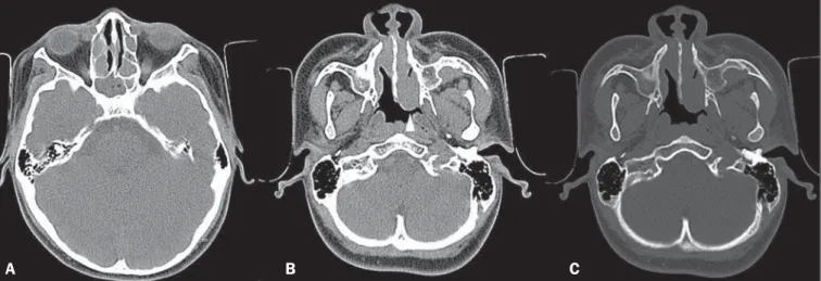
Here, we report the case of a 17-year-old male who presented with a three-month history of nasal obstruction, asthenia, and febrile episodes. Physical examination revealed bilateral enlarge-ment of cervical and axillary lymph nodes, all of which were pain-less on palpation. Laboratory tests showed mild leukocytosis, an elevated increased C-reactive protein level, and a high erythrocyte sedimentation rate. The venereal disease research laboratory test and monospot test were both negative, as was serology for HIV, toxoplasmosis, and cytomegalovirus. Computed tomography (CT) of the sinuses showed multiple, homogeneous, hypointense, rounded polypoid masses, which effectively narrowed the nasal
passages, together with opacification of the ethmoid cells and sphenoid sinuses, with no evidence of bone erosion (Figure 1). Biopsies of a cervical lymph node and nasal lesions were negative for neoplasia and acid-fast bacilli, showing diffuse lymphoplas-macytic infiltration, foamy histiocytes, and emperipolesis. Immu-nohistochemistry showed positivity for S-100 protein, positivity for CD68, and negativity for CD1a. A diagnosis of Rosai-Dorfman disease was made, and corticosteroid therapy was started, result-ing in slow, progressive improvement.
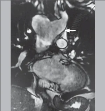
Recent studies in the radiology literature of Brazil have stressed the importance of CT and magnetic resonance imaging (MRI) in improving the diagnosis of head and neck masses(1–5). Rosai-Dorfman disease, also known as sinus histiocytosis with massive lymphadenopathy, is a rare, benign lymphoproliferative, usually self-limiting, condition characterized by bilateral, painless cervical Figure 1. A,B: Axial T2-weighted spin-echo magnetic resonance imaging
show-ing right aortic arch (arrow).
A
B
Figure 2. Coronal T2-weighted spin-echo magnetic resonance imaging showing Kommerell’s diverticulum (arrow).
Letters to the Editor
Radiol Bras. 2016 Jul/Ago;49(4):267–276
276
lymphadenopathy(6–11), with spontaneous resolution in approxi-mately half of all cases(7). The disease has a slight predilection for males and primarily affects children, adolescents, and young adults, 80% of cases occurring in individuals under 20 years of age(6). Extranodal involvement occurs in 30–40% of al cases(6,8,9), being most common in immunocompromised individuals and prefer-entially affecting the skin, respiratory tract, reticuloendothelial system, genitourinary tract, or bones(6,8,9). Although uncommon, enlargement of the mediastinal, hilar, axillary, and inguinal lymph nodes can occur.
The etiology of Rosai-Dorfman disease is unclear, although it could be related to changes in the immune response or to infec-tions caused by agents such as varicella-zoster virus and other herpes viruses, as well as Epstein-Barr virus, cytomegalovirus,
Brucella spp., and Klebsiella spp.(6,7,9,11).
Imaging tests such as CT and MRI are useful for evaluating the extent of Rosai-Dorfman disease, although there are no spe-cific characteristics. When it affects the paranasal sinuses, it typi-cally manifests as polypoid masses, mucosal thickening, with or without bone erosion, with preferential involvement of the maxil-lary sinuses and ethmoid cells(9,10). The diagnosis is established by histopathology(8).
The differential diagnoses include several types of lympho-reticular malignancy, such as lymphoma, malignant histiocyto-sis, and monocytic leukemia, which have histopathological fea-tures similar to those of Rosai-Dorfman disease but present aty-pia and a rapid, aggressive evolution. Another major differential diagnosis is Kikuchi-Fujimoto disease (histiocytic necrotizing lym-phadenitis), the clinical profile of which resembles that of Rosai-Dorfman disease, with cervical lymphadenopathy, although the former predominantly affects females and manifests as necrotiz-ing histiocytosis on histopathology(12,13).
In Rosai-Dorfman disease, the treatment modality of choice and the timing of treatment are controversial. Nevertheless, the choice of treatment strategies depends on the severity of the dis-ease, mild cases being managed with observation only, whereas cases that are more severe are typically managed with corticoster-oid therapy, chemotherapy, radiotherapy, or surgery(6–11).
In conclusion, although Rosai-Dorfman disease does not present specific imaging characteristics, it should be considered among the diagnostic possibilities in cases of painless bilateral cervical lymphadenopathy, particularly in children and adolescents.
REFERENCES
1. Niemeyer B, Marchiori E. Giant pilomatrixoma: conventional and diffu-sion-weighted magnetic resonance imaging findings [Letter]. Radiol Bras. 2015;48:63–4.
2. Niemeyer B, Salata TM, Antunes LO, et al. Desmoplastic fibroma with perineural spread: conventional and diffusion-weighted magnetic reso-nance imaging findings [Letter]. Radiol Bras. 2015;48:266–7. 3. Alfenas R, Niemeyer B, Bahia PRV, et al. Parry-Romberg syndrome:
findings in advanced magnetic resonance imaging sequences – case report. Radiol Bras. 2014;47:186–8.
4. Feres MFN, Hermann JS, Sallum AC, et al. Radiographic adenoid evalu-ation: proposal of an objective parameter. Radiol Bras. 2014;47:79–83. 5. Santos D, Monsignore LM, Nakiri LM, et al. Imaging diagnosis of dural
and direct cavernous carotid fistulae. Radiol Bras. 2014;47:251–5. 6. Pinto DCG, Vidigal TA, Castro B, et al. Doença de Rosai-Dorfman como
diagnóstico diferencial de linfadenopatia cervical. Rev Bras Otorrinola-ringol. 2008;74:632–5.
7. Oliveira CD, Gonçalves ACP, Moura FC et al. Acometimento orbitário na doença de Rosai-Dorfman. Rev Bras Oftalmol. 2011;70:46–50. 8. Pradhananga RB, Dangol K, Shrestha A, et al. Sinus histiocytosis with
massive lymphadenopathy (Rosai-Dorfman disease): a case report and literature review. Int Arch Otorhinolaryngol. 2014;18:406–8. 9. Akyigit A, Akyol H, Sakallioglu O, et al. Rosai-Dorfman disease
originat-ing from nasal septal mucosa. Case Reports in Otolaryngology. 2015; 2015:232898.
10. La Barge DV 3rd, Salzman KL, Harnsberger HR, et al.Sinus histiocy-tosis with massive lymphadenopathy (Rosai-Dorfman disease): imaging manifestations in the head and neck. AJR Am J Roentgenol. 2008;191: W299–306.
11. Maia RC, Meis E, Romano S, et al. Rosai-Dorfman disease: a report of eight cases in a tertiary care center and review of the literature. Braz J Med Biol Res. 2015;48:6–12.
12. Sah RP, Wilson ME, Seningen J, et al. Relapsing fevers and lymphad-enopathy in a young woman. BMJ Case Rep. 2013;2013. pii: bcr 2013200237.
13. Ramkumar A. Kikuchi-Fujimoto disease as a differential diagnosis for cervical lymphadenopathy in India: a case report and review of litera-ture. Indian J Otolaryngol Head Neck Surg. 2011;63(Suppl 1):110–2.
Bruno Niemeyer de Freitas Ribeiro1, Edson Marchiori2 1. Instituto Estadual do Cérebro Paulo Niemeyer, Rio de Janeiro, RJ, Brazil. 2. Universidade Federal do Rio de Janeiro (UFRJ), Rio de Janeiro, RJ, Brazil. Mailing address: Dr. Bruno Niemeyer de Freitas Ribeiro. Instituto Estadual do Cérebro Paulo Niemeyer – Serviço de Radiologia. Rua do Rezende, 156, Centro. Rio de Janeiro, RJ, Brazil, 20231-092. E-mail: [email protected].
Figure 1. A: Axial CT section without contrast, showing opacification of the sphenoid sinuses and ethmoid cells by hypointense material. B: Axial CT section without contrast, showing a homogeneous, hypointense polypoid formation (arrowhead) in the left nasal cavity. C: Axial CT section, with a bone window setting, showing that there is no associated bone erosion.