Letters to the Editor
Radiol Bras. 2016 Jan/Fev;49(1):56–64
57
http://dx.doi.org/10.1590/0100-3984.2014.0056 REFERENCES
1. Bioque JC, Feu N, Rubio JM, et al. Tracheobronchopathia osteochondro-plastica – clinical study and follow-up in nine cases. Journal of Bron-chology. 2001;8:78–83.
2. Pinto JA, Silva LC, Perfeito DJP, et al. Osteochondroplastic tracheo-bronchopathy: report on 02 cases and bibliographic review. Braz J Otorhinolaryngol. 2010;76:789–93.
3. Williams SM, Jones ET. General case of the day. Tracheobronchopathia osteochondroplastica. Radiographics. 1997;17:797–9.
4. Faig-Leite FS, Defaveri J. Traqueobroncopatia osteocondroplástica em portador de tumor de Klatskin: relato de caso e revisão da literatura. J Bras Patol Med Lab. 2008;44:459–62.
5. Sá JM, Almeida J, Amado J, et al. Traqueobroncopatia osteocondroplás-tica – experiência de uma unidade de broncologia. Rev Port Pneumol. 2002;VIII:329–39.
6. Webb EM, Elicker BM, Webb WR. Using CT to diagnose nonneoplas-tic tracheal abnormalities: appearance of the tracheal wall. AJR Am J Roentgenol. 2000;174:1315–21.
7. Prince JS, Duhamel DR, Levin DL, et al. Nonneoplastic lesions of the tracheobronchial wall: radiologic findings with bronchoscopic correla-tion. Radiographics. 2002;22 Spec No:S215–30.
8. Khan AM, Klapper P, Jain VR, et al. Tracheobronchopathia osteochondro-plastica: an entity diagnosed on bronchoscopy. Journal of Bronchology. 2006;13:99–101.
9. Kwong JS, Müller NL, Miller RR. Diseases of the trachea and main-stem bronchi: correlation of CT with pathological findings. Radio-graphics. 1992;12:645–57.
10. Marchiori E, Pozes AS, Souza Junior AS, et al. Alterações difusas da traquéia: aspectos na tomografia computadorizada. J Bras Pneumol. 2008;34:47–54.
11. Grenier PA, Beigelman-Aubry C, Brillet PY. Nonneoplastic tracheal and bronchial stenoses. Radiol Clin North Am. 2009;47:243–60.
Gabriela Maria Ribeiro e Ribeiro1, Marcelo Ricardo Canuto Natal1, Eduardo Felipe Silva1, Sabrina Cardoso Freitas1, Waldete Cabral Moraes1, Fernanda Cunha Maciel1
1. Hospital de Base do Distrito Federal (HBDF), Brasília, DF, Brazil. Mailing Address: Dra. Gabriela Maria Ribeiro e Ribeiro. Rua Gomes de Carvalho, 1005, ap. 3110, Vila Olímpia. São Paulo, SP, Brazil, 04547-004. E-mail: [email protected].
Giant pedunculated hemangioma of the liver
Hemangioma hepático gigante pedunculado
Dear Editor,
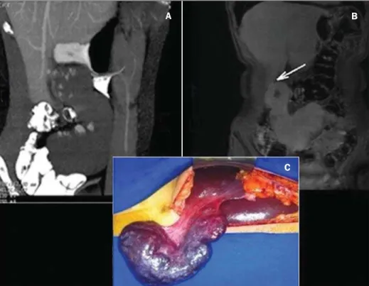
A previously healthy, 28-year-old woman presenting a palpable mass in the right hypochondrium for 3 years, evolving with local discomfort over the last 20 days. Ultrasonography (US) demon-strated an expansile mass best characterized by computed tomog-raphy (CT) and magnetic resonance imaging (MRI) which showed a well defined solid mass in continuity with the liver by a thin pedicle originating from the segment V and caudally extending towards the pelvis, measuring 18.0 × 9.4 × 5.2 cm, with features and pat-tern of enhancement suggestive of hemangioma (Figures 1A and 1B). Surgical resection was the treatment of choice because of the patient’s symptoms and the risks of torsion. The anatomo-pathological analysis confirmed the diagnosis (Figure 1C).
Hemangioma is the most common benign liver tumor(1–8),
with a prevalence of 0.4–20% in necropsies(1,5–8). In most cases,
hemangiomas are small, asymptomatic and incidentally found at imaging studies(1,2,5,6).
In spite of the lack of consensus about the dimensions to define a giant hemangioma, ranging from 4 to 10 cm according to the literature, it is known that the exophytic presentation, par-ticularly those pedunculated, are very rare (1–3,5,6). The first case
was reported by Ellis et al. in 1985; and up to 2013, only 24 cases were described in the literature(1,4).
In almost 50% of cases, pedunculated hemangiomas are symptomatic at the diagnosis(1) and, likely any giant lesion, may
determine compression of the intrahepatic biliary ducts, vascular structures or adjacent organs, manifesting with pain, early sati-ety, hemorrhage, jaundice, nausea and vomiting(1,2,5,6,8). Main
complications include torsion due to a long and mobile pedicle,
Figure 1.A: Contrast-enhanced total abdominal CT (oral and intravenous contrast-enhancement), sagittal section, venous phase showing a well defined mass in the right hypochondrium/flank in continuity with the liver, presenting with a pattern of peripheral, globuliform and centripetal enhance-ment, with a thin pedicle originating from the seg-ment V. B: Coronal MRI, T1-weighted SPGR, at delayed phase showing homogenization of the lesion and identifying a pedicle contiguous with the liver parenchyma (arrow). C: Surgical speci-men of the reddish blue pedunculated lesion with cirrhotic appearance, showing pedicle contiguous with the liver parenchyma.
B
Letters to the Editor
Radiol Bras. 2016 Jan/Fev;49(1):56–64
58
http://dx.doi.org/10.1590/0100-3984.2014.0057
infarction(5,6), spontaneous or traumatic rupture, congestive heart
failure, and Kasabach-Merritt syndrome(2,6,7).
A correct diagnosis of the pedunculated lesion may be diffi-cult, despite the typical radiological presentation, because of the limitation in define the origin of the mass, since a thin pedicle may be almost undetectable at images(1,4,5).
The most used modalities of imaging in diagnosis include US, CT and MRI(1–4,6,8). At US, the image is typically hyperechoic,
homogeneous, with well defined margins; and, in cases of giant lesions, central heterogeneity may be present(8). At CT, with a
certain frequency, giant hemangiomas do not present with the typical pattern of hypoattenuating lesion with centripetal enhance-ment and homogenization at delayed sections, due to the pres-ence of avascular areas of necrosis, fibrosis or hemorrhage(3,8).
MRI is the most sensitive and specific (> 90%) diagnostic method(4,6). The lesions are well defined, homogeneous, with low
signal intensity at T1-weighted sequences, and high signal in-tensity at T2-weighted sequences.
Biopsy is not recommended in such cases, due to the risk of hemorrhage(6).
There are reports in the literature describing pedunculated hemangiomas as gastric, adrenal tumor(1,4), retroperitoneal
mass(1), other pedunculated liver tumors such as hepatocellular
carcinoma, mesenchymal hamartoma, focal nodular hyperplasia or adenoma(4).
Surgical treatment is reserved for cases of giant or symp-tomatic lesions, uncertain diagnosis, lesions with complica-tions(1,2,4–7), and for cases of pedunculated hemangiomas due
to their tendency to torsion(5,6).
REFERENCES
1. Ha CD, Kubomoto SM, Whetstone BM, et al. Pedunculated hepatic he-mangiomas often misdiagnosed despite their typical findings. The Open Surgery Journal. 2013;7:1–5.
2. Moon HK, Kim HS, Heo GM, et al. A case of pedunculated hepatic he-mangioma mimicking submucosal tumor of the stomach. Korean J Hepatol. 2011;17:66–70.
3. Choi BI, Han MC, Park JH, et al. Giant cavernous hemangioma of the liver: CT and MR imaging in 10 cases. AJR Am J Roentgenol. 1989;152: 1221–6.
4. Liang RJ, Chen CH, Chang YC, et al. Pedunculated hepatic heman-gioma: report of two cases. J Formos Med Assoc. 2002;101:437–41. 5. Ersoz F, Ozcan O, Toros AB, et al. Torsion of a giant pedunculated liver
hemangioma mimicking acute appendicitis: a case report. World J Emerg Surg. 2010;5:2.
6. Guenot C, Haller C, Rosso R. Hémangiome caverneux pédiculé géant du foie: à propôs d’un cas et revue de la littérature. Gastroenterol Clin Biol. 2004;28:807–10.
7. Acharya M, Panagiotopoulos N, Bhaskaran P, et al. Laparoscopic resec-tion of a giant exophytic liver haemangioma with the laparoscopic Habib 4× radiofrequency device. World J Gastrointest Surg. 2012;4:199–202. 8. D’Ippolito G, Appezzato LF, Ribeiro ACR, et al. Apresentações incomuns do hemangioma hepático: ensaio iconográfico. Radiol Bras. 2006;39:219– 25.
Paula de Castro Menezes Candido1, Izabela Machado Flores Pereira1, Breno Assunção Matos1, Mario Henrique Giordano Fontes1, Teófilo Eduardo de Abreu Pires1, Petrônio Rabelo Costa1
1. Hospital Felício Rocho, Belo Horizonte, MG, Brazil. Mailing Address: Dra. Paula de Castro Menezes Candido. Hospital Felício Rocho – Setor de Radiologia. Avenida do Contorno, 9530, Barro Preto. Belo Horizonte, MG, Brazil, 30110-934. E-mail: [email protected].
Posterior reversible encephalopathy syndrome following immunoglobulin therapy in a patient with Miller-Fisher syndrome
Síndrome encefálica reversível posterior em paciente com síndrome de Miller-Fisher pós-tratamento com imunoglobulina
Dear Editor,
A 54-year-old female patient presenting with ophthalmopa-resis, ataxia and areflexia for one week. The patient denied fever, muscle weakness, and did not report any previous comorbidity. At physical examination, she was normotensive, oriented, with bilat-eral flexor cutaneous-plantar reflex and preserved superficial/deep sensitivity. Human immunodeficiency virus, Epstein-Barr virus, cytomegalovirus, HTLV-1 and VDRL serologies were negative. Considering such findings, the hypothesis of Miller-Fisher
syn-drome was raised, and liquor cerebrospinalis analysis demonstrated hyperproteinorachia, confirming the diagnosis.
Within 24–48 hours after immunoglobulin therapy initiation, the patient presented with intense headache followed by tonic-clonic seizures and later decreased level of consciousness, with no asso-ciation with hypertensive peaks. Magnetic resonance imaging (MRI) (Figure 1A,B,C) showed sparse hyperintense areas in the white substance, bilaterally on T2-weighted and FLAIR sequences, predominantly in the parieto-occipital regions, without diffusion restriction and without gadolinium enhancement, demonstrat-ing an imagdemonstrat-ing pattern suggestive of posterior reversible encepha-lopathy syndrome (PRES). After the therapy suspension and adop-tion of support measures, the patient progressed satisfactorily, with no sequelae and reversion of the MRI findings (Figure 1D).
Figure 1.A: Axial MRI FLAIR sequence demonstrating hyperintensity in the occipital lobes white substance bilaterally and symmetrically (arrows). B: Axial diffusion-weighted MRI does not demonstrate any alterations (arrows). C: Contrast-enhanced T1-weighted sequence revealing absence of gadolinium-enhanced areas (arrows).
D: Axial FLAIR sequence acquired after four weeks demonstrating resolution of the alterations in the occipital lobes white substance (arrows).