STREPTOZOTOCIN- INDUCED DIABETES DUR
STREPTOZOTOCIN- INDUCED DIABETES DURA
ATION IS
TION IS
IM POR
IM POR T
TANT TO DETERM INE CHANGES IN THE NUM BER
ANT TO DETERM INE CHANGES IN THE NUM BER
AND BASOPHIL
AND BASOPHILY OF M YENTERIC NEURONS
Y OF M YENTERIC NEURONS
LUZMARINA HERNANDES* , ROBERTO BARBOSA BAZOTTE* * , PATRÍCIA GAMA* * * , MARCÍLIO HUBNER DE MIRANDA-NETO*
ABSTRACT - The aim of present study was to evaluate the number and basophily of cell bodies of myenteric neurons in the ileum of rats with diabetes mellitus induced by streptozotocin. Four groups of rats were used: diabetes was induced in two (D) whereas the other two worked as controls (N). Animals were sacrificed six (6N, 6D) or nineteen (19N, 19D) weeks after diabetes induction. A segment of the terminal portion of the ileum of each rat was obtained and stained with Giemsa’s solution, for whole-mount preparation studies. Forty fields were analyzed in each animal, and the number and basophily intensity of cell bodies were recorded. After counting, the following mean numbers of neurons/mm2 were obtained: 6N=593.1 ± 95.75, 6D=639.1 ± 130.8, 19N=580.1 ± 175.6 and 19D=402.0 ± 144.8. The analysis of basophily shown that highest frequency of neurons with weak/ intermediary basophily was verified in 6D group (55.3%), whereas the groups 6N, 19N e 19D presented 38%, 36% e 40% respectively. The statistical analysis showed that a long period is necessary to decrease the number of neurons/mm2 in the rat ileum after diabetes induction, and that there was a reduction in basophily intensity in diabetic rats after 6 weeks of treatment, and such cells do not recover after a longer period (19 weeks).
KEY WORDS: myenteric plexus, streptozotocin, diabetes mellitus, ileum.
A duração do diabetes induzido por estreptozootocina é importante para determinar as mudanças no número e basofilia dos neurônios mientéricos
RESUMO - O objetivo deste estudo foi avaliar o número e basofilia dos corpos celulares dos neurônios mientéricos no íleo de ratos com diabetes mellitus induzido por estreptozootocina. Quatro grupos de ratos foram usados. O diabetes foi induzido em dois grupos (D), enquanto outros dois eram controles (N). Os animais foram sacrificados 6 (6N, 6D) ou dezenove (19N, 19D) semanas após a indução do diabetes. Preparados totais de um segmento do íleo terminal, de cada rato, foram corados com solução de Giemsa. Foram contados 40 campos em cada animal, e o número e a intensidade de basofilia citoplasmática foram registrados. Após a contagem, as seguintes médias no número de neurônios/mm2 foram obtidos: 6N=593,1 ± 95,75, 6D=693,1 ± 130,8, 19N=580,1 ± 175,6 e 19D=402,0 ± 144,8. A análise da basofilia mostrou que a maior frequência de neurônios com basofilia fraca e intermediária foi verificada no grupo 6D (55,3%), enquanto os grupos 6N, 19N e 19D apresentaram 38%, 36% e 40% respectivamente. A análise estatística mostrou que um longo período é necessário para que ocorra a redução no número de neurônios/mm2no íleo de ratos, após a indução do diabetes. Também demonstrou uma redução na intensidade da basofilia citoplasmática 6 semanas de tratamento com estreptozootocina, e que estas células não se recuperam após um longo período de tempo (19 semanas).
PALAVRAS-CHAVE: plexo mientérico, estreptozootocina, diabetes mellitus, íleo.
Among the many effects of diabetes in the human gastrointestinal tract, those which affect the intestine are of major importance to the patient1. Diabetic diarrhea, and intestinal constipation
*Professor, Department of Morphophysiological Sciences, Universidade Estadual de Maringá; **Professor, Department of Pharmacology, Universidade Estadual de Maringá; ***Professor, Department of Histology and Embriology, Universidade de São Paulo. Aceite: 13-julho-2000.
are the most frequent symptoms, and autonomic neuropathy is a probable cause1-3. However, no clear association has been established between gastrointestinal dysfunction and diabetes4. Usually, the morphological alterations of the autonomic nervous system in diabetes, either in rats or humans, are reported to be related to degenerative processes observed in the nervous fibers and their myelin sheaths5-7. Electrophysiological and morphological studies suggest that the axon is the initial site of damage in the peripheral nerve8.
The morphology of the intramural plexus has been poorly studied in diabetic patients9-11. Ultrastructural analysis did not show significant changes in the acute stage of the disease in diabetic rats. However, degenerative alterations were reported in axons, from 7 days to 6 weeks of diabetes induction. By the end of this period, some axonal regeneration was observed5.
In response to axonal injury, the perikaryon of the lesioned neuron undergoes a cascade of biochemical and physiological processes, and one of these cellular reactions is chromatolysis, i.e. the dispersion, and later redistribution of Nissl bodies. Morphologically, such process represent the disintegration of the granular endoplasmic reticulum, and functionally, the transformation of the polyribosome signal to transmiter-related proteins and membrane-associated or cytoskeletal proteins12. In simpathetic ganglia of diabetic rats, chromatolysis becomes apparent as a degenerative change, two weeks after the induction of the disease by streptozotocin5. Immunohistochemical techniques have revealed differential responses to diabetes in nerves containing neuropeptides in enteric plexuses of streptozotocin-diabetic rats4,13. Such results have not yet elucidated the real origin of the neuropathy, but demonstrated that the diabetes-induced neuropathy “is not selective, and it involves other factors, besides neurotransmitters” 13.
Taking into account the diabetic complications on sympathetic and parasympathetic divisions of the autonomic nervous system and peripheral nerves, as well as the lack of studies on the morphological abnormalities of the myenteric plexus in rats with induced diabetes, this study aims to evaluate the number and the cytoplasmic basophily of the cell bodies of myenteric neurons in rats with diabetes mellitus induced by streptozotocin, 6 and 19 weeks after its induction.
M ETHOD M ETHOD
Animal treatment
Twenty male Wistar rats (70-d-old, around 200-250 g) were used from the Central Biotery of Universidade Estadual de Maringá. Ten animals received a single i.v. streptozotocin injection (35 mg/Kg b.w., Sigma, USA) and ten animals were used as control counterparts. They were all distributed in 4 groups of five rats as follows: 6D (sacrificed 6 weeks after diabetes induction) 6N (control group); 19D (sacrificed 19 weeks after diabetes induction; 19N (control group). Animals received water and chow (Nuvital®) ad libitum.
All animals were anesthetized with thyonembutal (35 mg/kg) (Abbott®). Blood glucose was measured by the glucose-oxidase method14, which was performed immediately after sacrifice, in order to confirm that a diabetic condition had been induced.
A midline abdominal incision was made and the small intestine was removed. After washing the lumen with 0.9% saline, the intestine was distended for 1 minute in Giemsa’s fixative solution. For that procedure, a tension of 5g was applied at distal end and the intestine was filled with a fixative solution. The volume was enough to induce a mild distension of the intestinal wall, similar to that promoted by the alimentary bolus. Afterwards, the ileum was taken (based on ileum-caecal junction), and the final 3cm were discarded.
Analysis of cytoplasmic basophily
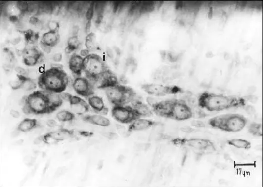
Six hundred neurons (150 from each group) were classified according to cytoplasmic staining intensity, which is determined by the affinity of Nissl bodies to Giemsa’s liquid. Neurons presenting dark homogenous blue staining were classified as deeply basophilic; those which showed many basophilic granules interspersed with weakly stained areas were considered to have intermediary basophily; neurons which presented few basophilic granules were classified as weakly basophilic.
The chi-square test was used to compare the basophily in neurons. In this analysis, the intermediary and weakly basophilic neurons were grouped in a single category, which was tested against the deeply basophilic group. Significance level was set at p<0.05.
Quantitative analysis
The cell bodies of myenteric neurons were counted by sampling under light microscope (Olympus CBB, using a Bausch & Lomb WF x10/18 eyepiece and x40 objective). To determine the microscopic area, we considered the retraction calculated and shown above. Each preparation was visually divided into quadrants, in which fields were counted at random. Myenteric neurons were identified and counted in these fields. Half-seen neurons were counted on alternate fields. In that way, forty fields (0.166 mm2 each) were quantified in different ileum whole-mount preparations, making up a total area of 6.64 mm2 for each animal. Results are expressed as neurons/mm2. Student’s t test was used to compare the number of neurons between the groups and significance level was set at p<0.05.
RESUL
RESULTSTS
Diabetic condition - The concentration of glucose in blood was around 130mg/dl in control
rats, whereas in the streptozotocin-treated group it was at 400 mg/dl, assuring the diabetic condition.
Cytoplasmic basophily - The basophily in the cell bodies of myenteric neurons was visualized
as shown in Figure 1. Table 1 presents the results on the frequencies of deeply and intermediary/
weakly basophilic neurons. The χ2 test showed that the 6D group is significant different from the others.
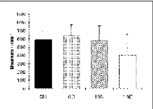
Number of myenteric neurons - Figure 2 shows the mean number of cell bodies of ileum
myenteric neurons. Rats from 6N group showed a mean number of 593.1 ± 95.75 neurons/mm2, whereas group 6D had 639.1 ± 130.8 neurons/mm2. In the 19N and 19D groups, the mean numbers of neurons were 580.1± 175.1 and 402.0 ± 144.8 neurons/mm2, respectively. Significant differences (P<0.05) were found between groups 6D and 19D.
DISCUSSION DISCUSSION
The Giemsa’s staining technique used in this study allowed us to classify the cell bodies of myenteric neurons according to cytoplasmic staining intensity, which was determined by the affinity of the rough endoplasmatic reticulum to the stain. Thus, we found the frequency of neurons based on the cytoplasmic basophily in the different experimental groups.
The frequency of basophilic neurons in 6D group was significantly different when compared to the other groups. Our results indicated that these neurons have probably begun a chromatolysis process, without going through degeneration, as shown by the maintainance of the number of neurons per mm2. The disappearance of the chromatolytic substance could indicate a decline of cell metabolism in the acute stage of the disease16. Although chromatolysis typical features have not been observed in the 6th week of diabetes, the neurons possibly did not re-establish their Nissl bodies, as shown by the cytoplasmic basophily. On the other hand, the low frequency of intermediary/weakly basophilic neurons in the 19D group should not be regarded as a recovery of cytoplasmic basophily, since we have to consider that by this age, the total number of neurons per mm2 has been reduced by the treatment.
The statistical analysis of our results did not show significant differences in the number of neurons of 6N and 6D groups, suggesting that the streptozotocin-induced diabetes did not alter the number of neurons per mm2 in the ileum of the rats. These results also indicated that there was no neuronal loss related to the toxicity of streptozotocin in this period, although it is a critical stage when the diabetes inducing drug could lead to neuron degeneration5.
By comparison of the mean number of neurons from 6N group to the 19N group, no significant difference was found. This result suggests that the aging process in non-diabetic animals is not enough to induce a decrease in the number of myenteric neurons per mm2 in the ileum after 19 Table 1. Frequency of basophilic myenteric
neurons in the ileum of diabetic (6D, 19D) and control rats (6N, 19N). Basophily was classified as weak/intermediary and deep (100%=22.6 neurons/mm2).
Basophily
Groups Weak/Intermediary Deep
6N 38.0% 62.0%
6D* 55.3% 44.7%
19N 36.0% 64.0%
19D 40.0% 60.0%
* P< 0.05 when compared to 6N and 19D groups
Fig 2. Number of myenteric neurons/mm2 observed in the ileum
weeks. On the other hand, when 19D group was compared to 6D group, there was a significant reduction in the number of myenteric neurons per mm2 in the ileum, showing that the pathophysiological mechanisms of this disease probably accelerate the neuronal loss in the myenteric plexus17.
It should be considered that the diabetes mellitus induces pathological complications such as cardiovascular diseases and arteriosclerosis, which are similar to what happens during aging18. Aging itself is correlated with a decrease in the number of neurons in both small and large intestines19-21. We found a 30-37% reduction in myenteric neurons in the ileum, by comparing 19D group to the others. In rats, comparative values of 40, 64 and 42% were observed, in the small intestine, colon and rectum, respectively21, whereas reductions of 40 and 60% were found in the small intestine of old guinea-pigs, in relation to the young ones20.
Although the difference in the number of myenteric neurons per mm2 in the ileum of the 19N and 19D rats did not reach the significance level, it is very close to it. This fact suggests that the smaller number of neurons is related to the pathological condition, and not to the age.
We conclude that: a) the streptozotocin-induced diabetes leads to the decrease of cytoplasmic basophily of myenteric neurons in ileum of the 6D group; b) intermediary and weakly basophilic neurons are major lost along the treatment; c) chronic diabetes leads to a significant reduction in the number of neurons per mm2 in the ileum.
We believe that the current results can be added to other studies, which worked with different techniques, but that also aimed to find the major factors that contribute to the development of neuropathy, either in clinical or experimental diabetes.
REFERENCES REFERENCES
1. Katz LA, Spiro HM. Gastrointestinal manifestations of diabetes. N Engl J Med 1966;275:1350-1361.
2. Clements RS Junior, Bell DSH. Diabetic neuropathy: peripheral and autonomic syndromes. Diabetic Neuropathy 1982;71:50-67.
3. Hosking DJ, Bennet T, Hampton JR. Diabetic autonomic neuropathy. Diabetes 1978;27:1043-1055.
4. Lincol J, Bokor JT, Crowe R, Griffith SG, Haven A J, Burnstock G. Myenteric plexus in streptozotocin-treated rats: neurochemical and histochemical evidence for diabetic neuropathy in the gut. Gastroenterology 1984;86:654-661. 5. Monckton G, Pehowich E. Autonomic neuropathy in the streptozotocin diabetic rat. J Can Sci Neurol 1980;7:135-142. 6. Powell HC, Lee S, Orloff MJ, Lampert PW. Alloxan diabetic neuropathy: electron microscopic studies. J Neuropathol Exp
Neurol 1976;35:335.
7. Yasaki S, Dyck PJ. Duration and severity of hypoglycemia needed to induce neuropathy. Brain Res 1990;531:8-15. 8. Clements RS Junior. Diabetic neuropathy: new concepts of its etiology. Diabetes 1979;28:604-611.
9. Berge KG, Sprague RG, Bennett WA. The intestinal tract in diabetic diarrhea. Diabetes 1956;5:289-294.
10. François R, Mouriquand C. Diarrhée incoercible et fatale chez un jeune diabétique: étude des plexus myentériques. Sem Hop Paris1958;34:1526-1531.
11. Hensley GT, Soergel KH. Neuropathologic findings in diabetic diarrhea. Arch Pathol 1968;85:587-597.
12. Liberman AR. The axon reaction: a review of the principle features of responses to axonal injury. Int Rev Neuro 1971;14:49-124.
13. Belai A, Burnstock G. Changes in adrenergic and peptidergic nerves in the submucous plexus of streptozotocin-diabetic rat ileum. Gastroenterology 1990;98:1427-1436.
14. Bergmeyer HU, Bernet E. Determination of glucose with glucose-oxidase and peroxidase. In. Methods of enzymatic analysis, New York: Verlag Chemie-Academic Press, 1974.
15. Barbosa AJA. Técnica histológica para gânglios nervosos intramurais em preparados espessos. Rev Bras Pesq Méd Biol 1978;11:95-97.
16. Spence AP. Biology of human aging. New Jersey: Prentice-Hall, 1995.
17. Belai A, Facer P, Bishop A, Polak JM, Burnstock G. Effect of streptozotocin-diabetes on the level of VIP mRNA in myenteric neurones. NeuroReport 1993;4:291-294.
18. Kristal BS, Yu BP. An emerging hypothesis: synergistic induction of aging free radicals and maillard reactions. J Gerontol 1992;47:107-114.
19. Gabella G. Neuron size and number in the myenteric plexus of the newborn and adult rat. J Anat 1971;109:81-95. 20. Gabella G. Fall in the number of myenteric neurons in aging guinea-pigs. Gastroenterology 1989;96:1487-1493. 21. Santer RM, Baker DM. Enteric neuron numbers and sizes in Auerbach¢s plexus in the small and large intestine of adult and